

![]()
Hemoglobin adducts in paint industry workers: An electrophoretic analysis
Sumera Qureshi1*, Sikander Ali Memon1, Allah Bux Ghanghro1, Muhammad Faisal Qureshi2, Moina Akhtar Mughal2, Tahira Qureshi2
Adv. life sci., vol. 1, no. 4, pp. 208-214, August 2014
*- Corresponding Author: Sumera Qureshi (Email: s.dr582@gmail.com)
Author Affiliations
2- Mining Engineering department, Mehran University of Engineering & Technology, Jamshoro – Pakistan
Abstract![]()
Introduction
Methods
Results
Discussion
References
Abstract
Background: Hemoglobin (Hb) has a significant role among other blood proteins vital for carrying nutrients to blood cells. Being a conjugated protein, Hb is prone to be captured by compounds of low molecular weight like organic acid anhydrides (OAAs) which are prominent industrial/occupational hazards. Hindered or lowered availability of Hb to blood cells can cause anemia, thalassemia and porphyria. Along with these disorders, workers exposed to OAAs can also acquire like type-I allergy, type-IV allergy, skin problems, rhinitis and asthma. Revelation of Hb-OAAs compounds prior to appearance of actual symptoms could be important for subsequent therapy.
Methods: The Hb separation was achieved successfully by SDS-PAGE electrophoresis on 10-15% gels of different concentration, stained with CBB-R250 Blue. Total of 66 blood protein samples were used for the comparative study of exposed workers of paint industry workers with control (normal) group to detect proteins, which might serve as marker for the early disease diagnosis.
Results: The better Hb separation resolution was achieved on 12% gel as shown in electrograms. The electrograms of paint workers exposed to OAAs showed bands at 12, 48, 66, 78, 128 and 132 KDa in most of cases. In normal cases the bands were found at 13, 30, 48, 67, 76, 125 and 155 KDa in majority of control samples for Hb electrophoresis.
Conclusion: This study supports the association between Hb and OAAs adducts among the exposed paint workers from hypersensitive effects like fever (rhinitis) leading to asthma, skin allergies and major clinical effects.
Key words: Hemoglobin (Hb), Organic acid anhydrides (OAAs), SDS-PAGE electrophoresis
Introduction
Oxygen carries hemoglobin (Hb) protein in red blood cells of all livings [1]. The vital role of iron in Hb is related with metabolism of several neurotransmitters, synthesis of gamma amino-butyric acid (GABA), serotonin and dopamine [2,3]. Iron deficiency anemia is a common global disorder (found in almost every age group from children to elders) in Pakistan [4-6]. Hence Hb protein plays a vital role in metabolism is expected to be disturbed by exposure of low organic acid anhydrides upon industrial workers by forming adducts (complex) reported earlier [7-9]. Proteins like Hb-OAAs conjugates adducts found immunologically active in previous studies done. Such conjugation of Hb adducts formed in vitro by OAAs was even confirmed with N-terminal valine and lysine residues of α and the β-chains characterize the sites of adduct formation [10,11]. Although Rosqvist S et al studied total plasma protein adducts of workers exposed for about months by GC-MS analysis in the negative ion chemical ionization mode [9].
These findings suggest a possible role of Hb in blood to find out impact of iron deficiency anemia in relation to workers compared with control group. It is even reported earlier that more than 60% of exposed workers to these reactive chemicals may develop airways infections and allergy. Different types of allergy like type-I allergy and type IV allergy are most common forms induced by such small reactive organic chemicals. These low molecular weight organic chemicals have adverse health effects even at lower exposure [12-14].
The main aim of the study was to find out the relation between level of Hb protein and OAAs by analyzing blood of workers exposed to anhydrides in industrial settings. The blood samples for Hb of both workers and control (normal) groups were compared for this purpose.
Methods
The study included a total of 66 blood samples in which 36 blood samples were taken from workers employed at local paint manufacturing industry, SITE Area, Kotri, using OAAs and 30 from normal (control) of same age group. These workers were exposed to phtalic anhydride (PA), succinic anhydride (SA) and acetic anhydride (AA) mainly by vapors inhalation. This research work was carried out to analyze Hb protein separated by SDS-PAGE and stained with Coomassie brilliant blue, to detect proteins, which could possibly serve as markers for the early detection of allergy and hypersensitivity problems among exposed workers to OAAs. All of the workers had been exposed for about 10-12 years as mentioned in questionnaire taken by physician.
The main objective of this study was to fractionate and identify blood plasma proteins responsible for work related sensitization and respiratory problems after forming adducts (complex) as reported earlier by Rosqvist et al., [9]. Average age of the 36 paint industry workers was 28.8 years while group of 30 normal persons had average age 28.6 years.
A. Blood Sample Collection:
This study was conducted from January 2013 to March 2013. The paint industry workers gave consent for giving their samples by their own free will and recorded in confidential files. The intravenous blood samples (10 ml) were collected in EDTA tubes and stored at 400C prior to SDS-PAGE Hb analysis.
B. Reagents for SDS-PAGE:
All reagents are standard chemicals of high purity analytical graded, and purchased from Sigma Aldrich and Merck (Germany).
Preparation of Solutions: All of the solutions of above mentioned chemicals were freshly prepared according to the standard protocol method of Lammeli gel preparation [15].
Sample preparation for hemoglobin SDS-PAGE
A. Serum Separation
Mix 100 µl of whole blood with 400 µl of an isotonic solution (0.100 M NaCl) in a 500 µl microcentrifuge tube and centrifuge for 2 minutes at 6500 RCF (approximately) to pellet the red blood cells. The blood serum is supernatant, which is transferred to another 500 µl microcentrifuge tube and saved.
B. Hb extraction from Red Blood Cells
The pellet of red blood cells is re-suspended in 400 µl of a hypotonic solution (0.065 M KCl) and vortexed vigorously to lyse all cells. The lysed red blood cell solution was then centrifuged at 6500 RCF (approximately) for 10 minutes to remove any undissolved materials.
Sample preparation for protein samples:
20 µl of protein sample was added into 20 µl of sample buffer (0.5 M tris HCl, pH 8), 30% Glycerol, 5% SDS, 0.06% BPB and 2-Mercaptoethanol was boiled in tube for about 5 min. 10 µl of sample was applied on sample per well along with molecular weight marker sample.
Gel staining and decolorization
Gel was placed into staining gel solution (0.25%, CBB-R 250) for approx. 35-40 minutes with constantly shaking the gel. Finally the gel was washed with water and poured into decolorizing solution (7% acetic acid, 5% Methanol) for decolouring the gel.
SDS-PAGE electrophoresis for Hb adducts
Electrophoresis apparatus was fixed with glass plate and filled the upper tank with SDS running buffer. Then sample was loaded with molecular weight marker (10-180 KDa) Fermentus pre staining protein ladder. After loading samples the power supply at 30 mA (300V Constant current) samples were running for about 6 hrs. SDS-PAGE Electrophoresis will be stopped running when PBS line reached up to lower part of the gel.
Data analysis
Gel reading and calculations was done by Gel documentation system of Bio-Rad for studying variations between workers and normal protein samples.
Results
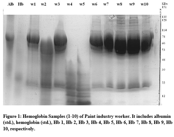
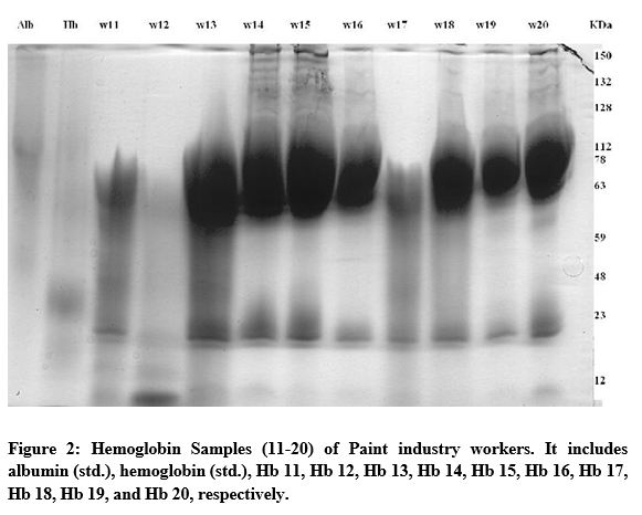
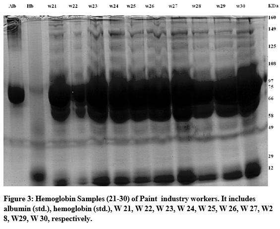
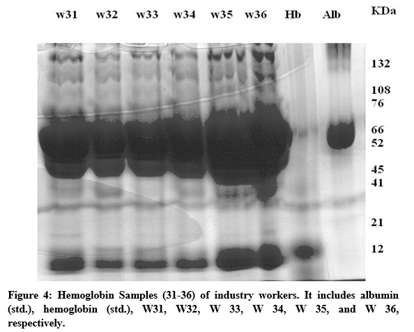
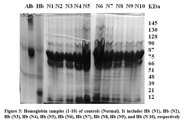
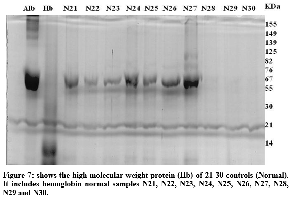
In case of Hb electrophoresis, includes a total of 66 samples, 36 exposed workers to OAAs samples and 30 controls, aged (20-60 years) for both groups (4-15%) on SDS-PAGE Electrophoresis. Our study was simple, reliable as confounding by paint industry worker class (exposed to OAAs) compared with the control group for explanation of differences in Hb protein status. The better Hb separation resolution was achieved on 12% gel as shown in electrograms (figures 1-4). The Protein ladder of Marker comes as 10.17, 26, 34, 43, 55, 72, 95, 130 and 170 bands range as reference (standard marker) in each case. The plasma protein resolved into clear broad bands and smears at 10-160 KDa in both groups were studied along with Hb standard (as shown in figures 1-7).The electrograms of paint workers exposed to OAAs (figure1-3) showing bands at 12, 48, 66, 78, 128 and 132 KDa in most of cases. In case of normal cases the bands were appeared at 13, 30, 48, 67, 76, 125 and 155 KDa (as shown in figures 5-7).
Data & Figures

Discussion
This study has established the fact the workers in paint industry in Pakistan are prone OAA-Hb adducts, which could have serious effects on their health. Study of these chemical adducts cases would produce significant information on allergenic proteins. However, many studies were done previously just to investigate human Hb adducts formed with strong Hexahydrophthalic anhydride (HHPA) in-vitro reaction for chemical structures [13].
It is suggested that detection of Hb variants may also be performed by other methods of protein study like Capillary Electrophoresis (CE) and CE-HPLC for discovery of new variants of clinical interest, may give better explanation of several Hb abnormalities by interaction with low molecular weight haptens like OAAs. The blood iron Hb status is also need to be checked out with Hb disorder such as anemia leading to other diseases among exposed workers. Our electrophoretic study supports the association between Hb and OAAs among the exposed paint workers, but pathophysiological mechanism involved is still unknown [15]. In our research observation, the population of paint workers exposed to OAAs chemicals were with iron deficiency anemia as compared to the control group that never exposed to hazardous OAAs. It was even noted that the workers with iron deficiency anemia suffering from hypersensitive effects like fever (rhinitis) leading to asthma, skin allergies and major clinical effects [10].
Our electrophoretic study supports the association between Hb and OAAs among the exposed paint workers, but pathophysiological mechanism involved is still unknown. It was even noted that the workers with iron deficiency anemia suffering from hypersensitive effects like fever (rhinitis) leading to asthma, skin allergies and major clinical effects as in accordance with previous studies [13].
References
- Kakar S, Hoffman FG, Storz JF, Fabian M, Hargrove MS. Structure and reactivity of hexacoordinate hemoglobins. Biophysical chemistry, (2010); 152(1): 1-14.
- Amiri M, Farzin L, Moassesi ME, Sajadi F. Serum trace element levels in febrile convulsion. Biological trace element research, (2010); 135(1-3): 38-44.
- Bianco L, Unger E, Beard J (2010) Iron deficiency and neuropharmacology. Iron Deficiency and Overload: Springer. pp. 141-158.
- Baker RD, Greer FR. Diagnosis and prevention of iron deficiency and iron-deficiency anemia in infants and young children (0–3 years of age). Pediatrics, (2010); 126(5): 1040-1050.
- Huma N, Salim-Ur-Rehman, Anjum FM, Murtaza MA, Sheikh MA. Food fortification strategy—preventing iron deficiency anemia: a review. Critical reviews in food science and nutrition, (2007); 47(3): 259-265.
- Beard JL. Iron biology in immune function, muscle metabolism and neuronal functioning. The Journal of nutrition, (2001); 131(2): 568S-580S.
- Kristiansson MH, Jönsson BA, Lindh CH. Mass spectrometric characterization of human hemoglobin adducts formed in vitro by hexahydrophthalic anhydride. Chemical research in toxicology, (2002); 15(4): 562-569.
- Richter E, Branner B. Biomonitoring of exposure to aromatic amines: haemoglobin adducts in humans. Journal of Chromatography B, (2002); 778(1): 49-62.
- Rosqvist S, Johannesson G, Lindh CH, Jönsson BA. Total plasma protein adducts of allergenic hexahydrophthalic and methylhexahydrophthalic anhydrides as biomarkers of long-term exposure. Scandinavian journal of work, environment & health, (2001); 133-139.
- Zhang X-D, Welinder H, Jönsson BA, Skerfving S. Antibody responses of rats after immunization with organic acid anhydrides as a model of predictive testing. Scandinavian journal of work, environment & health, (1998); 220-227.
- Nielsen J, Welinder H, Jönsson B, Axmon A, Rylander L, et al. Exposure to hexahydrophthalic and methylhexahydrophthalic anhydrides—dose-response for sensitization and airway effects. Scandinavian journal of work, environment & health, (2001); 327-334.
- Nielsen J, Welinder H, Bensryd I, Rylander L, Skerfving S. Ocular and airway symptoms related to organic acid anhydride exposure–a prospective study. Allergy, (2006); 61(6): 743-749.
- Wernfors M, Nielsen J, Schütz A, Skerfving S. Phthalic anhydride-induced occupational asthma. International Archives of Allergy and Immunology, (1986); 79(1): 77-82.
- Venables KM. Low molecular weight chemicals, hypersensitivity, and direct toxicity: the acid anhydrides. British journal of industrial medicine, (1989); 46(4): 222-232.
- Cleveland DW, Fischer SG, Kirschner MW, Laemmli UK. Peptide mapping by limited proteolysis in sodium dodecyl sulfate and analysis by gel electrophoresis. Journal of Biological Chemistry, (1977); 252(3): 1102-1106.


