

Full Length Research Article
Rhizobacterial mediated antimetabolites and their significant effect on human pathogens
Unaiza Malik, Atia Iqbal*, Sadia Batool
Adv. life sci., vol. 7, no. 3, pp. 140-145, May 2020
*- Corresponding Author: Khushnood ur Rehman (Email: atia.iqbal@wum.edu.pk)
Authors' Affiliations
Abstract![]()
Introduction
Methods
Results
Discussion
References
Abstract
Background: The emergence of antibiotic resistant bacteria is increasing globally. Therefore, new strategies and exploration of new metabolites are need of the day to combat various diseases. The aim of the current study is to isolate the rhizobacterial strains from medicinal plants to examine their antibacterial activity against human pathogens.
Methods: The total 239 rhizobacterial isolates were screened with agar plug and well diffusion methods, characterized biochemically and identified by Bergey’s manual scheme. In chemical screening, metabolic extracts of rhizobacterial isolates were separated in thin layer chromatography (TLC) and further identified by HPLC. The ability of rhizobacterial strains for optimum metabolite production was checked in vitro under different environmental conditions.
Results: Results showed that 31% isolates had antibacterial activity against Bacillus cereus, Bacillus subtilis, E. coccus, E. coli, Salmonella, Klebsiella, Staphylococcus aureus ATCC20737, Staphylococcus aureus ATCC25923 and Neisseria. Gonorrhea ATCC19424, methicillin resistant Staphylococcus aureus 1, methicillin resistant Staphylococcus aureus 6 and methicillin resistant Staphylococcus aureus 8. Maximum rhizobacterial strains showed best antibacterial activity at 35ᵒC and at pH7. ST5 strain showed significant results against E. coli ETCC and methicillin resistant Staphylococcus aureus-7 (25mm zone of inhibition). The bands of ST5 and ST2 extracts on a TLC plate were detected at Rf value 4.7 and 4.8 respectively. In HPLC, different peaks at different retention times with maximum peak at 2.90 and 2.92 indicating the production of antimetabolites.
Conclusion: These rhizobacterial isolates are valuable inputs in natural consortium to produce specific antibacterial metabolites. Demonstrating the structure and purification of these bioactive compounds may enhance the antimicrobial activities which further can be helpful in development of antibiotics.
Keywords: Antibiotics; Rhizobacteria; Antimicrobial agents; MRSA; HPLC
Introduction![]()
Soil is the most challenging environment that helps in understanding ecological system that how community structure relates to its functions [1]. Bacteria present in soil usually contain two types of metabolites, primary and secondary metabolites. Primary metabolites play an important role in bacterial growth and reproduction while secondary metabolites are related to bacterial metabolism and also contain antimicrobial properties, so they can be used in defense mechanism [2]. Out of 1000 different bacterial species including filamentous fungi and bacteria, only 100 of bacterial species contain antimicrobial properties which are used to treat different bacterial infections related to plants, humans and animals [3]. Antibiotics are one of the most important and broadly employed secondary metabolites that are being produced by bacteria. The soil microbes are a major source of antibiotics. Various bacterial strains are selected for antibiotics production as its isolation, maintenance and strain improvements is easy [4]. Since long time, antibiotics are still widely used today’s treatment against infections caused by microorganisms [5]. Six major pathogens have been recognized by Infectious Diseases Society of America i.e. MRSA, VRE, and ESBL-EP, ESBL-KP, and Pseudomonas aeruginosa as notorious pathogens. Therapies against these pathogens with effective advance antimicrobials are urgently required [6]. The alternative treatments for these pathogens are extremely limited. Expensive or previously discarded drugs are forcibly used by physicians. These drugs have severe aftereffects to the health of patient [7]. Antibiotic resistance is frequently increasing worldwide, and advances that pursuing presently accessible antimicrobial agents are now insufficient to control microbial infection [8]. In regards to the threatening prevalence of resistance of antibiotics in bacteria, medicinal plants with their antimicrobial properties are gaining importance as new active metabolites bio resources [9]. Medicinal plants play a significant role in preventing different human diseases. According to a survey of World Health Organization (WHO), in the economically developing nations, about 70–80% of populations use plant-derived medicine [10]. The aim of current study is to explore rhizobacterial mediated antimetabolites from medicinal plants. And the objectives are to screen, investigate these beneficial bacteria for antimetabolite production, optimization and detection of extracted metabolites by HPLC.
Methods![]()
Isolation of bacterial strains from rhizosphere
Soil sampling was done from different areas of Pakistan including Multan, Ahmad Pursial, Khanewal and Muzaffargarh. Serial dilutions were carried out in which 1gm of soil was diluted in 9ml of distilled water and this process was carried out up to 10-6 dilution. Next step was the spreading in which dilution of each of the sample was spread with the help of spreader on LB-agar plates separately. These plates were then allowed to incubate at the temperature of 37oC for the time period of 24 hours. These purified colonies were further stored on separate LB-agar plates and were kept fresh after every 15 days’ test [11].
Morphological, physiological and biochemical characterization
Those bacterial colonies that were obtained were further studied for morphological and biochemical characterization. Bacterial colonies were observed visually and stereo-microscopically. In order to observe cells shape, size and arrangements, gram staining was performed in which fresh bacterial colonies were taken and smear was made on a slide and then observed under a microscope. For biochemical characterization, different biochemical tests were performed which included carbohydrate fermenting test, citrate test, oxidase test and catalase test [11].
Preparation of bacterial cell extracts
After the isolation of rhizospheric bacterial strains, the strains were inoculated into 100ml of L-broth. The flasks were then allowed to incubate at the temperature of 28-37oC for 24- 48 hours. After the incubation of bacterial strains, 100ml of ethyl acetate was added in each flask and the mixture was allowed for sonication for the time period of half an hour. After the sonication was done, the suspension was poured into a separating funnel. The mixture was vigorously shaken for the time period of about 15 minutes. The separating funnel was then allowed to stand without any disturbance so that two layers were obtained. Upper layer was organic layer and the lower layer was inorganic layer. So carefully, the upper layer of ethyl acetate was removed and then allowed to evaporate on the rotary evaporator in order to get the purified cell extract. At the end, 4ml of methanol was added in order to get purified methanolic bacterial cell extracts. The extracts that were prepared were further utilized for biological and chemical screening. These extracts were also analyzed for HPLC-UV screening [12].
Biological screening
Antimicrobial activity by agar plug method
Agar plug method is the most commonly used method to study the antagonistic effects among different microorganisms. The strains of interest were streaked on the surface of L-agar plate and were allowed to incubate for 1 hour. After incubation, bacterial cells released certain kind of molecules that diffused into the agar medium. The agar was then cut cylindrically with the help of sterilized cork borer and then placed on the agar plate that was previously inoculated with the pathogenic test strain. The plate was then allowed to incubate at the temperature of 37oC. The bacterial strains released certain antimicrobial molecules into the agar medium and a cleared zone was observed around the agar plug [12].
Antimicrobial activity by well diffusion method
This method has similarity to that of agar disc diffusion method. L-agar plates were inoculated with test microorganism. The holes were made on the surface of agar plate with the diameter of 6-8mm. Freshly prepared rhizobacterial strains were taken and then added into L-broth and allowed to incubate in shaking incubator. After removing from shaking incubator, supernatant of bacterial strains was taken and then poured into the wells. The agar plate was then allowed to incubate in order to ensure the maximum diffusion of supernatant into the agar medium. After incubation, a clear zone of inhibition was observed [13].
Antimicrobial activity by disc diffusion method
For this experiment, filter paper discs were cut with the suitable size of 6-8mm and autoclaved. These discs were inoculated with the antimicrobial compounds extracted from rhizobacterial strains that showed activity against pathogenic strains. Agar plates were prepared previously inoculated with test microorganisms. These discs were then placed on the surface of agar plates. These plates were then allowed to incubate at the temperature of 37oC. After incubation, a clear zone of inhibition was observed around the discs which were then carefully measured [14].
Chemical screening
Thin layer chromatography
The bacterial extract was prepared as mentioned above and then spotted on the TLC plate in the form of superimposed manner. The plate was then allowed to dry. A solvent system was developed with methylene chloride in 10% methanol (CH2Cl2 /10%MeOH). The air dried developed plates were then observed under ultraviolet light (UV) under the wavelength of 254nm and 366nm. The components that showed fluorescence and ultraviolet absorbance were scanned and were also marked. Spraying agents were also sprayed on the TLC plate with Ehrlich’s reagent and anisaldehyde in sulphuric acid separately [15].
High performance liquid chromatography (HPLC)
The crude extract that was prepared was also analyzed through HPLC system. Reverse phase C18 column was used with the dimension of 30 cm length. Methanol and water was used as a mobile phase in the ratio of 95:5 and the solvent flow rate was adjusted at 1ml per minute. First the prepared bacterial extract was allowed to dissolve in methanol. Micro syringe injector was used for injecting 50uL of crude extract. The sample was allowed to run for 15 minutes. Ultraviolet absorbance (UV) was determined at the wavelength of 254nm. Each of the sample started giving peaks. Peaks of each sample were then observed by comparing Rf value of secondary metabolites of each sample with their respective standard ultraviolet absorption [15].
Results![]()
Isolation and morphological characterization
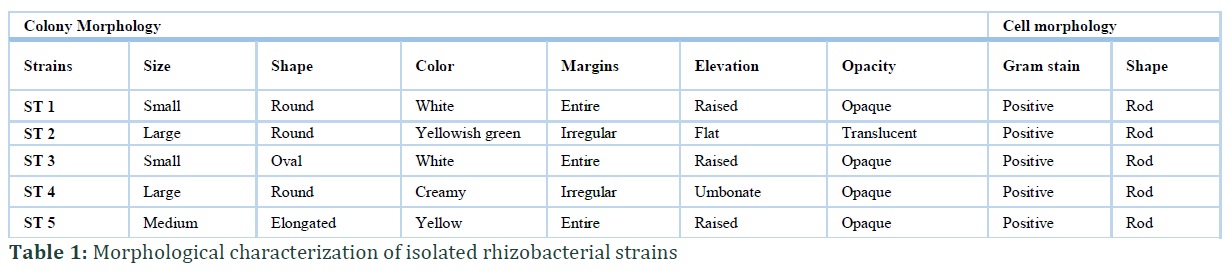
The total of 239 strains of rhizobacteria was isolated from rhizosphere of different medicinal plants jasmin, Aloe vera, onion, date palm, lemon grass, ginger, garlic and marigold from different regions of Punjab. Majority of colonies were circular in shape and other were irregular. Mostly were pigmented. Some were convex; some were raised in their elevation. Most of colonies were smooth mucoid in texture. Many strains were showed yellowish and creamy colored colonies (Table-1). All strains were gram positive and rods in shape. All strains had the ability to produce spores. All strains were able to produce catalase, urease and oxidase enzymes and ferment glucose, mannitol, sucrose and maltose. All strains had the capability to produce citrase enzyme except ST5. All strains were indole positive except ST2. ST1, ST2 and ST5 strains were showed positive starch hydrolysis but ST3 and ST4 strains were showed negative starch hydrolysis. ST3 and ST4 strains showed negative arginine hydrolysis. Whereas ST1 strain showed negative gelatin hydrolysis test (Table-2).
Biological screening
Out of 239 strains, 31% strains showed antimicrobial activity against different pathogenic strains as Bacillus cereus, Bacillus subtilis, E. coccus, E. coli, MRSA, Salmonella, Klebsiella, Staphylococcus aureus ATCC20737, Staphylococcus aureus ATCC25923 and Neisseria. Gonorrhea ATCC19424, MRSA6, MRSA7 and MRS8 in preliminary biological screening. Out of these strains, ST1 strain showed 3mm, 10mm, 15mm and 6mm zone of inhibition against MRSA1, E. coli ATCC, E. coli AK477, MRSA6 respectively. ST2 strain showed 15mm, 10mm, 7mm, 20mm, and 5mm zone of inhibition against B. cereus, MRSA1, E. coli ETCC, Bacillus subtilis, MRSA6 respectively. ST3 strain showed 12mm, 2mm, 17mm zone of inhibition against MRSA1, E. coli AK477, and Bacillus subtilis respectively.ST4 strain showed 4mm, 8mm, 15mm and 20mm against Bacillus cereus, E. coli ETCC, E. coli AK477, Bacillus subtilis respectively. ST5 showed 20mm,5mm,25mm,20mm,15mm and 25mm against P. aeruginosa, E. coccus, E. coli ETCC, Bacillus subtilis 6, MRSA7 respectively (Table No-3).
Chemical screening
Thin layer chromatography and HPLC of bacterial cell extracts
TLC of these different strains was also carried out separately with the respective bacterial cell extracts. In TLC of bacterial cell extract, strain ST1 showed one band with Rf value of 0.578. Strain ST2 and ST3 showed three bands with their respective Rf value (Table-4). Strain ST2 showed Rf values of 0.33, 0.5 and 0.8. Strain ST3 showed Rf values of 0.3215, 0.546 and 0.625. Strain ST4 showed one band with Rf value of 0.3125. Strain ST5 showed two bands with Rf values of 0.33 and 0.783 (Table-4).
HPLC of bacterial cell extracts
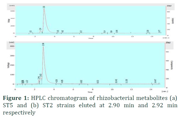
In this chromatogram of sample ST5 showed different peaks at different retention times. Each peak showed different Rf values against specific absorption unit. Here, three different peaks were observed with different retention times of 2.44, 2.63 and 2.90. However, maximum peak was observed at 2.90 with the absorption voltage of 400 mV (fig 1). In this chromatogram of sample ST2 showed different peaks at different retention times. Each peak showed different Rf values against specific absorption unit. Here, two different peaks were observed with different retention times of 2.54 and 2.92. However, maximum peak was observed at 2.92 with the absorption voltage of 650 mV (Figure-1).
Figures & Tables


Discussion![]()
Antibiotic resistance (AR) is becoming a major threat worldwide because of antibiotic-resistant pathogenic bacterial strains emergence [16, 17]. Previous study demonstrated the usefulness of medicinal plants that contain medicinal substance which can be used as antifungal, antibacterial, and anticancer. Due to their medicinal values plants are primary source of medicine [18]. Therefore, the purpose of this research is to evaluate the antibacterial activity of rhizobacterial strains from a various medicinal plants in Jasmin, Aloe vera, onion, date palm, lemon grass, ginger, garlic and marigold against human pathogens. In this study the rhizobacterial strains of Jasmin and Aloe vera plants showed significant impact against human pathogens. 239 rhizobacterial strains were isolated and characterized morphologically and biochemically. All strains were gram positive and rod shaped and spore formers. Antimetabolites produced by the different isolates showed variable zones of inhibition against bacterial pathogens after incubation period. The bacillus and Pseudomonas inhibited the growth of S. aureus and P. vulgaris with zone having diameter of 11 and 12 mm respectively in previous studies [19] but in current study out of 31% strains showed antimicrobial activity against different human pathogens in preliminary biological screening by agar well diffusion and disc diffusion methods. These strains are unique in this region to have such a remarkable activity (zone of inhibition 25mm) against human pathogens. ST5 strain showed highest activity against MRSA7 and E. coli ATCC. In a previous study, HPLC were used for detecting antibacterial compounds in methanol extracts [20]. The strain ST5 showed three peaks while strain ST2 produced two peaks. Either the two or three peaks that were showen in figure (2) had bioactive compounds that exhibited antimicrobial activity may play their role as a combined effect for observed significant antimicrobial activity which can be confirmed only by subsequent extraction and purification of all the four compounds using different column chromatographic techniques.
Due to emergence of antibiotic resistant bacteria, the effective treatment of infectious diseases becomes a major challenge now days. The current study explored the significant impact of rhizobacterial mediated antimetabolites on human pathogens condition. These compounds after purification and molecular identification may be used in future pharmaceutical industries to eradicate human pathogens.
Authors' Contribution
Batool S and Malik U performed experimental work. Iqbal A supervised the work. Batool S and Malik U prepared the manuscript and Iqbal A checked the final draft.
All the authors declare that they have no competing interest that can negatively affect the current study.
References![]()
- Bardgett, Richard D, and Wim H. Van Der Putten. Belowground biodiversity and ecosystem functioning. Nature, (2014);752: 7528-505.
- Köberl M, Schmidt R, Ramadan EM, Bauer R, Berg G. The microbiome of medicinal plants: diversity and importance for plant growth, quality and health. Frontiers in microbiology, (2013); 4: 400.
- Mahler HC, Friess W, Grauschopf U, Kiese S. Protein aggregation: pathways, induction factors and analysis. Journal of pharmaceutical sciences,(2009); 9: 2909-2934.
- Pandey A, Malviya T. Production of antibiotics isolated from soil bacteria from rhizospheric and non-rhizospheric region of medicinal plants. Indian journal of applied research, (2014);8: 25-32.
- Abada EA, El-Hendawy HH, Osman ME, Hafez MA. Antimicrobial activity of Bacillus circulans isolated from rhizosphere of Medicago sativa. Life Science Journal,(2014); 8: 711-719.
- Marasini, B. P, Baral, P, Aryal, P., Ghimire, K. R., Neupane, S., Dahal, N, and Shrestha, K. Evaluation of antibacterial activity of some traditionally used medicinal plants against human pathogenic bacteria. BioMed research international, (2015);6. https://doi.org/10.1155/2015/265425
- Boucher HW, Talbot GH, Bradley JS, Edwards JE, Gilbert D. Bad bugs, no drugs: no ESKAPE! An update from the Infectious Diseases Society of America. Clinical infectious diseases, (2009);1: 1-12.
- Aslam S, Sajid I. Antimicrobial potential of Halophilic actinomycetes against multi drug resistant (MDR) ventilator associated pneumonia causing bacterial pathogens. Pakistan journal of pharmaceutical sciences,(2016); 2:367-374.
- Köberl M, Schmidt R, Ramadan EM, Bauer R, Berg G. The microbiome of medicinal plants: diversity and importance for plant growth, quality and health. Frontiers in microbiology,(2013); 4: 400.
- Bibi F. Diversity of antagonistic bacteria isolated from medicinal plant Peganum harmala L. Saudi journal of biological sciences,(2017); 6: 1288-1293.
- Cappuccino, James G., Natalie Sherman, and A. Microbiology. A laboratory manual. (1983): 315-317.
- Aftab U, Zechel DL, Sajid I. Antitumor compounds from Streptomyces sp. KML-2, isolated from Khewra salt mines, Pakistan. Biological research, (2015); DOI:10.1186/s40659-015-0046-3
- El Euch IZ, Frese M, Sewald N, Smaoui S, Shaaban M, Mellouli L. Bioactive secondary metabolites from a new terrestrial Streptomyces sp. TN262. Applied biochemistry and biotechnology, (2010); 2: 579-593.
- Balouiri M, Sadiki M, Ibnsouda SK. Methods for in vitro evaluating antimicrobial activity: A review. Journal of pharmaceutical analysis,(2016); 2: 71-79.
- Hahn-Deinstrop, E. Applied thin-layer chromatography: best practice and avoidance of mistakes. John Wiley and Sons (2007);.1-15.
- Banin E, Hughes D, Kuipers OP. Bacterial pathogens, antibiotics and antibiotic resistance. FEMS microbiology reviews,(2017);3: 450-452.
- Piña B, Bayona JM, Christou A, Fatta-Kassinos D, Guillon E, et al. On the contribution of reclaimed wastewater irrigation to the potential exposure of humans to antibiotics, antibiotic resistant bacteria and antibiotic resistance genes–NEREUS COST Action ES1403 position paper. Journal of Environmental Chemical Engineering, (2018); 102-131.
- Kumar S, Das G, Shin HS, Patra JK. Dioscorea spp. (a wild edible tuber): a study on its ethnopharmacological potential and traditional use by the local people of Similipal Biosphere Reserve, India. Frontiers in pharmacology, (2017); doi: 10.3389/fphar.2017.00052
- Raj Y, Chauhan VS. Isolation, characterization and screening of novel antibiotic producing bacteria from natural habitats of Western Himalayas and industrial waste soil samples. International Journal of Computer Systems,(2019); 3: 3282-3288.
- Damle S, Sharon K. Antioxidant activity, TLC and HPLC-ESI-Q-TOF-MS fingerprinting of Catunaregam spinosa (Thunb.) triveng. Journal of Pharmacognosy and Phytochemistry,(2018);4: 2119-2124.
This work is licensed under a Creative Commons Attribution-Non Commercial 4.0 International License. To read the copy of this license please visit: https://creativecommons.org/licenses/by-nc/4.0


