

Full Length Research Article
Investigation on Possibility of Rodents to Transmit Human Ascaris lumbricoides eggs from Contaminated Soil
Mehru Nisha*, Athira Mohd Ghozi, Aina Syahirah Ali Baderudin, Pang Jyh Chyang
Adv. life sci., vol. 9, no. 1, pp. 80-84, May 2022
*- Corresponding Author: Mehru Nisha (Email: mehrunisha@unikl.edu.my)
Authors' Affiliations
Abstract![]()
Introduction
Methods
Results
Discussion
References
Abstract
Background: Ascaris lumbricoides is commonly found in soils among communities living with poor sanitation facilities. Commonly human helminths do not survive in animals’ guts and vice versa. However, the high burden of Ascariasis among indigenous communities could be transmitted by rodents as a vector. Hence, this study was carried out to address this hypothesis.
Methods: The eggs of Ascaris lumbricoides were isolated from soil samples from an indigenous village in Malaysia, the eggs were isolated using the floatation technique and cultured in vitro using 0.1% of sulphuric acid dish and embryonation of the parasite eggs was observed daily under a light microscope till day 28. For the in vivo method, the embryonated eggs around 400 epg and 800 epg were feed to the rats for four weeks. Finally, stool samples were collected for Ascaris egg shedding.
Results: The loamy eggs collected near toiled areas using the floatation technique were found to contain Ascaris lumbricoides eggs. The cultured eggs in sulphuric acid developed from the first-cell stage until the fourth stage of development. Discharge of Ascaris eggs were found in stool samples among rats fed with 200epg of Ascaris eggs.
Conclusion: It was found that 0.1% sulphuric acid was ideal for developing Ascaris lumbricoides eggs for cultivating in vitro condition, and there was the possibility of rats carrying the Ascaris eggs throughout the village. In future, these findings can be used to propagate the eggs for testing anthelminthic drugs inhibiting the developmental stages.
Keywords: Animal model; Ascaris lumbricoides; Sulphuric acid; In vivo; In vitro culture; Rat
Introduction![]()
More than a quarter of the world’s population have higher chances of getting infection with soil-transmitted helminths (STHs) like roundworm Ascaris lumbricoides, hookworms and Trichuris trichiura (whipworm) which can cause various diseases [9, 8, 13].
Ascaris lumbricoides (roundworm) is a nematode worm that multiplies inside the gastrointestinal tract (GI tract) of humans, commonly in the small intestine, and it is transmitted through direct contact with the eggs present in contaminated soil [15]. Ascariasis is widespread and often reported from warm tropical and sub-tropical regions. The most prevalence of Ascaris infection is reported from the countries in the tropical area because of the warm and humid environment that could favor the transmission of the Ascaris infection [7]. Furthermore, prevalence of both ascariasis infections is reported to be alarmingly high in poor, developing and highly populated countries [1,4,10,12].
There are two types of eggs in Ascaris lumbricoides that are produce by the female worm, fertilized eggs and unfertilized eggs. The fertilized eggs are produced when a female worm is inseminated by male worm; then embryonation and development will take place. Meanwhile, the un-inseminated female produces non-embryonated eggs and for the eggs that do not undergo fertilization and it is known as unfertilized eggs. The female worm can lay about 200,000 eggs per day and the eggs are passed with faeces [17]. Fertilized eggs and unfertilized eggs both have some common and differences in terms of shape, color, size, etc. Fertilized eggs are round oval. It measured as 45-75 µm in length while for breadth is 30-50 µm. There are stained with brown for mamillated eggs while clear stained for decorticated eggs [13].
Besides, the eggs are surrounded by two layers, thicker outer shell and thinner inside the shell. The viable eggs have spaces between the inner cells of the eggs and the outer shell of the Ascaris eggs. The eggs are smooth, have a decorticated shell and also bumpy, mamillated shell [13]. Ascaris lumbricoides eggs can grow under favorable conditions into 1-cell egg. The eggs are then advance into larva for about 2 to 3 weeks, where it needs a host, which is human and eventually it become infective when the eggs being ingested through fecal–oral route of transmission [6]. The ova retrieved from soil evince soil pollution and sanitation condition of the community leaving nearby the areas. The contaminated soil might be harmful for people that live in poor sanitation area as they may fortuitously ingest the eggs and be infected with ascariasis [5, 16]. Fertilized eggs float in the saturated solution of common salt. Published literature reveal that high-density sugar or salt solution can be used to float up the eggs/ova of helminths [5].
Despite enormous reports are available on Ascaris cases as well as drug treatment, scarcely studies has been conducted on culturing the eggs in vitro and in vivo. Hence, the aim of this research is to optimize culturing methods of Ascaris lumbricoides eggs in laboratory by using sulphuric acid from various soil type and in various acidic conditions as well as culturing Ascaris lumbricoides in vivo using rat model.
Methods![]()
Study design and location
The soil sampling was conducted at Kampung Orang Asli Sungai Lalang Baru, Ulu Semenyih, Selangor, Malaysia. The study location is about 22 Kms from the university’s laboratory (UniKL MESTECH, Malaysia). Lack of proper latrine facilities causes high risk of parasite transmission. Some of the villagers practiced open defecation even now which resulted in soil contamination and soil transmitted helminths (STH) propagations. The collections of soil samples were at the riverside, near the toilet area, at the pond areas, outdoor residential areas and alongside the street.
Sample collection
The samples were collected by using a shovel to transfer the soil into a plastic bag prior transporting to the laboratory and used. It was placed inside the ice box in order to preserve the soil. Approximately 200-300 gram of soil were collected with a depth of 4-6 inch (optimized) in the area; common toilet area, near the riverside, near the pond area, residential area and alongside the road by using plastic bags and a small shovel. Upon reaching the laboratory the soil type was clearly labelled based on the location and stored in a refrigerator between 4ºC to 10 ºC until used. The soil was kept up until 30 days. Sieving analysis was done to remove any unwanted debris that could interfere with the isolation process. Identification of type was carried out as previous described by Nisha et al, 2019 [11].
Ascaris eggs isolation and culturing process
The first method for eggs isolation from soil sample is via flotation technique. The floatation technique was used to float up the eggs at the rim of the centrifuge tube.
For this, around 3 grams of the collected soil was mixed with distilled water (15ml) in a centrifuge tube and centrifuged at 2500 rpm for 5 minutes. The supernatant was discarded into the sink and filled up with 15 ml of the floatation fluid which was high density salt and/or sugar solution (specific gravity; 1.28). The ratio for both solutions were similar. Granulated sugar and fine salt were used.
Upon centrifugation, the centrifuge tubes were examined for any floating eggs. The eggs were observed at the edge of the centrifuge tubes, and they were collected using a disposable pipette. Later, the eggs were transferred into glass slide and observed under the light microscope for the egg morphology.
Culturing Ascaris lumbricoides in vitro using petri dish
Around 1ml of 0.1% sulphuric acid was used to culture the Ascaris lumbricoides. Upon confirmation of the egg’s morphology on the glass slides, quantification was done using McMaster chamber. Whereby, approximately six Ascaris eggs were transferred into six different glass petri dishes each contained one egg. The ratio of Ascaris eggs in floatation fluid to volume of sulphuric acid was 5:1. Around 5 ml of floatation solution containing eggs were shifted first followed by 1 ml of 0.1% sulphuric acid. The isolation methods were repeated with the same three different types of soil samples. But this time around, the 10 ml of floatation solution with the eggs were shifted instead of 5 ml. In addition, the sulphuric acid was doubled; 2 ml of the acid was used. Three glass petri dishes were used with each of the samples were in each glass petri dishes. This was the optimized part of the culturing method using different volumes of sulphuric acid. Glass petri dishes were used instead of the normal plastic petri dishes because the concentration of sulphuric acid (0.1%) was highly corrosive therefore the plastic petri dishes were not recommended to due to reactivity. Next, the glass petri dishes were incubated at 37°C for a few days and microscopic observation were carried out on daily basis up to 28 days, attributing to the life cycle of Ascaris lumbricoides embryonation. Few drops of 0.95% sodium chloride (NaCl), about 2 to 3 ml of distilled water or normal saline were added once in a while to avoid drying.
Culturing Ascaris lumbricoides in vivo using rat model
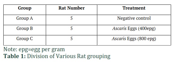
Fifteen (n=15) male Wistar rats were used for the in vivo studies. The rats were divided into three different groups (Group A – C). Group A was used as negative control, Group B were fed with 400 epg of Ascaris eggs and group C were fed with 800 epg of Ascaris eggs (Table 1) in every feed. Group B and Group C were fed with Ascaris eggs continuously for four weeks. The feces were collected every day for the entire four weeks and a floatation technique was done to find the Ascaris eggs from the feces.
Results![]()
Isolation of Ascaris eggs from soil
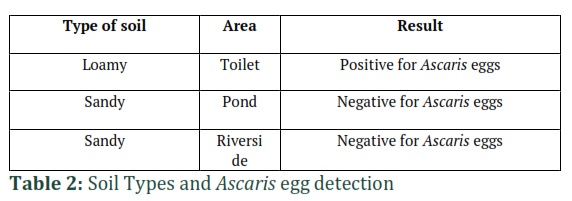
The soils were collected from there different location containing various soil types as in Table 2. The sandy soil near the pond area and riverside showed negative result for the presence of Ascaris eggs. Meanwhile, the loamy soil from toilet areas showed positive result for Ascaris eggs (Table 2).
Culturing and observation of Ascaris lumbricoides eggs in vitro
The cultured eggs were spotted to develop from first cell-stage to larva development (Figure 1). The egg was covered with corticated layer and thick chitin shell was presence. This egg appeared approximately on day 2 (A). Over time, the cell began to cleave into two developing cells which could be observed on day 10 (B). On day 18, the cells were in fourth cell-stage and the cells were still in developing process (C). For the final stage (D), a tiny larva formed inside the egg. The outermost layer of the eggs was irregular on the surface. The outer layer of the egg was thin proteinaceous membrane also a middle protein and chitin layer which actually gave a structural strength for the egg. Apart from that, there was presence of ascarocide layer at the innermost layer.
Culturing Ascaris lumbricoides eggs in vivo using rat model
There was presence of Ascaris eggs in the rats’ stool for direct oral administration groups on third and fourth week while no presence of any eggs from negative test group for four weeks. The vitamin D levels of studied population was also determined. The level of vitamin D from 20 to 29 ng/mL was considered to be deficient and the vitamin D level was less than 12 ng/ml, it was considered to be an indication of severe vitamin D deficiency. Out of 79 female participants, only 20 (25.31 %) females had normal serum vitamin D levels. Overall, 59 (74.68%) females had vitamin D deficiency as indicated in figure 5.3. When the vitamin D deficiency was correlated with thyroid dysfunction, it was observed that vitamin D levels were non-significantly (p = 0.35) associated with hypothyroidism.
Figures & Tables
Discussion![]()
Total three soil samples that were collected from Kampung Orang Asli Sungai Lalang: loamy and two sandy soils. The soil samples collected from different locations were between 200-300 grams. The loamy soil consists of the combination of sandy particles, silt, and clay soil. Loamy soil conditions revealed high Ascaris egg density as previous reported by Nisha et al. [11] and it provided favorable conditions for the STHs growth as the eggs needs warm, moist soil and the temperature should be over 18°C to inhabit.
On the other hand, sandy soil type showed the absence of Ascaris eggs. This can be due to the sandy soil lack of characteristics features needed for the eggs to fertilize. The sandy soil composed of high proportion of sand and a little clay inside. These elements did not support embryonation of the helminths eggs, hence the eggs were not be seen in a sandy type soil. All the soils were kept inside the refrigerator for a few days before isolation procedure technique as they tend to become dried after if it is kept for too long, the samples need to be moist for better yield. For the optimization technique, about 5 to 10 grams were kept in the incubator and incubated for 37°C. The soil sample with viable eggs lasted up to one month in the incubator in moist conditions (distilled water was added at regular intervals to avoid dryness). For the isolation of egg, floatation technique was used using high density floatation fluid in combination of salt/sugar solution. However, it was very difficult to get a very clear viewed of Ascaris eggs when observed under microscope as the soil samples contained a lot of debris and some small particles, despite sieving the soil samples few times prior to experiment.
The glass petri dishes were incubated for 37°C and the embryonation stage were observed daily starting with the next day of the incubation. Subsequently, to avoid the dryness effect such as the solutions in the petri dishes was dried, also to maintain the humid environment for the eggs to develop, 2 to 3 ml ditilled water and few drops of NaCl was added into the glass petri dishes. This technique was supported by the previous research by Bessat and Dewair in 2019 [2]. Only distilled water was added to maintain moisture in the petri dishes containing the Ascaris eggs and the addition of sulphuric acid should be avoided since it can lead to hyper acidity affecting the developmental process. When distilled water was added to the culture in petri dishes, the pH was slightly changed but it would not cease the cultivation process.
The fully developed Ascaris eggs was on observed on 28th day. Each stage was observed for at least 2 days before the cells began to develop into more specialized form which at the end, the cell turned into larvae. From the previous research by Cruz et al. [6], each of the cell stage took at least 3 days to be observed except for the 3-cell stage. Temperature could be one of the factors of the viability of the eggs, from the previous study had mentioned that it could speed up the development of embryo if the temperature was higher compare to low temperature [6]. The morphology of the eggs observed was corticated layer, the thick chitin shell and undeveloped embryo of the eggs. The corticated layer was a layer that surrounded the eggs. Next, for the second cell stage, the cells began to develop, and the cleavage could be seen in this stage. The second cell stage was observed between day ten and fifteen, and the eggs could be possibly turned into cleavage as early as day eight, but this could be overlooked because the eggs were observed at the same time under the microscope. Subsequently, the decorticated eggs that undergo the embryonation could be seen at day eighteen and finally the egg had completed the embryonation period. Also, the larvae development was seen in the last stage on day twenty-eight. The larvae was nicely captured and observed under microscope.
Apart from that, some of the eggs that were being cultivated by using sulphuric acid were incapable to develop as early as stage 2. This can be due to some errors during cultivating process for instance; the eggs were too fragile that the eggshells broke probably during isolating process. Next, the eggs could be immotile while cultivation because the ratio of acid was too high for example 1:1 ratio of sulphuric acid and floatation fluid. The amount of sulphuric acid volumes was optimized and the other eggs managed to survive with the right proportion of sulphuric acid which was 2 ml of sulphuric acid with 10 ml of the floatation fluid that suitable for aiding in cultivating process. Furthermore, the distilled water that was mixed together with the sulphuric acid during cultivating process could be contaminated and cause the eggs to not be able to develop. Nonetheless, the errors were managed to overcome successfully and Ascaris egg propagation were monitored. Limitation in this project was lack of previous research studies on this topic. If there is enough research studies, more data can be obtained for better result.
The feeding process took place almost four weeks. Group B and Group C were fed with 400 epg and 800 epg of Ascaris eggs respectively. The feces were collected every day for four weeks and floatation technique was done to find the presence of Ascaris eggs in the feces. Aside from mites and Enterobius vermicularis (pinworm), there were no presences of Ascaris eggs in the feces of Group A (negative control) and during the first and second week in Group B and Group C. However, during third and fourth week, around 200 epg of unfertilized Ascaris eggs were found in Group C which was fed with 800 epg of Ascaris eggs.
From the studies, it was found that 0.1% sulphuric acid was ideal for the development of Ascaris lumbricoides eggs for cultivating in vitro condition. Next, the result indicated there are possibility of rats in spreading the infection as there were positive discharges of Ascaris eggs in the rats’ faeces. Hence, we recommend a proper pest control for rodents in this village by removing the food sources, water and places that can provide as the rodents’ shelter. In future, these in vitro and in vivo methods could be used to propagate the eggs for testing anthelminthic drug inhibiting the developmental stages.
Authors' Contribution
All authors contributed equally to this work and no conflict of interest identified.
The authors declare that there is no conflict of interest in this paper.
References
- Alelign T, Degarege A, Erko B. Soil-Transmitted Helminth Infections and Associated Risk Factors among Schoolchildren in Durbete Town, Northwestern Ethiopia. Journal of Parasitology Research, (2015); 2015: 641602.
- Bessat M, Dewair A. Assessment of the inhibitory effects of disinfectants on the embryonation of Ascaridia columbae eggs. PLoS One, (2019); 14(5): e0217551.
- Chammartin F, Guimarães LH, Scholte RG, Bavia ME, Utzinger J, Vounatsou P. Spatio-temporal distribution of soil-transmitted helminth infections in Brazil. Parasit Vectors, (2014); 7: 440.
- Chammartin F, Scholte RG, Malone JB, Bavia ME, Nieto P, Utzinger J, et al. Modelling the geographical distribution of soil-transmitted helminth infections in Bolivia. Parasites & Vectors, (2013); 6: 152.
- Cranston I, Teoh PJ, Baker SM, Sengupta ME, Ensink JH. Evaluating the efficacy of a centrifugation-flotation method for extracting Ascaris ova from soil. Transactions of the Royal Society of Tropical Medicine and Hygiene, (2016); 110(7): 400-7.
- Cruz LM, Allanson M, Kwa B, Azizan A, Izurieta R. Morphological changes of Ascaris spp. eggs during their development outside the host. Journal of Parasitology, (2012); 98(1): 63-8.
- Gupta S, Kumar S, Satapathy A, Ray U, Chatterjee S, Choudhury TK. Ascaris lumbricoides : an unusual aetiology of gastric perforation. Journal of Surgical Case Reports, ( 2012); (11):rjs008
- Izurieta R, Reina-Ortiz M, Ochoa-Capello T. Trichuris trichiura. In: Rose JB, Jiménez-Cisneros B, Robertson L, editors. Section 4: Helmint; 2018.
- Jourdan PM, Lamberton PHL, Fenwick A, Addiss DG. Soil-transmitted helminth infections. Lancet. 2018;391(10117):252-65.
- Karshima SN. Prevalence and distribution of soil-transmitted helminth infections in Nigerian children: a systematic review and meta-analysis. Infectious Diseases of Poverty, (2018); 7(1): 69.
- Nisha M, Amira NA, Nadiah N, Davamani F. Detection of Ascaris lumbricoides and Trichuris trichiura in various soil types from from an indigenous village in Malaysia. Tropical Biomedicine, (2019); 36(1): 201-8.
- Salam N, Azam S. Prevalence and distribution of soil-transmitted helminth infections in India. BMC Public Health, (2017); 17(1): 201.
- Steinbaum L, Kwong LH, Ercumen A, Negash MS, Lovely AJ, Njenga SM, et al. Detecting and enumerating soil-transmitted helminth eggs in soil: New method development and results from field testing in Kenya and Bangladesh. PLoS Neglected Tropical Diseases, (2017); 11(4): e0005522.
- Steinbaum L, Njenga SM, Kihara J, Boehm AB, Davis J, Null C, et al. Soil-Transmitted Helminth Eggs Are Present in Soil at Multiple Locations within Households in Rural Kenya. PLoS One, (2016); 11(6): e0157780.
- Walker M, Hall A, Basáñez M-G. Ascaris lumbricoides : New Epidemiological Insights and Mathematical Approaches. Ascaris: The Neglected Parasite, (2013); 155–201.
- Weatherhead JE, Hotez PJ. Worm Infections in Children. Pediatrics Review, (2015); 36(8): 341-52.
- Zierhut M, Pavesio C, Orefice F, Ohno S, Rao NA. In: Intraocular inflammation. 1st ed. Springer; 2016. p. 1609.
This work is licensed under a Creative Commons Attribution-Non Commercial 4.0 International License. To read the copy of this license please visit: https://creativecommons.org/licenses/by-nc/4.0


