

Full Length Research Article
The effects of vitamin D on Immunoresponsive gene 1 and Krüppel-like Factor 2 protein expression in the lung due to the cadmium poisoning
Nasim Kardan1, Zohre Eftekhari2*, Saeed Ozmaie1, Hesam-Uddin Hoseinzadeh3
Adv. life sci., vol. 9, no. 4, pp. 526-533, December 2022
*- Corresponding Author: Zohre Eftekhari (Email: Z_eftekhari@pasteur.ac.ir)
Authors' Affiliations
2. Biotechnology Department, Pasteur Institute of Iran, Tehran -Iran
3. Department of Clinical Sciences, Faculty of Veterinary Medicine, Islamic Azad University, Karaj Branch, Karaj – Iran
[Date Received: 27/07/2022; Date Revised: 20/09/2022; Date Published: 31/12/2022]
Abstract![]()
Introduction
Methods
Results
Discussion
References
Abstract
Background: Cadmium, a well-known hazardous heavy metal and non-essential component, has several negative health effects. The long-term use of cadmium toxin to develop a pulmonary model, evaluation of Irg1 gene expression and KLF-2 protein and serum IL4 levels following model induction, and evaluation of vitamin D's therapeutic effects in reducing pulmonary and hepatic complications in a mice model have not been studied.
Methods: A total of 40 healthy female C57 black 6 mice weighing 20–25 g and approximately 6–8 weeks’ old were purchased from animal husbandry, Pasteur Institute of Iran. After induction of the model, the mice were assigned to the following groups such as Group 1 (G1): mice were euthanized the day after induction. Group 2(G2): mice were ethically killed 21 days after induction. Group 3(G3) mice were treated with vitamin D and euthanized 5 days after treatment. Group 4(G4): mice were treated with vitamin D and euthanized 21 days after treatment. Vitamin D3 with a concentration of 300,000 units per milliliter, which is equivalent to 7.5 mg per 1 microliter, and 13.5 μl of the main stock is equivalent to 100 ng, which is used for 1 kg of mice. Blood samples were collected to measure serum glutamic-oxaloacetic transaminase, serum glutamic-pyruvic transaminase, and alkaline phosphatase to evaluate liver toxicity.
Results: Based on the results obtained, serum SGPT levels in vitamin D treatment groups did not show a significant decrease compared to cadmium groups (p >0.001). The ALP biomarker in the groups treated with vitamin D was decreased significant in comparison to untreated model groups (p<0.001). While any significant differences were not observed between the Cd model and the Cd model treated with Vitamin D.
Conclusion: This study proved that administration vitamin D to some extent reduces the toxic effects of cadmium on the liver and lung.
Keywords: Ameliorated; Vitamin D; Protein expression; Lung; Cadmium
Introduction![]()
Respiratory system is vital organ, which consists of small particles such as dust, smoke, dirt, pollen, and aerosols. While, the lungs must provide protection, in addition to the mechanical forces of spontaneous respiration, at the same time inhaled viruses, bacteria, and different respiratory pathogens can impair the protection lines. The special environment of lung is protected by complicated immune interactions [1]. In the developmental and industrial countries, air pollution is the most frequent reason for respiratory diseases that influence on the structure and the function of the respiratory system [2].
Cadmium, a well-known hazardous heavy metal [3] and non-essential component [4], has several negative health effects. Smoking, tainted water or food, contaminated air from fossil gasoline burning, or occupational pollution are all sources of Cadmium exposure. Cd is harmful to both the environment and living things [3]. The kidneys, bone, vasculature, and lungs are Evidence shows that cadmium increases cytokine production and lung inflammation, impairs macrophage-mediated immune function, disrupts tight junction integrity in human airway epithelial tissue at the level of the air-liquid-interface airway, and potentiates pulmonary infection. These are some of the reasons why cadmium burden may predispose individuals to worse pulmonary disease from respiratory infections. Cadmium has been found to build up in human lung tissue. Cd binds to the sulfhydryl group of thiols such as glutathione and metallothionine, disrupting the antioxidant defence mechanism and causing oxidative stress. Localized cadmium deposition has been related to weakened innate immunity [5].
Vitamin D is a prohormone. Since vitamin D receptors (VDRs) are found in many cells and tissues throughout the body, several studies suggest the importance of vitamin D in a variety of physiological activities other than bone and muscle health. Vitamin D is a fat-soluble vitamin that comes in two forms: ergocalciferol (vitamin D2) and cholecalciferol (vitamin D3). The main distinction between these two types is their side chain structure. While D2 is mostly derived from plants and vegetables, D3 is primarily generated in the skin in response to UVB radiation from 7 dehydrocholesterol. Vitamin D is also available as a dietary supplement in both forms [6].
In respiratory system, the airway epithelium has a high quantity of 1-hydroxylase, which converts inactive Vitamin D into an active form. The active form of Vitamin D stimulates cathelicidin secretion and the production of other peptides in epithelial cells that protect against bacterial and viral infections. As a result, vitamin D produced locally promotes innate immunity and regulates inflammation and tissue damage in the respiratory system. It also stimulates the adaptive immune response by activating macrophages and the antigen-recognition cells, T- and B-lymphocytes. Active Vitamin D is produced by alveolar macrophages and performs an important intracrine function in the macrophage response to infection. It has been discovered that vitamin D has a direct effect on T- and B-cells. Vitamin D aids in the production of the cytokine interleukin (IL)-10, which is important in anti-inflammation and immunosuppression. Thus, by removing the microorganisms, locally created Vitamin D in the airways and lungs will reduce tissue damage and inflammation [7].
KLF2 or lung Kruppel-like factor (LKLF) belongs to the zinc-finger transcription factor subfamily known as Kruppel-like family (KLFs) [8]. Cellular differentiation and tissue development are regulated by this family.[9] KLF2 or LKLF is a human and mouse-specific gene with the chromosomal location of 19p13 [10]. LKLF was first reported as a lung-specific transcription factor. LKLF is mostly expressed in the fetal and adult lungs, with limited expression in other organs, and KLF2 is required for proper lung development. LKLF is recognised to have an important role in cell proliferation, differentiation, and apoptosis. [11] Reports on T cell and monocyte function, as well as endothelial proinflammatory activation, have all suggested that KLF2 plays a regulatory role in the immune system [12].
Irg1 (Immune-Responsive Gene 1) is a mitochondrial enzyme [13] that is highly expressed in mammalian macrophages during inflammation [14]. Irg1 was discovered as a 2.3-kb cDNA from a library made from mRNA extracted from a murine macrophage cell line following LPS stimulation.[14] Inflammatory stimuli stimulate the expression of immunoresponsive gene 1, which catalyses the synthesis of itaconate by diverting cis-aconitate away from the tricarboxylic acid cycle. The immunoregulatory function of the immunoresponsive gene 1/itaconate axis in lipopolysaccharide-activated mouse and human macrophages has recently been reported [15].
Interleukin-4 (IL-4) is a protein involved in numerous immunological regulatory and pro-inflammatory activation processes, including atopic inflammation and asthma [16]. This cytokine belongs to the γc family. This group of cytokines has extensive pleiotropic effects on the immune system, including the innate and adaptive immune systems, with these cytokines collectively contributing to the development of T, B, natural killer (NK), and innate lymphoid cells (ILCs), promoting either cell survival or cell death of immune populations depending on the context, and significantly modulating the differentiation of cellular populations into more terminally differentiated cells [17]. Some of the characteristics of IL-4 and IL-13 are similar. Both IL-4 and IL-13 are released into the plasma by Th2 lymphocytes and mast cells. These cytokines are overproduced and play essential roles in atopic, asthma, and allergic reactions by stimulating IgE production in naïve B-lymphocytes [16].
Because of the importance of infectious lung diseases in recent years and the relationship between exacerbation of these diseases and air pollution and the effect of toxins on accelerating the pathophysiological process of lung diseases, developing an animal model to evaluate this relationship and the treatment process with supplements can be helpful. The long-term use of cadmium toxin to develop a pulmonary model, evaluation of Irg1 gene expression and KLF-2 protein and serum IL4 levels following model induction, and evaluation of vitamin D's therapeutic effects in reducing pulmonary and hepatic complications in a mice model have not been studied.
Methods![]()
A total of 40 healthy female C57 mice weighing 20–25 g and approximately 6–8 weeks’ old were purchased from animal husbandry, Pasteur Institute of Iran. The mice were housed in a controlled environmental condition with a 12 h light/dark cycle, temperature (23±3°C) and humidity (50± 10%), and free access to autoclaved commercial food and water [18]. All animal studies were carried out in accordance with the instructions of the research ethics committee. Research Ethics Committees of Islamic Azad University (Science and Research Branch) has reviewed the study protocol and approved this research (approval number: IR.IAU.SRB.REC.1400.331).
Animal Study Design
The mice were randomly divided into 4 groups with 10 mice in each group. Based on previous research, the cadmium toxin first dissolved in normal saline. Then, according to the ethical principles of 2 mg per kg of mouse weight for 28 days by injecting IP 5 days a week with two days of rest in the experimental group, the disease was induced. After induction of the model, the mice were assigned to the following groups:
Group 1 (G1): Mice were euthanized the day after induction
Group 2(G2): Mice were ethically killed 21 days after induction
Group 3(G3): Mice were treated with vitamin D and euthanized 5 days after treatment
Group 4(G4): Mice were treated with vitamin D and euthanized 21 days after treatment. Vitamin D3 with a concentration of 300,000 units per milliliter, which is equivalent to 7.5 mg per 1 microliter, and 13.5 μl of the main stock is equivalent to 100 ng, which is used for 1 kg of mice.
In this study, each group was placed in a box containing CO2 for about 2 minutes until they were completely euthanized. Then, after euthanasia, liver and lung tissues were collected in two forms of fix (10% formalin) and freeze for further studies.
Assessment the Liver Biomarkers
Before euthanizing the animal, blood samples were collected to measure serum glutamic-oxaloacetic transaminase (SGOT), serum glutamic-pyruvic transaminase (SGPT), and alkaline phosphatase(ALP) to evaluate liver toxicity.
Real-time Polymerase Chain Reaction (RT-PCR)
RNA Extraction of Lung Tissue
The Irg1 genes expression level in different examined groups was measured. After extracting the cells from the homogenized lung tissue, a specified amount of trisol (1 ml of trisol per 50 to 100 mg of tissue) was added for cell lysis. For 1 ml of trisol added in the first phase, 200 μl of chloroform was added to the samples and shaken for 15 seconds to mix (without vortex). The microtubes were incubated for 15 minutes at room temperature and then centrifuged at 12000 RPM for 4 minutes at 4 ° C. The RNA from the clear superior phase was extracted and transferred to another microtube. Isopropanol was then added to these microtubes and incubated for 10 minutes at -20 ° C after mixing the contents. In order to precipitate RNA, the samples were centrifuged at 12000 RPM for 4 minutes at 4 ° C. The superior phase was drained, and the precipitates were washed with 75% ethanol before vortexing for a few seconds. The samples were then recentrifuged at 7500 RPM for 5 minutes at 4 ° C. At this stage, after draining the superior phase, the precipitate was semi-dry for 15 minutes. Then, 20 to 30 microliters of DEPC or RNase-free water was added to each microtube to dissolve the RNA precipitate.
cDNA Synthesis
Complementary DNA (cDNA) was prepared from the extracted RNA. Components for making cDNA include 5µl of RT mix (RT buffer and RT enzyme), 1µl of Oligo dT primer, and 3 µl of DEPC water are mixed together and then distributed in volumes of 9 µl in 0.2 ml microtubes. The mixture was incubated at 25 °C for 10 minutes and 47 °C for 60 minutes. It was then cooled for 2 minutes at 4 °C. The process was stopped after five minutes of heating at 85 °C, and the mixture was kept on ice until use.
Primer Design and Quantitative real time PCR (qPCR)
Quantitative real-time PCR (Q-PCR) analysis was performed by Real-time PCR Thermocycler (ABI Stepone) and the SYBR RT-PCR kit (addbio). The Oligo dt was used to design primers for Irg1 and GAPDH (as a housekeeping gene). Table 1 shows the forward and reverse primer sequences for the aforementioned genes.
Enzyme-linked Immunosorbent Assay (ELISA)
Serum was isolated from the blood samples then, IL-4 were detected in the collected sera by ELISA (MBS2504956).
Western Blot Analysis of KLF-2 Protein Levels
We lysed the tissue in a microtube, added 300 microliters of Protease + RIPA inhibitor solution, and chilled it for one hour (4 ° C) to extract the protein. The samples were then centrifuged (4 ° C, 7000 rpm, 10 min) and Lowry's technique was used to collect the superior phase in order to assess the total protein content. Tissue lysed samples were loaded on PAGE-SDS gel and electrophoresed at 90 volts. Following the electrophoresis stage, the gel was shaken for 2 hours at room temperature with Coomassie Blue (Germany, Merck). Then the gel was placed in the dye solution (Destain) and finally the gel is washed with distilled water. In this way, the proteins were separated on the PAGE-SDS gel based on the size of the molecular weight, according to the standard Ladder. The electrophoresis steps were performed using a RAD-BIO device. For one and a half hours, the dissociated proteins on the gel at 100 volts were constantly and wet transferred to PVDF paper having pores of 0.45 μm. Using a 12.5 percent Sodium Dodecyl Sulfate-Poly Acrylamide electrophoresis (PVDF), proteins were transported to a polyvinylidene fluoride membrane from 50 g of homogenized samples. PVDF paper was placed in the blocking buffer on the shaker for 2 hours at room temperature. Then, TBST (Tween20) was used for washing. The PVDF paper was then placed in the original antibody dilution in the refrigerator overnight. The next day, the membranes were washed three times with Tween 20 saline buffer tris (TBST). BA1054-2, a secondary antibody linked to horseradish peroxidase (HRP), was then added to the shaker for 120 minutes at room temperature, and protein expression was confirmed. The bands were detected using an enhanced chemiluminescence (ECL) technique. The band intensities obtained from each protein extract were normalized against comparable values for the GAPDH household protein bands, and protein band density was assessed using Image J software (National Institute of Health, USA).
Histopathology Evaluation of Liver
After extracting the liver samples from the mice, they were fixed in a 10% formalin solution for 24-72 hours. Samples were cut by a 5 μm thick microtome machine and placed on a silane slide. The liver tissue was then stained with Masson's trichrome stain, and the lung tissue with hematoxylin and eosin (HE). The pathological structural alternations were observed under an optical microscope (200× and 100×).
Statistical Analysis
GraphPad Prism 5.04 software was used for two-way ANOVA and multiple Tukey comparison test analysis. Data were expressed as the mean± SE compared to the command. In all studies, a p-value of less than 0.05 was considered a significant difference. The asterisks replicate significant differences with* for p<0.05, ** for p<0.01, and *** for p<0.001, with a 95% CI.
Results
![]()
Evaluation of SGOT, SGPT, and ALP Biomarkers Activity
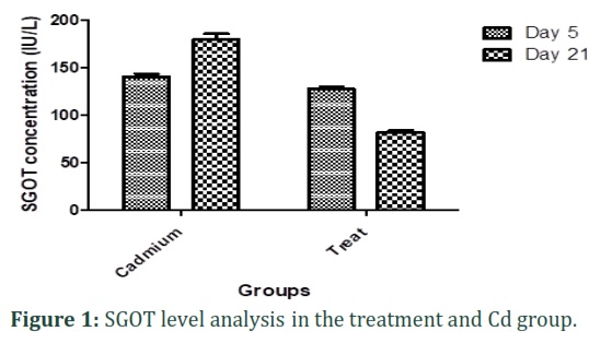
Based on the results, SGOT level in sera isolated from treatment groups was not statistically significant in comparison with the cadmium groups (p >0.001).
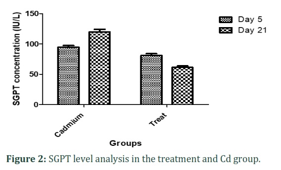
Serum SGPT levels in vitamin D treatment groups did not show a significant decrease compared to cadmium groups (p >0.001).
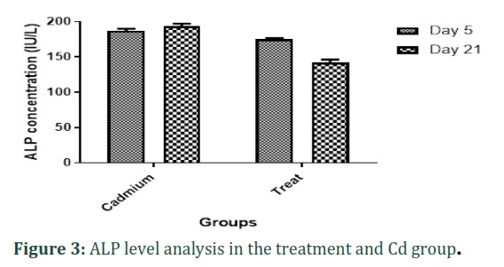
The ALP biomarker in the groups treated with vitamin D was decreased significant in comparison to untreated model groups (p<0.001).
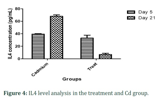
Estimation of IL-4 Level by ELISA
In the study of IL-4, any significant differences were not observed between the Cd model and the groups treated with vitamin D.
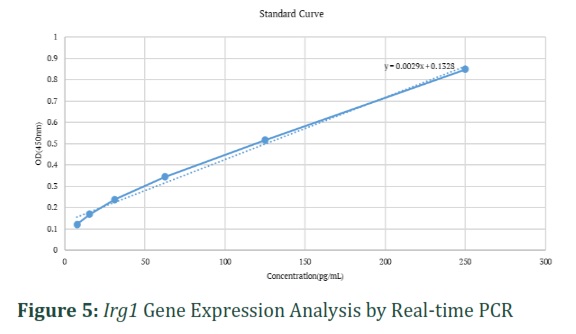
Irg1 Gene Expression Analysis by Real-time PCR
The expression of Irg1 gene, there was a statistically significant decrease in the group treated with Vitamin D (p<0.05). While any significant differences were not observed between the Cd model and the Cd model treated with Vitamin D.
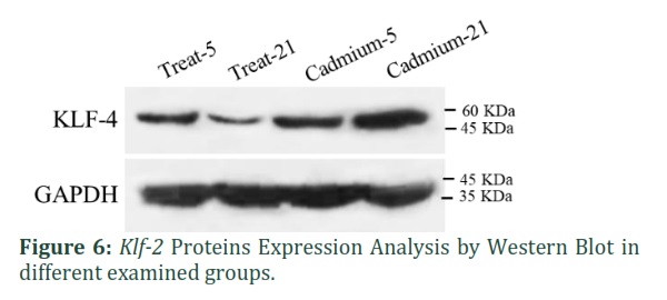
Klf-2 Proteins Expression Analysis by Western Blot
Based on Klf-2 proteins band results, the protein expression in the treatment groups was significantly lower than the Cadmium group.
Liver Histopathology Analysis
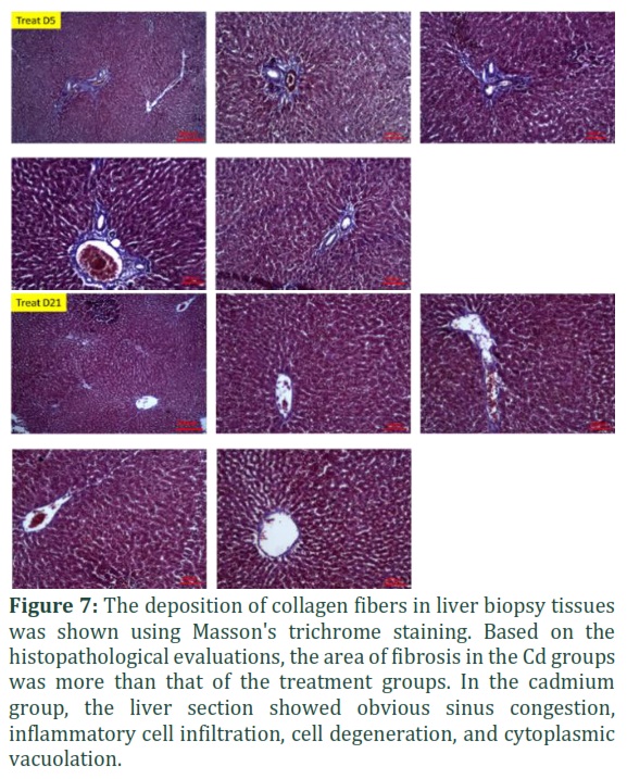
Figure 7 shows Masson's trichrome stained cross-section of liver tissue in Cadmium D5, Cadmium D21, Treat D5 and Treat D21 groups. The deposition of mature collagen fibers in typical liver biopsy tissues was demonstrated using Masson's trichrome staining. Based on the histopathological evaluations in the Cd groups, the fibrosis area was more than the treatment groups. As can see in figure 7, in the groups treated with Vitamin D, there was a statistically significant decrease in fibrosis area compared to the Cd groups (p<0.001), respectively. Based on histopathological evaluations, in the Cd group, the liver section of showed evident sinusoidal congestion, inflammatory cell infiltration, cellular degeneration and cytoplasmic vacuolization.
Figures & Tables
The lung is a vital organ that is extremely sensitive and vulnerable to infections, damage, and particles in the air (or airborne). According to the book "The Global Impact of Respiratory Disease" published by the Assembly of International Respiratory Associations (2017), respiratory diseases are the world's leading cause of death and impose significant health costs [19]. Air pollution has become a serious problem in recent years, and related researches on respiratory health are increasing.
Cadmium is a hazardous non-essential transition metal that can have harmful effects on the human body [20] and it is absorbed mostly through the respiratory system and to a lesser extent through the gastrointestinal system, while skin absorption is rather infrequent [21]. This element accumulates in organs such as the lungs and affects intracellular signaling, which in turn may lead to dysfunction of the host defense, including innate immunity and inflammatory response, impaired lung function and the incidence of lung disease, and enhanced susceptibility to bacterial and viral infections [22, 23]. The latter item emphasizes the need to manage respiratory disorders such as COVID 19. As a result, reduction of this toxic metal exposure might be viewed as a potential strategy for lowering the susceptibility and severity of viral diseases affecting the respiratory system, such as COVID-19 [24].
Several studies have shown cross-sectional links between vitamin D deficiency and chronic respiratory disorders. According to certain research, vitamin D insufficiency may be a precursor to the development of respiratory disorders, and an adequate amount of Vitamin D in the body is associated with better lung function [25]. Vitamin D builds up the innate immune system by increasing the amount of beneficial immunological proteins in the body. It also functions as an immune system regulator, preventing excessive inflammatory cytokine production [7].
The mechanism of cadmium-induced toxicity occurs in two mechanisms: The first stage is caused by metal ions, which induce ischemia and ionic problems, while the second stage is caused by inflammation caused by free radicals and oxidative stress [26].
In this study, we investigated the therapeutic effects of vitamin D in reducing the pulmonary and hepatic effects of cadmium in mice.
The levels of SPGT (ALT) and SGOT (AST) in the blood have been utilized as key markers of clinical liver function, and they increase when liver function is compromised [27]. In present study, these indications were all significantly higher in Cd-groups, indicating clear hepatic damage caused by Cd, which was consistent with previous research findings [27, 28]. While in comparison between Cd groups and vitamin D-treated groups, there was not a significant change.
In a study, the effect of progesterone as an anti-inflammatory and antioxidant agent in cadmium-induced toxicity was studied. The effect of cadmium on liver function was studied using liver enzymes such as ALP and bilirubin, which are indications of hepatic injury. Exposure to cadmium increases ALP activity in tissues. Progesterone therapy significantly reduced cadmium-induced hepatic damage by reducing ALP levels [29]. In our study, ALP levels were significantly higher among the control groups. However, in the treatment groups with vitamin D, they significantly decreased. By lowering ALP levels, vitamin D therapy mitigated cadmium-induced liver damage.
Ren et al., studied the expression of Irg1 gene during Syncytial Respiratory Virus infection. Research suggests that reactive oxygen species (ROS) production plays a crucial role in RSV disease lung inflammation and tissue damage. ROS production was increased by RSV infection or IRG1 overexpression. As a result, inhibiting IRG1 induction prevented RSV-induced ROS production and proinflammatory cytokine gene expression. In RSV-infected mice, blocking IRG1 induction decreased immune cell infiltration and prevented lung damage [30]. On the other hand, Nair et al. (2018) found in a study that the expression of Irg1 in myeloid cells prevents immune damage during the bacterial infection in M.tuberculosis . This gene plays a key role in regulating neutrophil-dependent inflammation during Mtb infection in the lung [13].
Cadmium increases ROS production and the imbalance in antioxidants, leading in oxidative stress [31]. In our study, in the cadmium groups, the expression of the Irg1 gene and lung damage increased. However, in the treatment groups, the expression of this gene decreased due to the administration of vitamin D, which, according to research, plays an important role in regulating oxidative stress, inflammation, and energy metabolism [32].
Hu et al., investigated the effect of dietary cadmium on lung injuries in mice with respiratory syncytial virus (RSV). This study showed that low-dose of Cd can cause low-grade of inflammation and exacerbate RSV-induced inflammation via IL-4 [33].
Hyun Lim et al., studied the therapeutic and anti-allergic effects of vitamin D on asthma. This vitamin has a preventive and inhibitory effect on the development and exacerbation of asthma. Low levels of this vitamin in patients with asthma are associated with decreased lung function and increased airway overreaction. The effect of vitamin D on the penetration of eosinophils may be due to the effect of this vitamin on interleukin-4 [34].
Zahedi et al. (2021) examined the concentrations of metals in fine particulate matter PM2.5 in the outdoor air around the home sites of 123 male children from Ahvaz, average age 7, along with their blood samples to measure pro-inflammatory responses. These children, who lived in an industrial region with high metal concentrations in PM2.5, had high metal levels in their blood as well as high levels of IgE, IL-4, and IL-13, indicating immunological dysregulation and rapid inflammatory reactions [16].
Based on previous studies, the IL-4 levels of the cadmium groups in the present study were high. However, treatment with vitamin D reduces this amount. Zahlten et al. in a study investigated the role of Klf2 in the innate immune responses of lung epithelial cells infected with Streptococcus pneumoniae [12]. The results showed that it may act as a counterregulatory transcription factor in the pro-inflammatory response during pneumococcal lung infections, preventing lung hyperinflammation and subsequent organ failure.
Numerous studies have been performed on the cadmium and its toxic effect on different organisms of body. Due to the importance of infectious lung diseases in recent years and the exacerbation of these diseases with air pollution and the effect of toxins on accelerating the pathophysiological process of lung diseases, creating an animal model to evaluate this relationship and the treatment process with supplements can be helpful. This study proved that administration vitamin D to some extent reduces the toxic effects of cadmium on the liver and lung.
![]()
Author Contributions
Nasim Kardan: Conceptualization, Methodology, Validation, Formal analysis, Investigation, Resources, Data curation, Writing – original draft, Writing – review & editing. Zohre Eftekhari: Conceptualization, Methodology, Resources, Writing – review & editing, Supervision. Saeed Ozmaie & Hesam-uddin Hoseinzadeh: Conceptualization, Methodology, Formal analysis, Resources, Data curation, Writing – review & editing, Supervision, Project administration
![]()
The authors declare no known competing financial interests or personal relationships.
Acknowledgment
The authors would like to acknowledge the contributions of the Pasteur Institute of Iran research team, the Zakaria Razi Laboratory (Islamic Azad University, Science and Research Branch, Tehran, Iran).
References
- Shahsavari S, Sarkar S, Jyoti Sen D, Kumar Mandal S.Determination of the Total Antioxidant Activity of Methanolic Extract of Falcaria Vulgaris. J Biochem Phytomed, (2022);1(1): 8-12. doi: 10.34172/jbp.2022.3
- Koopsamy Naidoo SV. Oral exposure to cadmium and mercury alone and in combination causes damage to the lung tissue of Sprague-Dawley rats. Environmental Toxicology and Pharmacology, (2019); 69: 86-94.
- Razooqi M. Risk exposure of dairy cows to environmental contamination of brick kilns on milk content of antioxidants and heavy metals in Al-Nahrawan region, Iraq. Caspian Journal of Environmental Sciences, (2022); 20(3): 491-502.
- Jiang YL, et al. Serum cadmium positively correlates with inflammatory cytokines in patients with chronic obstructive pulmonary disease. Environmental Toxicology, (2022); 37(1):151-160.
- Park SK. Environmental Cadmium and Mortality from Influenza and Pneumonia in U.S. Adults. Environmental health perspectives, (2020); 128(12):127004-127004.
- Mohammed Z, Rasool K, Ahmed M. Relationship between Helicobacter pylori infections and vitamin D level and lipid profile in some obese Iraqi women. Caspian Journal of Environmental Sciences, (2021); 19(5): 801-807.
- Poorna R, Biswal N. Respiratory infections: Role of Vitamin D and surfactant proteins A and D. Lung India : official organ of Indian Chest Society, (2020); 37(5): 421-424.
- Fan Y. Atorvastatin Combined with Low-Dose Dexamethasone Treatment Protects Endothelial Function Impaired by Chronic Subdural Hematoma via the Transcription Factor KLF-2. Drug design, development and therapy, (2020); 14: 3291-3299.
- SenBanerjee S. KLF2 Is a novel transcriptional regulator of endothelial proinflammatory activation. The Journal of experimental medicine, (2004) 199(10): 1305-1315.
- Kaczynski JT. Cook, and R. Urrutia, Sp1- and Krüppel-like transcription factors. Genome biology, (2003); 4(2): 206-206.
- Dastyar N, Mahdeyeh Ahmadi M. An Ethnobotanical Study of Medicinal Plants Administered for the Treatment of Hyperlipidemia in Bushehr, South Iran. J Biochem Phytomed, (2022); 1(1): 26-30. doi: 10.34172/jbp.2022.7
- Zahlten J. TLR2- and Nucleotide-Binding Oligomerization Domain 2-Dependent Krüppel-Like Factor 2 Expression Downregulates NF-κB–Related Gene Expression. The Journal of Immunology, (2010); 185(1): 597.
- Nair S. Irg1 expression in myeloid cells prevents immunopathology during M. tuberculosis infection. The Journal of experimental medicine, (2018); 215(4): 1035-1045.
- Michelucci A.Immune-responsive gene 1 protein links metabolism to immunity by catalyzing itaconic acid production. Proceedings of the National Academy of Sciences, (2013); 110(19): 7820-7825.
- Kuo PC.Immunoresponsive gene 1 modulates the severity of brain injury in cerebral ischaemia. Brain Communications, (2021); 3(3): 187.
- Zahedi A. Effect of ambient air PM2.5-bound heavy metals on blood metal(loid)s and children’s asthma and allergy pro-inflammatory (IgE, IL-4 and IL-13) biomarkers. Journal of Trace Elements in Medicine and Biology, (2021); 68: 126826.
- Leonard WJ, Lin JX, O'Shea JJ. The γ(c) Family of Cytokines: Basic Biology to Therapeutic Ramifications. Immunity, (2019); 50(4): 832-850.
- Castillo EC.Effects of Vitamin D Supplementation during the Induction and Progression of Osteoarthritis in a Rat Model. Evidence-based complementary and alternative medicine : eCAM, 2012. (2012);156563-156563.
- Societies FR. The Global Impact of Respiratory Disease. , ed. S. Edition. 2017, Sheffield: European Respiratory Society, (2017): 3.
- Hayat MT. Chapter 7 – Environmental Hazards of Cadmium: Past, Present, and Future, in Cadmium Toxicity and Tolerance in Plants, M. Hasanuzzaman, M.N.V. Prasad, and M. Fujita, Editors, (2019); Academic Press: 163-183.
- Genchi G. The Effects of Cadmium Toxicity. International journal of environmental research and public health, (2020); 17(11): 3782.
- Ganguly K. Cadmium in tobacco smokers: a neglected link to lung disease? European Respiratory Review, (2018) 27(147): 170122.
- Knoell DL, Wyatt TA. The adverse impact of cadmium on immune function and lung host defense. Seminars in Cell & Developmental Biology, (2020); 1.
- Skalny AV. Toxic metal exposure as a possible risk factor for COVID-19 and other respiratory infectious diseases. Food and chemical toxicology : an international journal published for the British Industrial Biological Research Association, (2020); 146: 111809-111809.
- Chen L. Identification of vitamin D sensitive pathways during lung development. Respiratory research, (2016); 17: 47-47.
- Kamenova K. Ameliorative effect of the anticancer agent salinomycin on cadmium-induced hepatotoxicity and renal dysfunction in mice. Environmental Science and Pollution Research, (2018); 25(4): 3616-3627.
- Chen, Z. Exploration of the optimal strategy for dietary calcium intervention against the toxicity of liver and kidney induced by cadmium in mice: An in vivo diet intervention study. PloS one, (2021); 16(5): e0250885-e0250885.
- He Q. Hepatoprotective and antioxidant potential of radish seed aqueous extract on cadmium-induced hepatotoxicity and oxidative stress in mice. Pharmacognosy Magazine, (2019); 15(61): 283-289.
- Alese MO, Bamisi OD, Alese OO. Progesterone modulates cadmium-induced oxidative stress and inflammation in hepatic tissues of Wistar rats. International journal of clinical and experimental pathology, (2021); 14(10): 1048-1055.
- Ren K. Suppression of IRG-1 Reduces Inflammatory Cell Infiltration and Lung Injury in Respiratory Syncytial Virus Infection by Reducing Production of Reactive Oxygen Species. Journal of virology, (2016); 90(16): 7313-7322.
- Unsal V.The Role of Natural Antioxidants Against Reactive Oxygen Species Produced by Cadmium Toxicity: A Review. Advanced pharmaceutical bulletin, (2020); 10(2): 184-202.
- Wimalawansa SJ. Vitamin D Deficiency: Effects on Oxidative Stress, Epigenetics, Gene Regulation, and Aging. Biology, (2019) 8(2): 30.
- Hu X, et al. Environmental Cadmium Enhances Lung Injury by Respiratory Syncytial Virus Infection. The American journal of pathology, (2019); 189(8): 1513-1525.
- Cho SW, et al. Preventive and therapeutic effects of vitamin D in a mouse model of allergic asthma. Asian Pacific Journal of Allergy and Immunology, (2019); 37(3):130-137.
This work is licensed under a Creative Commons Attribution-Non Commercial 4.0 International License. To read the copy of this license please visit: https://creativecommons.org/licenses/by-nc/4.0


