

Full Length Research Article
Serological detection of important new viruses infecting pepper crop in Saudi Arabia
Zaheer Khalid1, Mahmoud Ahmed Amer1, Muhammad Amir1, Muhammad Zaman1, Khadim Hussain1, Ibrahim Mohammed Al-Shahwan1, Mohammed Ali Al-Saleh1,2*
Adv. life sci., vol. 12, no. 2, pp. 378-383, May 2025
*– Corresponding Author: Mohammed Ali Al-Saleh (Email: malsaleh@ksu.edu.sa)
Authors' Affiliations
2. King Saud University, Chair of Date Palm Research, Center for Chemical Ecology and Functional Genomics, College of Food and Agriculture Sciences, Riyadh 11451 – Saudi Arabia
[Date Received: 20/10/2024; Date Revised: 05/01/2025; Date Available Online: 31/08/2025]
Abstract![]()
Introduction
Methods
Results
Discussion
References
Abstract
Background: Pepper (Capsicum annum L.) is a popular spice and vegetable crop belonging to the family Solanaceae, grown in a wide range of climates and environments around the world. Pepper is an economically important crop playing a key role in the agriculture sector of Saudi Arabia and its production is limited by viral diseases.
Methods: During the growing seasons 2021-2022, a survey was conducted in different regions of Saudi Arabia. A total of 319 plant samples were collected showing virus-like symptoms (mottling, mosaic, chlorosis, leaf distortion, dwarfing, stunted growth, and necrotic lesions of leaves and fruits). These samples were tested against the important pepper viruses using double antibody sandwich enzyme-linked immunosorbent assay (DAS-ELISA) kits.
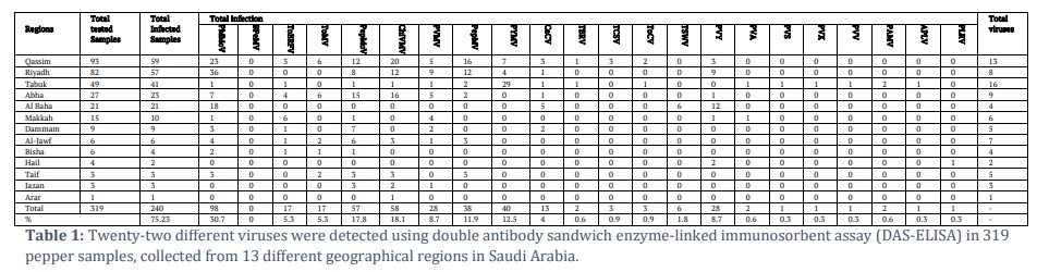
Results: According to ELISA results, two hundred and forty samples were infected with at least one of the tested viruses with a percentage of 75.23%. Infection rates varied significantly by region, with the highest prevalence observed in Al-Baha (95%) and Dammam, Al-Jawf, Taif, and Jazan (100%). A total of twenty-one viruses were detected, with PMMoV, ChiVMV, and PepMoV being the most widespread.
Conclusion: This study reports the first detection of twenty-two viruses in pepper in Saudi Arabia. Notably, three of these viruses (APLV, PLRV, and PVV) are reported for the first time infecting pepper worldwide.
Keywords: DAS-ELISA; Pepper; viruses; Saudi Arabia.
Introduction![]()
Peppers (Capsicum spp.) of both types pungent and non-pungent are importantly increasing vegetable and spice crop throughout the world [1]. There are 27 species in the Capsicum genus, which are native to tropical Central and South America, but only five of them are domesticated. [2].
Of these, Capsicum annum is the one that is most frequently produced for commercial purposes and has plenty of fruit market varieties that are both pungent and non-pungent. Pepper fruits are primarily produced for food coloring and flavoring, dried spices (paprika, chili powder), fresh vegetables (hot and sweet peppers), and different compound extraction for industrial or medical use. [1].
According to the FAO production data (http://faostat.fao.org/), the annual global fresh pepper output rose around four times between 1982 and 2012, from around 8.2 to about 31.2 million tons. Pepper production has steadily increased in the past 20 years, in terms of the amount of growing space and fruit yield. Over 35 million tons of fresh peppers were produced worldwide in 2017 from over two million hectares. With almost 17 million tons production in 2017, China was the top producer, with Mexico, Europe, Turkey, and Indonesia following closely behind with one to 3.3 million tons each.
Viruses are the most important factors threatening pepper production throughout the world including Australia [3], Africa [4], Asia [5] and Europe [6]. Viral diseases not only lower the fruit output and quality but also raise the price of taking preventative measures and making sanitary planting materials. Additionally, due to the enormous genetic diversity of viral strains and the accumulation of these strains in propagation materials, unaffected areas are easily infected by them [1]. The widespread consensus is that viruses tend to infect cultivated peppers quite easily [7].
About 68 virus species have been reported to infect pepper [8]. In the Mediterranean Basin region mostly pepper infecting viruses include tobamoviruses (TMV, ToMV, TMGMV, and PMMoV), the aphid-transmitted potyvirus (PVY and TEV), thrips-transmitted tospoviruses (TSWV) and cucumovirus (CMV) [9].
In Saudi Arabia, during the visits to several farms in 2021-2022, virus-like symptoms such as mosaic, mottling, chlorotic and necrotic lesions, blistering, yellowing of leaves and fruits, and plant stunting were observed in different Regions of Saudi Arabia. Based on observed symptoms, the present study was designed to detect viruses infecting pepper crops.
![]() Methods
Methods
Field Survey and serological detection
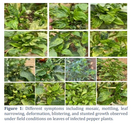
During the growing season of 2021-2022, a total of 319 symptomatic and asymptomatic plant samples showing virus-like symptoms including mottling and mosaic on leaves, puckering of leaves, vein thickening, vein chlorosis, leaf curling, dwarfing, stunted growth, deformed fruits, and leaves upward cupping (Figure 1) were collected from the following different regions in Saudi Arabia: Qassim (93) Riyadh (82), Tabuk (49), Abha (27), Al-Baha (21), Makkah (15), Dammam (9), 6 samples from each Al-Jawf and Bisha , Hail (4), 3 samples from each Taif and Jazan, and one sample from Arar.
All the collected samples were tested by double antibody sandwich enzyme-linked immunosorbent assay (DAS-ELISA) against important viruses infecting pepper using specific antibodies for the following viruses: pepper mild mottle virus (PMMoV), tomato brown rugose fruit virus (ToBRFV), tomato mosaic virus (ToMV), pepper mottle virus (PepMoV), chili veinal mottle virus (ChiVMV), pepper veinal mottle virus (PVMV), pepino mosaic virus (PepMV), pepper yellow mosaic virus (PYMV), tomato black ring virus (TBRV), Tomato Chlorotic Spot Virus (TCSV), tomato chlorosis virus (ToCV), Tomato Spotted Wilt Virus (TSWV), Potato Virus Y (PVY), Potato Virus A (PVA), Potato Virus S (PVS), Potato Virus X (PVX), Potato Virus V (PVV), Potato Aucuba Mosaic Virus (PAMV), Andean potato latent virus (APLV) and Potato Leaf Roll Virus (PLRV) available from the manufacturers LOEWE®, Biochemica, Germany, whereas Bell-Pepper Mottle Virus (BPeMV) and Capsicum Chlorosis Virus (CaCV) were obtained from DSMZ, Germany. The ELISA procedure was performed according to the manufacturer’s instructions, both companies as demonstrated by [10, 11].
Briefly, commercial polyclonal antibodies were utilized at the concentrations recommended by their producers (LOEWE Biochemica GmbH, and DSMZ, Germany). IgG from the original vial was diluted in a Coating Buffer and mixed gently. In each well of the ELISA plate, 0.2 ml of the IgG working solution was added. Plates were covered with sealing tape tightly and the plates were kept in the refrigerator overnight. To remove the IgG, plates were washed four times using a washing buffer. A sample buffer in a ratio of 1:20 w/v was used to homogenize the samples.
The sample buffer was used to dissolve positive and negative controls. In each well of the ELISA plate, 0.2 ml sample extract and control solution were added. Sealing tape was used to cover the plates tightly. All the plates were incubated overnight. A washing buffer was used four times to remove the unbound extracted samples. IgG from the original vial was diluted in a conjugated buffer and mixed gently. A conjugate working solution (0.2 ml) was added in each well of the ELISA plate. Sealing tape was used to cover the plates then kept at 37 °C for 2- 4 hours or in the refrigerator overnight. The washing buffer was used four times to remove the conjugate buffer. The substrate solution was prepared by mixing para-nitrophenyl- phosphate tablets (1mg/ml) into substrate buffer, after that 200 μL solution was added in ELISA plates.
To acquire a clear reaction, plates were covered and incubated for 30-60 min at 37 °C in a dark place. BioTek Instruments, ELx 808 reader, USA was used to record the absorbance at 405 nm. In each microtiter test plate, ELISA test samples were deemed positive when their absorbance values were more than double the average of suitable healthy controls.
Field observations and sampling
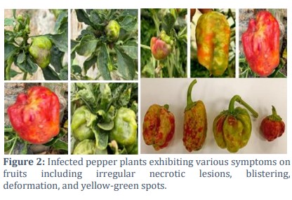
Three-hundred and nineteen symptomatic and asymptomatic samples were collected from different locations in-growing areas in the Qassim, Riyadh, Al-Baha, Abha, Tabuk, Al-Jawf, Makkah, Dammam, Arar, Jazan, Taif, Hail and Bisha of Saudi Arabia. Symptomatic leaf samples exhibited complex symptoms including clear mottling on the leaf and becoming narrow, deformed evidently, intermingled dark to light green patches referred to as mosaic with mottling, yellow mosaic, vein chlorosis, leaf curling, blistering symptoms causing deformation of leaves and stunted growth (Figure 1). While the fruit symptoms showed blistering and irregular necrotic lesions, deformation, and yellowish green spots. (Figure 2).
Serological detection of important pepper viruses
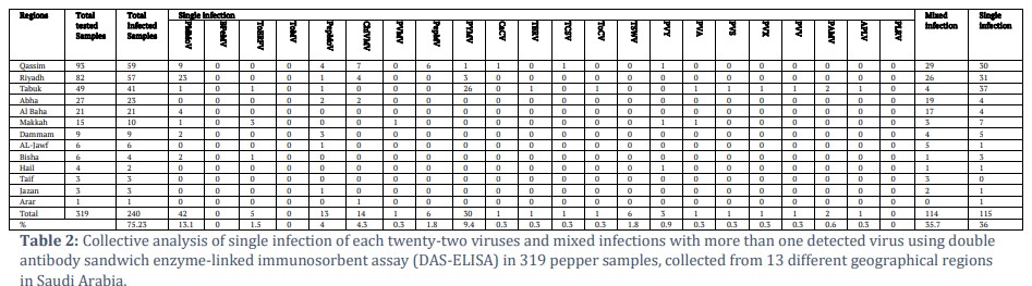
ELISA results for the Qassim region showed that thirteen tested viruses were present in 59/93 (63.4%) of the samples including PMMoV 23 (24.7%), ToBRFV 3 (3.2%), ToMV 6 (6.4%), PepMoV 12 (12.9%), ChiVMV 20 (21.5%), PVMV 5 (5.3%), 16 PepMV (17.2%), PYMV 7 (7.5%), CaCV 3 (3.2%), TBRV 1 (1%), TCSV 3 (3.2%), ToCV 2 (2.1%) and PVY 3 (3.2%) samples respectively (Table 1). The obtained results showed that 30/59 samples (50.8%) were found to be singly infected with PMMoV (9.6%), PepMoV (4.3%), ChiVMV (7.5%), PepMV (6.4%), and 1 % for each PYMV, TCSV, PVY, respectively. However, 29/59 samples had mixed infections with at least more than one virus in the tested samples, while the remaining samples were negative (Table 2). For Riyadh region, ELISA results showed that 57/82 (69.5%) samples were infected with eight viruses including: PMMoV 36 (43.9%), PepMoV 8 (9.8%), ChiVMV 12 (14.6%), PVMV 9 (11%), PepMV 12 (14.6), PYMV 4 (4.87%), CaCV 1 (1.2%) and PVY 9 (11%) and however the remaining viruses were not detected in tested samples (Table 1). The obtained results of ELISA revealed that 26 samples were singly infected with PMMoV (28%), PepMoV (1.2%), ChiVMV (4.8%), and PYMV (3.6%). However, 31 samples had mixed infections with at least more than one virus.. Whereas the remaining samples were found to be negative against any of the viruses tested (Table 2).
For Tabuk region, according to the ELISA results, 41/49 (83.6%) samples were infected with 16 viruses including: PMMoV 1 (2%), ToBRFV 1 (2%), PepMoV 1 (2%), ChiVMV 1 (2%), PVMV 1 (2%), PepMV 2 (4%), PYMV 29 (59%), CaCV 1 (2%), TBRV 1 (2%), ToCV 1 (2%), PVA 1 (2%), PVS 1 (2%), PVX 1 (2%), PVV 1 (2%), PAMV 2 (4%) and APLV 1 (2%), while BPeMV and ToMV was not detected in any tested samples (Table 1). The obtained results showed that 31/49 (63%) samples were found to be singly infected with PYMV (53%) and PAMV (4%), while PMMoV, ToBRFV PepMoV, TBRV, ToCV, PVS, PVX, PVA, PVV and APLV had 2% single infection for each virus separately. Whereas 11/49 samples (22%) were detected with mixed infections with at least more than one virus, however, the rest of the samples were found to be negative (Table 2).
For Abha region, ELISA results showed that 23/27 (85%) samples were infected with the following nine viruses including: PMMoV 7 (25.9%), ToBRFV 4 (14.5%), ToMV 6 (22.2%), PepMoV 15 (55.5%), ChiVMV 16 (59.2), PVMV 5 (18.5%), PepMV 2 (7.4%), CaCV 1 (3.7%) and PVY 1 (3.7%) were found in the tested samples during the ELISA test (Table 1). Single infections with PepMoV and ChiVMV were each detected in 2 of the 27 samples (7.4%). Mixed infections were found in 4 samples (14.8%), while 4 samples were negative for all tested viruses. (Table 2).
For Al-Baha region, 20/21 (95%) samples were positive with four viruses including PMMoV 18 (85.7%), CaCV 5 (23.8%), TSWV 6 (28.5%), and 12 PVY (57.1%). The results obtained showed that single infection was found in 4/21 samples (19%) with only PMMoV. However, mixed infection in 16/21 samples of TSWV (28.5%), PVY (57.1%), and CaCV (23.8%) were found respectively (Table 1, 2). For Makkah region, six viruses (PMMoV, ToBRFV, PepMoV, PVMV, PVY, and PVA) were found in the tested samples and ten out of fifteen (66.6%) samples were positive. Single infection in PMMoV 1 (6%), ToBRFV 6 (40%), PepMoV 1 (6%), PVMV 4 (26%), PVY 1 (6%), and PVA 1 (6%) were detected, while 20% mixed infections were present in tested samples with at least more than one virus, however, 5 samples were negative during the test with any virus (Table 1, 2). Dammam region, all samples were positive with five viruses including PMMoV 3 (33.3%), ToBRFV 1 (11%), PepMoV 7 (77%), PVMV 2
(22.2%), and CaCV 2 (22.2%). Single infection in PMMoV (22%) and PepMoV (33%), was detected, and 100% mixed infections were found in tested samples with more than one virus, all samples give positive results (Table 1, 2).
For Al-Jawf region, ELISA results showed that all six samples were positive with the following seven viruses PMMoV 4 (66%), ToBRFV 1 (16%), ToMV 2 (33%, PepMoV 6 (100%), ChiVMV 3 (50%), PVMV 1 (16%), and PepMV 3 (50%). Single infection was found only in one sample (16%) with PepMoV, while 83% of mixed infection was present in tested samples with more than one virus (Table 1, 2).
For Bisha region, four viruses (PMMoV, ToBRFV, ToMV, and PepMoV) were found in 4/6 (66%). Single infection with PMMoV 2 (33%) while, ToBRFV, ToMV, and PepMoV 1 (16%) was detected, and 66% mixed infection was present in tested samples with at least more than one virus, however, 2 samples were negative during the test (Table 1, 2).
For Hail region, two viruses (PVY and PLRV) were found in the tested samples during the serological study and two out of four (50%) samples were positive. Single infection with PVY 1 (25%), was detected, and 50% of mixed infections were found in tested samples with at least more than one virus, however, 2 samples were negative during the test (Table 1, 2).
For Taif region, five viruses (PMMoV, ToMV, PepMoV, ChiVMV and PepMV) were detected in all 3 tested samples (100%). There was no single infection found with any of the tested viruses whereas 100% mixed infection with more than one virus was observed in all tested samples (Table 1, 2).
For Jazan region, three viruses PepMoV 3(100%), ChiVMV 2 (66%) and PVMV 1 (33%) were found in all the tested samples. Single infection in PepMoV (33%), was detected, and 100% mixed infection was found in tested samples with more than one virus (Tables 1,2). For Arar region, only one virus was found in the tested sample with ChiVMV 1(100%). Bell pepper mottle virus, the only virus which was not found to infect pepper crops in all tested samples collected from any of the regions. (Table 1, 2).
![]() Discussion
Discussion
Pepper is commercially a significant crop playing a major role in the agriculture sector of Saudi Arabia because of its usages as well as demand. In the Kingdom, viruses pose a threat to this crop and result in significant losses in pepper production every year [13] as well as throughout the world. There are about 68 viruses that are known to infect pepper crops globally, of which 20 species are the most harmful and cause significant losses to pepper crop [9, 14].
Wetter (1984) [15] used immunodiffusion test to detect PMMoV and ToMV from pepper plants, while Ayo-John and Odedara (2017) [16] detected PVMV and ToMV infecting pepper crop from Nigeria using DAS-ELISA. Topkaya (2022) [17] detected CMV, TSWV, AMV, and PMMoV through DAS-ELISA infecting pepper crop in Turkey.
Damayanti and Kurniawati (2022) [18] detected PMMoV infecting naturally pepper plants using dot-immunobinding assay from Indonesia. Because Tobamoviruses are seed transmitted [19] and had been detected from many countries PMMoV [20], this is the main source of due to which viruses are invading in Saudi Arabia through trade of infected seeds and propagated planting material. Comparing our results to aforementioned studies revealed that our study is more comprehensive and detailed, and there is a higher number of detected pepper viruses. The highest number of viruses identified are 16 in Tabuk region followed by Qassim having 12 viruses, while the lowest number is one from Arar region.
Prior to this study, only five viruses had been reported, namely Pepper Leafroll Chlorosis Virus (PLRCV) [13], Tomato Yellow Leaf Curl Virus (TYLCV) [21], Alfalfa Mosaic Virus (AMV) [22], Bean Yellow Mosaic Virus (BYMV) [23] and Pepper Mild Mottle Virus (PMMoV) [24,25] to infect pepper crop in Saudi Arabia.
Three hundred and nineteen samples were collected from different regions of Saudi Arabia were tested using DAS-ELISA and confirmed the presence of twenty-two viruses belonging to different virus groups including PMMoV, ToBRFV, ToMV, PepMoV, ChiVMV, PVMV, PepMV, PYMV, CaCV, TBRV, TCSV, ToCV, TSWV, PVY, PVA, PVS, PVX, PVV, PAMV, APLV and PLRV in two-hundred and forty samples with total infection of 75.2%. Interestingly from these twenty-two viruses, three viruses (APLV, PLRV and PVV) had never
been reported to infect pepper crop before throughout the world, while seventeen viruses have been reported for the first time in Saudi Arabia to infect pepper crop.
This work provides a detailed and comprehensive survey and diagnosis of important viruses infecting pepper crop in Saudi Arabia for the first time. Three viruses (APLV, PLRV and PVV) have been reported for the first time to infect pepper crop in world while seventeen viruses (ToBRFV, ToMV, PepMoV, ChiVMV, PVMV, PepMV, PYMV, CaCV, TBRV, TCSV, ToCV, TSWV, PVY, PVA, PVS, PVX, PAMV) has been reported for the first time to infect pepper crop in Saudi Arabia. BPeMV was the only virus from the tested virus which wasn’t reported in any of the regions.
Tables and Figures
Author Contributions
Amer MA, Hussain K, Al-Saleh MA: Conceptualization, sample collection, Khalid Z: Methodology and experiments, Amir M, Zaman M: Software and data analysis, Amer MA, Khalid Z, Al-Shahwan IM: Writing- Original draft preparation, Reviewing and Editing.
Acknowledgements
The authors extend their appreciation to the Deanship of Scientific Research, King Saud University, for funding through the Vice Deanship of Scientific Research Chairs, the chair of date palm research.
![]() References
References
- Kenyon L, Kumar S, Tsai W-S, Hughes Jd A. Virus diseases of peppers (Capsicum spp.) and their control, Advances in virus research, Elsevier, (2014); 90: 297-354.
- Ibiza VP, Blanca J, Cañizares J, Nuez F. Taxonomy and genetic diversity of domesticated Capsicum species in the Andean region. Genetic resources and crop evolution, (2012); 59 (8): 1077-1088.
- Solomon M, Edwards OR. First Complete Genome Sequence of Pepper vein yellows virus from Australia. Genome Announcements (2016); 4(3): e00450-16.
- Aliyu TH. The incidence, severity and occurrence of four viruses infecting pepper (Capsicum spp.) in the Southern Guinea Savannah Agro-ecological Zone of Nigeria. Agriculturae Conspectus Scientificus, (2014); 79(4): 233-237.
- Arogundade O, Ajose T, Osijo I, Onyeanusi H, Matthew J, et al. Management of viruses and viral diseases of pepper (Capsicum spp.) in Africa. Capsicum, IntechOpen,(2020); 6: 73-86.
- Svoboda J, Svobodová-Leišová L. Occurrence of viruses on pepper plantations in the Czech Republic. Horticultural Science, (2012); 39(12): 139-143.
- Green SK, Kim JS. Characteristics and control of viruses infecting peppers: a literature review (Vol. 18). Shanhua, Taiwan: Asian Vegetable Research and Development Center. Technical Bulletin, 1991; 18: 60
- Pernezny K, Roberts PD, Murphy JF, Goldberg NP. Compendium of Pepper Diseases. The American Phytopathological Society, St. Paul, Minnedota, (2003); p.73.
- Moury B, Verdin E. Viruses of pepper crops in the Mediterranean basin: a remarkable stasis. In Advances in virus research, Elsevier Academic Press, (2012); 84(1): 127-162).
- Clark MF, Adams A. Characteristics of the microplate method of enzyme-linked immunosorbent assay for the detection of plant viruses. Journal of general virology, (1977); 34(3): 475-483.
- Lommel SA, McCain AH, Morris TJ. Evaluation of indirect enzyme-linked immunosorbent assay for the detection of plant viruses. Phytopathology, (1982); 72(8):1018-1022.
- Daryono BS, Somowiyarjo S, Natsuaki KT. New source of resistance to Cucumber mosaic virus in melon. SABRAO Journal of Breeding and Genetics, (2003); 35:19-26.
- Kamran A, Lotos L, Amer MA, Al-Saleh MA. Alshahwan IM, et al. Characterization of pepper leafroll chlorosis virus, a new polerovirus causing yellowing disease of bell pepper in Saudi Arabia. Plant disease, (2018); 102(2): 318-326.
- Kumari N, Sharma V, Patel P, Sharma PN. Heterologous expression of pepper mild mottle virus coat protein encoding region and its application in immuno-diagnostics. Virusdisease, (2020); 31(3): 323-332.
- Fulton RW. Serological identification of four tobamoviruses infecting pepper. Plant Disease, (1984); 68: 597-599
- Ayo-John EI, Odedara OO. Serological detection of viruses infecting tomato and pepper in Southwest Nigeria and their distribution. Nigerian Journal of Biotechnology, (2017); 33: 78-82.
- Topkaya S. Serologic and molecular determination and phylogenetic analysis of some viruses infecting pepper in Turkey. Emirates Journal of Food and Agriculture, (2022); 34(11): 923-930.
- Damayanti TA, Kurniawati F. Pepper mild mottle virus infection in cayenne and sweet pepper in Indonesia, Australasian Plant Disease Notes, (2022);17(1): 4.
- Kaur SI, Kashyap PL, Kang SS, Sharma A. Detection and diagnosis of seed-borne viruses and virus-like pathogens. In: Kumar, R., Gupta, A. (eds) Seed-Borne Diseases of Agricultural Crops: Detection, Diagnosis & Management. Springer, (2020); 169-199.
- Svoboda J, Červená G, Rodová J, Jokeš M. First report of Pepper mild mottle virus in pepper seeds produced in the Czech Republic–Short communication. Plant Protection Science, (2006); 42(1): 34.
- Haq QM, Sohrab SS, Brown JK, Al-Harrasi A. Association of tomato yellow leaf curl virus-Oman strain with the leaf curl and yellow mosaic symptoms on papaya and wild poinsettia in Oman. Canadian Journal of Plant Pathology, (2022); 44(3): 465-472.
- Abdalla OA, Al-Shahwan IM, Al-Saleh MA, Amer MA. Molecular characterization of Alfalfa mosaic virus (AMV) isolates in alfalfa and other plant species in different regions in Saudi Arabia. European Journal of Plant Pathology, (2020); 156: 603-613.
- Al-Shahwan IM, Abdalla OA, Al-Saleh MA, Amer MA. Detection of new viruses in alfalfa, weeds and cultivated plants growing adjacent to alfalfa fields in Saudi Arabia. Saudi Journal of Biological Sciences, (2017); 24(6): 1336-1343.
- Al-Wabli Afaf S, Khattab Eman AH, Farag Azza G. Purification and production of antiserum against pepper mild mottle virus isolated from Saudi Arabia. Research Journal of Biotechnology, (2017); 12: 12.
- Khalid Z, Amer MA, Amir M, Hussain K, Al-Shahwan I, et al. Serological detection of important pepper viruses and characterisation of pepper mild mottle virus in Saudi Arabia. Australasian Plant Pathology, (2024); 53(1): 67-78.
This work is licensed under a Creative Commons Attribution-Non Commercial 4.0 International License. To read the copy of this license please visit: https://creativecommons.org/licenses/by-nc/4.0