

Full Length Research Article
Isolation and Screening of the Heavy Metals, Antibiotics Resistant and Acidophilic Profile of Bacterial strains from Lead Acid Batteries Repairing and Recycling Units
Sulaiman Faisal1, Gulzar Ahmad1, Zia Ur Rahman2, Muhammad Taimur Asad2, Muhammad Ijaz1, Adil Ahmad3, Sidra Ahmad4, Saqib Rauf1, Neelum Iqbal4, Sajjad Ullah2*
Adv. life sci., vol. 12, no. 2, pp. 451-459, May 2025
*– Corresponding Author: Sajjad Ullah (Email: sajjad.ullah@mlt.uol.edu.pk)
Authors' Affiliations 1. Institute of Integrative Bioscience (IIB) CECOS University Peshawar – Pakistan
2. University Institute of Medical Laboratory Technology, The University of Lahore – Pakistan
3. Institute of Nursing Science, Khyber Medical University Peshawar – Pakistan
4. Institute of Biotechnology and Genetic Engineering, The University of Agriculture Peshawar – Pakistan
2. University Institute of Medical Laboratory Technology, The University of Lahore – Pakistan
3. Institute of Nursing Science, Khyber Medical University Peshawar – Pakistan
4. Institute of Biotechnology and Genetic Engineering, The University of Agriculture Peshawar – Pakistan
[Date Received: 14/01/2024; Date Revised: 16/02/2025; Date Available Online: 31/08/2025]
Abstract![]()
Introduction
Methods
Results
Discussion
References
Abstract
Background: Heavy metal contamination from unregulated waste disposal poses serious risks to ecosystems, soil, water, and human health. Lead acid battery recycling sites are major sources of such pollution. This study aimed to isolate and characterize bacterial strains with potential for bioremediation from lead acid battery workshops in Shoba Bazaar, Peshawar.
Methods: Water samples were collected from contaminated sites and cultured in both nutrient agar and LB media. After incubation, 30 bacterial isolates were screened for tolerance to cadmium, lead, zinc, and chromium (50–300 mM), as well as resistance to commonly used antibiotics (ampicillin, amoxicillin, azithromycin, cefixime, and kanamycin). The most tolerant strains, designated LRB1, LRB2, and LRB3, were further analyzed for acid and temperature resistance. Morphological and molecular characterization included Gram staining, microscopic analysis, plasmid isolation and genomic DNA extraction.
Results: Isolates LRB1, LRB2, and LRB3 demonstrated high lead tolerance of 270 mM, 300 mM, and 270 mM, respectively. All three strains exhibited resistance to multiple antibiotics like ampicillin (10μg), amoxicillin (30μg), azithromycin (15μg), cefixime (5μg) and kanamycin (30μg), and LRB3 showed growth across a broad pH range (2–8). Plasmid DNA was successfully isolated, indicating potential plasmid-mediated resistance. Gram staining revealed that the isolates were Gram-positive bacilli and cocci. Furthermore, genomic DNA extraction and 16S rDNA PCR with universal primers were used for detection and identification of the bacterial isolates.
Conclusion: The isolated bacterial strains demonstrated remarkable tolerance to heavy metals, acids, and antibiotics, suggesting their potential role in the bioremediation of contaminated environments. Further molecular studies are required to elucidate the mechanisms underlying their resistance and to evaluate their suitability for biotechnological applications.
Keywords: Heavy metal resistance; Antibiotic resistance; Lead acid batteries; Acidophilic strains; Lead Resistant strains
Introduction![]()
Heavy metal contamination of soil and wastewater is a critical global environmental challenge, as these pollutants are non-biodegradable, highly persistent, and capable of bioaccumulation in food chains. Their presence poses serious threats to ecosystem stability and human health, adversely affecting animals, plants, and microbial communities [1]. The extensive use of heavy metals in industrial processes such as electroplating and paint manufacturing has resulted in their release into the environment, leading to contamination of the food chain and subsequent life-threatening health outcomes in humans, including cancer, metabolic disorders, and neurological disturbances such as insomnia. In addition to industrial activities, automobile emissions and intensive agricultural practices further contribute to the burden of heavy metal pollution [2,3].
Based on their biological functions and effects on the human body, metals can be classified into three categories: (i) essential metals with established physiological roles, such as Na, K, Mg, Fe, Co, Ni, Cu, and Zn; (ii) toxic metals, including Ag, Sn, Cd, Au, Hg, Ti, Pb, Al, Sb, and Se; and (iii) non-essential and non-toxic metals with no known biological function, such as Rb, Cs, and Sr. Among these, mercury (Hg) is considered the most toxic heavy metal, while other highly toxic metals include nickel, lead, zinc, cadmium, copper, and chromium. In particular, lead (Pb) is widely recognized as a major environmental pollutant, commonly detected in the atmosphere and associated with severe risks to human health [4-6]. Heavy metal toxicity is everywhere in the water bodies, in the soil and in the atmosphere causing serious damages to all forms of life. The industrial revolution has led to an unprecedented rise in environmental pollution due to the metal usage in the sector, in electroplating, in leather industries and in lead acid batteries among others[7]. Lead acid battery industries are growing very rapidly specially in the developing countries due to its utilization in solar panel systems, motor cars, motorcycles, and for storing electricity these lead batteries are mostly used[8]. Pakistan is facing a similar situation as electricity shortage in Pakistan is a major problem and to overcome this problem solar panel systems utilizing the lead acid batteries are being widely used. Although solar panels are very useful for overcoming the shortage of electricity but problem arises during the repair of these batteries as there is no proper way of repairing and recycling of lead acid batteries in workshops located in different cities; Lahore, Faisalabad, Peshawar [9,10]. During the repair of lead acid batteries, the expired raw material like cadmium plates, H2SO4 and lead plates are discarded directly to the environment and are extremely toxic to the ecosystem and can cause some serious problems. Lead, which is used in the lead acid batteries, is very toxic to human health and cause insomnia, fatigue, weakness and drowsiness. Exposure to exceedingly high levels of lead in the working environment cause adverse health effects [2]. In this study, we examined lead acid batteries repairing and recycling units located in Shoba bazaar, Peshawar Khyber Pakhtunkhwa Pakistan and isolated heavy metal resistance bacterial strains from the acidic water samples. Their tolerance towards heavy metal and antibiotics was checked because bacteria have adapted themselves to survive in environments contaminated with metals. These bacteria also show resistance towards antibiotics as bacteria harbor plasmids (circular DNA) that have antibiotic resistance genes and metal resistant genes [10]. These genes can be present either on plasmids or on chromosomal DNA of some bacterial species; in nature plasmids often carry genes that benefit the survival of the organism, such as by providing antibiotic resistance and metal resistance [11].
The role of the microbes in the removal of heavy metals from the environment is very important because different microbe utilized technologies are being used to remove the toxic metals from the environment i.e. bioremediation of contaminated sites. The traditional technologies used for removing toxic metals are ion exchange, chemical precipitation, reverse osmosis and evaporative recovery but these technologies are often inefficient and very expensive that's why different microbial technologies and genetically modified organisms are used for remediation processes. [12].These microbes adopt various mechanisms to interact and survive in the presence of heavy metals such as biotransformation, extrusion, use of enzyme, ion exchange, metal efflux pumps, intercellular, extracellular sequestration [13]. In this study, bacteria were isolated from lead-acid battery repair and recycling units in Shoba Bazar, Peshawar, and evaluated for their tolerance to cadmium (Cd) and lead (Pb). The isolates exhibited strong resistance to these metals as well as to multiple antibiotics. The research further explored their resistance mechanisms under heavy metal stress, antibiotic exposure, and varying pH conditions, highlighting their potential use in future bioremediation applications.
![]() Methods
Methods
Sample Collection
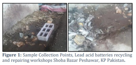
The two sampling sites decided for sampling were car batteries repair workshop and the battery acid waste water collection point in Shoba Bazaar, Peshawar. Water samples from these two sites were taken in sterile 50 mL Falcon tubes and stored in ice box. The GPS coordinates were LT 34.0068371 L71.5597000 for sample one and LT 34.0064124 L 71.5592058 for sample 2. The samples were brought to lab for further analysis the sampling points are shown in Figure 1.
Isolation and Screening of the bacterial strains
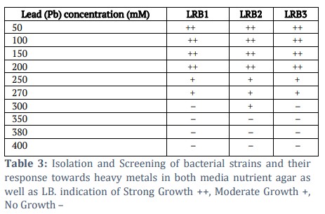
For the isolation and screening of metal-resistant bacterial strains, stock solutions of lead (Pb), cadmium (Cd), zinc (Zn), and chromium (Cr) salts were prepared. Working concentrations of 50, 100, 150, 200, 250, 270, 300, 350, and 380 mM were used to evaluate bacterial tolerance. A 100 µL aliquot of water sample was inoculated into LB broth supplemented with the respective metal salts and simultaneously spread onto LB agar plates containing the same concentrations. Control plates (without metal supplementation) were inoculated with 100 µL of the water sample to assess background growth. All inoculated broths and plates were incubated overnight at 37 °C. The three most resistant isolates, designated Lead-Resistant Bacteria (LRB1, LRB2, and LRB3), were selected and subsequently re-streaked for purification and further analysis.
Luria Bertani Media
For the preparation of 500 mL of LB broth, 5.2 g tryptone, 5.2 g NaCl, and 2.6 g yeast extract were dissolved in 400 mL of distilled water. The volume was then adjusted to 500 mL with distilled water, and the medium was sterilized by autoclaving. For solid LB agar medium, 15 g of agar was added per liter of LB broth prior to sterilization.
Optimum temperature and pH characterization of the isolates
To determine the optimum temperature and pH for the growth of the isolated strains, cultures were incubated under varying conditions. For temperature optimization, 100 µL of overnight culture was spread onto 20 mL LB agar plates and incubated at 20, 25, 30, 35, 37, 45, and 70 °C. For pH optimization, 100 µL of overnight culture was inoculated into 20 mL LB broth adjusted to pH 2, 4, 5, 6, 7, 8 and 9. Growth was monitored following incubation under the respective conditions [14].
Antibiotic Resistance via disc diffusion method
Antibiotic resistance of the isolates was evaluated using the standard disc diffusion method. LB agar plates 20 mL were prepared, and a bacterial lawn was generated by spreading 100 µL of overnight culture of each strain onto the surface. Antibiotic discs containing ampicillin (10 µg), amoxicillin (30 µg), azithromycin (15 µg), cefixime (5 µg), and kanamycin (30 µg) were aseptically placed onto the inoculated plates. The plates were incubated at 37 °C for 18–24 h, after which the diameters of inhibition zones were measured and interpreted according to Clinical and Laboratory Standards Institute (CLSI) guidelines [15].
Molecular analysis
Genomic DNA Extraction
Genomic DNA was extracted using the cetyltrimethylammonium bromide (CTAB) method. As a preparatory step, 2 µL of β-mercaptoethanol was added to 1 mL of CTAB buffer and preheated to 65 °C. A 1.5 mL aliquot of overnight bacterial culture was transferred to a microcentrifuge tube and centrifuged at 10,000 rpm for 3 min. The supernatant was discarded, and the pellet was resuspended in 500 µL of preheated CTAB buffer by pipetting. The suspension was incubated at 65 °C for 20 min. Following incubation, 400–500 µL of phenol–chloroform–isoamyl alcohol (25:24:1) was added, and the mixture was centrifuged at 12,000 rpm for 10 min. The aqueous phase was transferred to a fresh microcentrifuge tube, and genomic DNA was precipitated by adding two volumes of 95% ethanol. The mixture was either stored at –20 °C for later processing or centrifuged immediately at 12,000 rpm for 10 min. The resulting pellet was washed with 400 µL of 70% ethanol and centrifuged again at 12,000 rpm for 10 min. After discarding the supernatant, the pellet was air-dried for ~2 h. Finally, the dried pellet was resuspended in 50–100 µL of TE buffer, and the extracted DNA was stored at –20 °C until further use [16].
Plasmid DNA Extraction
Plasmid DNA from the bacterial isolates was extracted using a manual alkaline lysis method. A 1 mL aliquot of overnight culture was transferred into two labeled 1.5 mL microcentrifuge tubes and centrifuged at maximum speed for 1 min. The supernatant was discarded, and 100 µL of ice-cold GTE solution was added to each pellet. Cells were resuspended thoroughly by pipetting to avoid clumping. Subsequently, 200 µL of freshly prepared SDS/NaOH solution was added, and the tubes were gently inverted five times before incubation on ice for 5 min. Following this, 150 µL of ice-cold potassium acetate (KoAc) solution was added, and the tubes were inverted five times until a white precipitate appeared. After a further 5 min incubation on ice, the samples were centrifuged at maximum speed for 5 min. Approximately 400 µL of the clear supernatant was transferred to fresh microcentrifuge tubes, to which 400 µL of isopropanol was added. The tubes were inverted five times, incubated at room temperature for 2 min, and centrifuged for 5 min to precipitate plasmid DNA. The supernatant was carefully removed, and the pellet was washed with 200 µL of 100% ethanol by gentle inversion, followed by centrifugation at maximum speed for 2–3 min. After discarding the supernatant, the DNA pellet was air-dried for 10 min at room temperature. Finally, the pellet was resuspended in 15 µL of TE buffer, and DNA from both tubes was pooled into a single tube and stored at –20 °C until further use [17].
16S rDNA PCR
PCR amplification of the 16S rRNA gene was performed using universal primers 27F and 1392R. A PCR master mix was prepared for the DNA samples as well as for positive and negative controls. Each 50 µL reaction mixture contained 5 µL of 10× Taq buffer, 1 µL each of forward and reverse primers, 0.5 µL of dNTPs mix, 0.25 µL of Taq DNA polymerase, and nuclease-free water to make up the final volume. Thermocycling conditions were as follows: initial denaturation at 94 °C for 4 min; 30 cycles of denaturation at 94 °C for 1 min, annealing at 50 °C for 30 s, and extension at 72 °C for 1.5 min; followed by a final extension at 72 °C for 5 min. Amplified products were stored at –20 °C until further use [18].
Gel electrophoresis
DNA samples were analyzed by agarose gel electrophoresis. A 1% agarose gel was prepared by dissolving 0.5 g of agarose in 50 mL of 1× TAE buffer with heating until fully dissolved. The solution was cooled to ~50 °C, after which 0.5 µL of ethidium bromide (EtBr) was added for nucleic acid staining. The molten agarose was poured into a gel casting tray fitted with a comb pre-cleaned with 70% ethanol and allowed to solidify at room temperature for 30 min. The gel was then placed in an electrophoresis chamber containing 1× TAE buffer for subsequent DNA loading and separation [19].
Gel loading
A 1 kb DNA ladder was prepared by diluting 1 µL of the ladder with 9 µL of TE buffer, followed by the addition of 2 µL of 6× loading dye, and subsequently loaded onto the gel. DNA samples and control DNA samples were prepared by mixing 5 µL of DNA with 1 µL of 6× loading dye and loaded into adjacent lanes. Electrophoresis was carried out at 80 V for 60 min, and DNA bands were visualized using a gel documentation system.
MIC (Minimal Inhibitory Concentration)
The minimum inhibitory concentration (MIC) was defined as the lowest antibiotic concentration that visibly inhibited bacterial growth. For MIC determination, bacterial isolates were first sub-cultured on nutrient agar and incubated for 24 h. The inoculum was prepared in nutrient broth containing 0.45 g yeast extract and 0.75 g bacteriological peptone in 150 mL distilled water, and the turbidity was adjusted to match the McFarland standard. Serial dilutions of ampicillin, amoxicillin, azithromycin, cefixime, and kanamycin were prepared. Five concentrations of each antibiotic were tested, ranging from half to double the predefined CLSI (2023) cutoff value. Each well of a sterile 96-well microtiter plate was loaded with 195 µL of the antibiotic dilution and inoculated with 5 µL of bacterial suspension. Optical density (OD) was recorded at 600 nm using a spectrophotometer immediately after inoculation. Plates were then incubated at 37 °C for 24 h, after which OD was measured again to determine bacterial growth inhibition [20].
Statistical Analysis
Data analysis was performed using statistical software including Origin, GraphPad, and SPSS. A Student’s t-test was applied to assess differences, with statistical significance set at P ≤ 0.05. Both data processing and statistical analyses were conducted. For MIC determination, t-tests were specifically employed to compare initial and final bacterial growth readings in the presence of antibiotics.
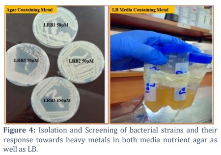
Isolation and Screening of bacterial strains and their response towards heavy metals
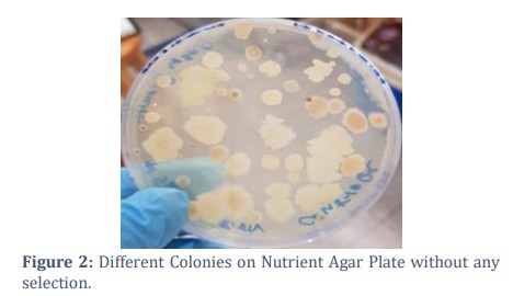
Screening was carried out against varying concentrations of lead, chromium, cadmium, and zinc salts. Three strains demonstrating the highest tolerance to lead were selected and designated LRB1 (Lead Resistant Bacteria), LRB2, and LRB3. LRB1 and LRB3 exhibited resistance up to 270 mM lead acetate, while LRB2 tolerated concentrations up to 300 mM Figure 5, Table 3. In contrast, the isolates were unable to withstand higher concentrations of zinc, cadmium, or chromium salts, with no growth observed beyond 50 mM. Control plates without metal supplementation showed approximately 30 colonies Figure 2.
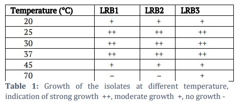
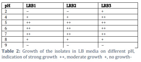
Optimum temperature and pH characterization of the isolates
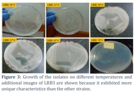
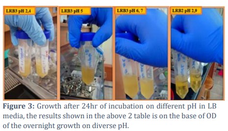
The optimum growth conditions for the isolates were determined by culturing them at different temperatures and pH values. All three strains exhibited maximum growth at 37 °C and a range of pH 5-7. Figure 4; Tables 1 and 2. Interestingly, LRB3 demonstrated minimal growth even at 70 °C, whereas no growth was observed for LRB1 and LRB2 at this temperature Figure 3. A similar trend was observed at pH 2, where LRB3 showed minimum growth, while LRB1 and LRB2 failed to grow under such highly acidic conditions.
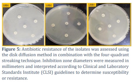
Antibiotic Resistance
Antibiotic susceptibility profiling revealed that strain LRB1 was resistant to all tested antibiotics except kanamycin. Strain LRB2 exhibited resistance to azithromycin, ampicillin, and cefixime, while showing susceptibility to kanamycin and intermediate susceptibility to amoxicillin.
Strain LRB3 was resistant to azithromycin, ampicillin, and cefixime, susceptible to amoxicillin, and demonstrated intermediate susceptibility to kanamycin. Resistance patterns were assessed using the disc diffusion method, and inhibition zones were measured in millimeters (mm) according to CLSI standards. The results are presented in Figure 6 and Table 4.
Minimal Inhibitory Concentration
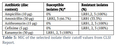
To evaluate the effect of different antibiotic concentrations on the bacterial strains, the minimum inhibitory concentration (MIC) was determined for ampicillin, amoxicillin, azithromycin, cefixime, and kanamycin using a 96-well microtiter plate assay. Optical density (OD) readings were recorded before and after 24 hours of incubation. Among the tested antibiotics, cefixime exhibited complete resistance (0% susceptibility, 100% resistance) across all isolates, whereas kanamycin showed the highest level of susceptibility at 100%. The OD results indicated resistance and susceptibility patterns, which are 0% susceptibility for ampicillin, 66.6 % for amoxicillin, and 0 % for azithromycin. The detailed results are presented in Table 5.
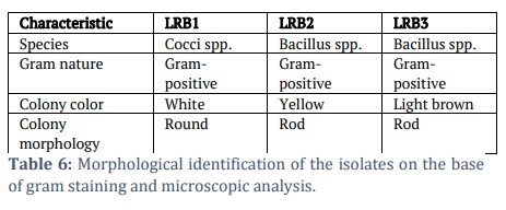
Morphological identification
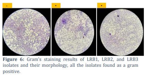
Gram’s Staining
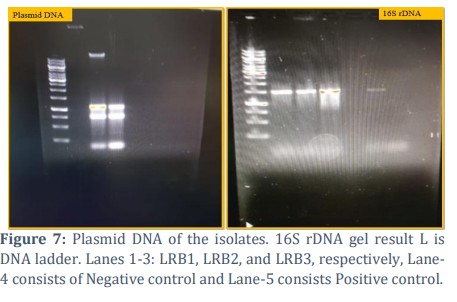
The Gram staining results of the three isolates are shown in Table 6 and Figure-7.
Molecular analysis
Genomic and plasmid DNA extraction
Genomic DNA of the isolates was successfully extracted through CTAB method. Similarly, Plasmid DNA was extracted and the DNA was run on 1% agarose gel at 80V for 60 minutes. Results are shown in Figure 8.
16S rDNA PCR
The 16S rRNA gene PCR of the isolates was performed, including positive and negative controls. The amplified DNA bands are shown in Figure 8. A distinct band of approximately 1500 bp was observed in the bacterial isolates, consistent with the expected product size amplified by the universal 16S primers.
![]() Discussion
Discussion
Rapid and alarming changes in the ecosystem are causing several environmental issues, leading to the extinction of various organisms and the loss of their natural habitats. These impacts are even more severe in developing countries such as Pakistan, Afghanistan, and regions of Africa, where factors like poor drainage systems, deforestation, and limited environmental management exacerbate the situation. Microbes are also affected by these environmental changes; however, they possess unique adaptive mechanisms. For instance, through genomic alterations and the presence of antibiotic resistance genes located on both plasmids and chromosomal DNA, microbes can survive and thrive under changing conditions [21]. Microbes play a vital role in the system they are sustaining toxic metals from the environment which are now as an essential process for many industries in the form of bioremediation, bioaccumulation, bioleaching [22,23]. the point is that the microbes and specially these acidophilic and heavy metal toxic strains or halophiles strains can play a vital role for maintaining the environment friendly for plants and animals and for that biotechnology and recombinant DNA technology should be brought to the field to make the ecosystem friendly[24]. On the other hand to understand the antibiotic mechanisms of these strains will overcome the therapeutics threats of (Multi Drug Resistance) MDR [22,25].
In this study water sample was collected from lead acid batteries repairing units located in Shoba bazar Peshawar KPK. Metal resistant bacteria were isolated by spread plate method on LB and LB Agar, plates supplemented with various metals (Pb, zinc, chromium, and cadmium). The isolates show highest resistance towards lead, three isolates were selected from the LB/Agar plate supplemented with 300mM, 270mM and 270mM lead acetate salt. Gram staining was performed for morphological and species-level identification. The isolates were identified as Bacillus species (LRB2 and LRB3) and a Cocci species (LRB1).
Lead toxicity occurs as a result of changes in the conformation of nucleic acids and proteins, inhibition of enzyme activity, disruption of membrane functions and oxidative phosphorylation, as well as alterations of the osmotic balance [26]. Lead (Pb) also shows a stronger affinity for thiol and oxygen groups than essential metals such as calcium and zinc. Despite the high toxicity of Pb, many micro-organisms have evolved mechanisms that enable them to survive Pb exposure[27]. Lead has a low biological available concentration due to its low solubility. Thus Pb is not extraordinarily toxic to microorganism [28]. Some forms of lead salt like lead acetate or nitrate, induced mutagenicity and DNA breaks at a toxic dose in some bacterial species [29]. Microorganisms have developed several mechanisms that allow them to survive exposure to Pb. The main mechanism of lead resistance involve adsorption by extracellular polysaccharides, cell exclusion, sequestration as insoluble phosphates, and ion efflux to the cell exterior [30]. A study by Uqab, B. et al., [31] conducted in Kashmir they also isolated lead Pb+ and other metal resistant bacterial species from environmental samples the study demonstrated the Staphylococcus equorum, Staphylococcus warneri, Bacillus safensis and Bacillus thuringiensis using MALDI-TOF assays [31] and 16s rRNA approach they found various strains which can be used for bioremediation purpose and removing toxic metals from the ecosystem, but our study has not just found the isolates which are resistant to metal but can also resistant to antibiotics, acids and even temperature. Tariq et al 2022, Isolated a novel strain from high saline environment from Peshawar area which are resistant up to 2M solution of salt and shown resistance to antibiotics as well[5]. These all strains need to characterize and sequencing analysis these strains will be very effective for bioremediation, bioleaching of toxic and essential metals and for agriculture sector as well, the plants which are susceptible to grown in the saline area with the help of these strains that’s area will be possible for any kind of crops and plants specially in Sindh and Balochistan areas of Pakistan [32].
In this study the lead resistant isolates were found to resistant Ampicillin, Amoxicillin, Cefixime, Kanamycin and Azithromycin. These genes imparting resistance to metal ions and antibiotics can co-exist on plasmid or on chromosomes [33]. This co-occurrence is caused by cross- and co-resistance Phenomena. Cross-resistance occurs when the same mechanism simultaneously reduces the susceptibility to metals and antimicrobial agents used in therapy, and co-resistance occurs when separate resistance genes are situated on the same genetic element [4,34]. The isolates were also grown under pH 2.0‐9.0, and the optimum pH of the isolates is 5-7. In summary, this study describes that the three bacterial isolates had a wide range of adoptability under different environmental condition based on the tolerance towards heavy metals, antibiotics, temperature and pH and the strains were useful for bioremediation prospects and can be use in the biotechnology applications like bioleaching of metals[35], removal of toxic metals, agriculture sector and also need a proper recycling programed to avoid these toxic metals into the ecosystem that will avoid healthcare issues of all the organisms because they are throwing these acids and waste products directly to the environment which is a hot concerns over all, unfortunately due to the unavailability of funding we could not sequence the selected isolates to check their genomic and plasmid genes responsible for this mechanisms these are the limitations of this study as well.
This study aimed to isolate and characterize bacterial strains from lead-acid battery repair and recycling workshops, with a focus on their tolerance to heavy metals, antibiotics, temperature, and pH. Three isolates, designated as Lead-Resistant Bacteria (LRB1, LRB2, and LRB3), exhibited high lead tolerance, with maximum growth observed up to 270 mM. Antibiotic susceptibility testing revealed varied resistance patterns: all isolates were resistant to cefixime and ampicillin, while differential susceptibility was observed for amoxicillin, azithromycin, and kanamycin. Notably, LRB3 demonstrated growth across a wide pH range (2–8), while all isolates grew optimally at pH 5–7 and 37 °C. Genomic and plasmid DNA were successfully extracted, and 16S rDNA PCR confirmed the isolates as Gram-positive bacteria, with LRB2 and LRB3 identified as Bacillus spp. and LRB1 as a Cocci species. These findings highlight the potential application of the isolates in bioremediation of heavy metal contaminated environments and suggest their relevance for future biotechnological exploitation. Further molecular characterization is required to elucidate the underlying mechanisms of metal and antibiotic resistance. Overall, this study underscores the significance of exploring bacterial diversity for addressing environmental contamination and advancing biotechnological and agricultural solutions because of their unique diverse properties.
Tables and Figures

Authors Contributions
Dr. Sulaiman Faisal and Dr. Zia Ur Rahman supervised the project under the kind correspondence of Dr. Sajjad Ullah. All authors contributed to the study’s conception and design, performed experiments, analyzed the data, and participated in the interpretation of results. They were collectively involved in drafting, reviewing, and editing the manuscript, approved the final version, and agreed to its submission.
Competing interest
All authors of this work have declared that there is no conflict of interest.
Funding Information
This work doesn’t receive any funding.
Data Availability
All relevant data are within the paper.
![]() References
References
- Dajani Maity S, Sarkar D, Poddar K, Patil P, Sarkar A. Biofilm-mediated heavy metal removal from aqueous system by multi-metal-resistant bacterial strain Bacillus sp. GH-s29. Applied Biochemistry and Biotechnology, (2023); 195(8): 4832-4850.
- Awad el Karim MA, Hamed AS, Elhaimi YA, Osman Y. Effects of exposure to lead among lead-acid battery factory workers in Sudan. Archives of Environmental Health, (1986); 41(4): 261-265.
- Ijaz M, Ahmad G, Anjum F, Zeb U, Muhammad N, et al. Morphological Identification and Resistance Profile of Antibiotic and Heavy Metals-Resistant Bacteria in Hospital Sewage of Peshawar. Advancements in Life Sciences, (2023); 10(3): 452-456.
- Gurave NA, Korde VV, Dhas SS, Disale M. Isolation and identification of heavy metal resistant bacteria from petroleum soil of Loni, Ahmednagar. European Journal of Experimental Biology, (2015); 5(12): 6-11.
- Hussain T, Usman S, Khan A, Hussain A, Aziz A, et al. Isolation and Characterization of Salt Tolerant Bacteria from Saline Areas of Khyber Pakhtunkhwa. Journal of Clinical and Medical Research, (2022); 4(4): 1-8.
- Khan A, Ahmad G, Ijaz M, Ahsan K, Akhtar H, et al. Prevalence and Antibiotic Resistance of Salmonella Species Isolated from Chicken Liver in Peshawar. Journal of Clinical and Medical Research (2021); 3(3): 1-11.
- Raja CE, Selvam G, Omine K. Isolation, identification and characterization of heavy metal resistant bacteria from sewage; International Joint Symposium on geodisaster and Geoenvirnement, 2009. pp. 205-211.
- Khan I, Zia NAM, Nisa SJP, Biology A. 06. Isolation and identification of heavy metal tolerant bacteria from volta battery effluent, Hattar, Haripur Pakistan. Pure and Applied Biology, (2021); 5(4): 719-726.
- Valasai GD, Uqaili MA, Memon HR, Samoo SR, Mirjat NH, et al. Overcoming electricity crisis in Pakistan: A review of sustainable electricity options. Renewable and Sustainable Energy Reviews, (2017); 72734-745.
- Ahmad G, Rizwan MM, Azam F, Ijaz M, Khan AU, et al. Prevalence of Staphylococcus aureus and Antimicrobial Drug Residues in Chicken Liver its Antibiogram Analysis and Implications on Public Health. Indian Journal of Microbiology, (2024); 1-9.
- de Silva AAL, de Carvalho MAR, de Souza SA, Dias PMT, da Silva Filho RG, et al. Heavy metal tolerance (Cr, Ag and Hg) in bacteria isolated from sewage. Brazilian Journal of Microbiology, (2012); 43(4): 1620.
- March-Rosselló GAJEiymc. Rapid methods for detection of bacterial resistance to antibiotics. Enfermedades Infecciosas y Microbiología Clínica, (2017); 35(3): 182-188.
- Martínez JL, Baquero F, Andersson DIJCoip. Beyond serial passages: new methods for predicting the emergence of resistance to novel antibiotics. Current Opinion in Pharmacology, (2011); 11(5): 439-445.
- Hmani I, Ghaderiardakani F, Ktari L, El Bour M, Wichard T. High-temperature stress induces bacteria-specific adverse and reversible effects on Ulva (Chlorophyta) growth and its chemosphere in a reductionist model system. Botanica Marina, (2024); 67(2): 131-138.
- Carvalho GM, Silva BA, Xavier RGC, Zanon IP, Vilela EG, et al. Evaluation of disk diffusion method for testing the rifampicin, erythromycin, and tetracycline susceptibility of Clostridioides (prev. Clostridium) difficile. Anaerobe, (2023); 80102720.
- Weerakkody LR, Witharana C. A rapid, inexpensive and effective method for the efficient isolation of genomic DNA from Gram-negative bacteria. Molecular Genetics and Genomics, (2024); 299(1): 26.
- Kodackattumannil P, Sasi S, Krishnan S, Lekshmi G, Kottackal M, et al. Protocol for the High-quality Plasmid Isolation from Different Recalcitrant Bacterial Species: Agrobacterium spp., Rhizobium sp., and Bacillus thuringiensis. Bio-protocol, (2023); 13(15).
- Faniyan O, Akpe V, Cock IE. Analyzing bacterial species from different environments using direct 16S rRNA gene sequencing methods. Pharmacognosy Communications, (2023); 13(1): 24-33.
- Dendani Chadi Z, Arcangioli M-A. Pulsed-Field Gel Electrophoresis Analysis of Bovine Associated Staphylococcus aureus: A Review. Pathogens, (2023); 12(7): 966.
- Dortet L, Niccolai C, Pfennigwerth N, Frisch S, Gonzalez C, et al. Performance evaluation of the UMIC® Cefiderocol to determine MIC in Gram-negative bacteria. Journal of Antimicrobial Chemotherapy, (2023); 78(7): 1672-1676.
- Rangasamy K, Athiappan M, Devarajan N, Samykannu G, Parray JA, et al. Pesticide degrading natural multidrug resistance bacterial flora. Microbial Pathogenesis, (2018); 114304-310.
- Sun J, He X, Le Y, Al-Tohamy R, Ali SS. Potential applications of extremophilic bacteria in the bioremediation of extreme environments contaminated with heavy metals. Journal of Environmental Management, (2024); 352120081.
- Gallo G, Aulitto M, Contursi P, Limauro D, Bartolucci S, et al. Bioprospecting of Extremophilic Microorganisms to Address Environmental Pollution. Journal of Visualized Experiments, (2021); (178).
- Kochhar N, I․K K, Shrivastava S, Ghosh A, Rawat VS, et al. Perspectives on the microorganism of extreme environments and their applications. Current Research in Microbial Sciences, (2022); 3100134.
- Varshney S, Bhattacharya A, Gupta A. Halo-alkaliphilic microbes as an effective tool for heavy metal pollution abatement and resource recovery: challenges and future prospects. Biotechnology, (2023); 13(12): 400.
- Macdonald C, Singh B. Harnessing plant-microbe interactions for enhancing farm productivity. Bioengineered, (2014); 5(1): 5-9.
- Yazdankhah S, Skjerve E, Wasteson Y. Antimicrobial resistance due to the content of potentially toxic metals in soil and fertilizing products. Microbial Ecology in Health and Disease, (2018); 29(1): 1548248.
- Nies DH. Microbial heavy-metal resistance. Applied microbiology and biotechnology, (1999); 51(6): 730-750.
- Tchounwou PB, Yedjou CG, Patlolla AK, Sutton DJ (2012) Heavy metal toxicity and the environment. Molecular, clinical and environmental toxicology: Springer. pp. 133-164.
- Jarosławiecka A, Piotrowska-Seget Z. Lead resistance in micro-organisms. Microbiology, (2014); 160(1): 12-25.
- Uqab B, Nazir R, Ahmad Ganai B, Rahi P, Rehman S, et al. MALDI-TOF-MS and 16S rRNA characterization of lead tolerant metallophile bacteria isolated from saffron soils of Kashmir for their sequestration potential. Saudi Journal of Biological Sciences, (2020); 27(8): 2047-2053.
- Oldfield E, Feng XJTiPS. Resistance-resistant antibiotics, (2014); 35(12): 664-674.
- Novick RP, Roth C. Plasmid-linked resistance to inorganic salts in Staphylococcus aureus. Journal of Bacteriology, (1968); 95(4): 1335-1342.
- Kalsoom, Batool A, Din G, Din SU, Jamil J, et al. Isolation and screening of chromium resistant bacteria from industrial waste for bioremediation purposes. Brazilian Journal of Biology, (2021); 83e242536.
- Ali K, Ahmad S, Ijaz-ul H, Khan S, Khan AJPJLPRR. Prevalence and Epidemiological Findings of MDR (Multi-Drug Resistant) Typhoid bacillus in District Swat KPK. Journal of Lung, Pulmonary & Respiratory Research, (2017); 4(5): 00142.
This work is licensed under a Creative Commons Attribution-Non Commercial 4.0 International License. To read the copy of this license please visit: https://creativecommons.org/licenses/by-nc/4.0