

Full Length Research Article
CT analysis of subcutaneous and visceral adipose tissue in normal BMI subjects: association with level of physical activity and hypertension
Hafiz Muhammad Rizwan1, Zareen Fatima1, Shoaib Rasool1, Iqra Ilyas1, Khalid Idrees2, Taiba Zulfiqar1, Maria Mohsan1, Arshad Jamal3,4, Ahmed Bilal Waqar5*
Adv. life sci., vol. 7, no. 3, pp. 164-169, May 2020
*– Corresponding Author: Ahmed Bilal Waqar (Email: drabwaqar@yahoo.com)
Authors' Affiliations
2. Department of Radiology, Amna Inayat Medical College, Lahore, Pakistan
3. Department of Biology, College of Science, University of Hail – Kingdom of Saudi Arabia
4. Department of Medical Laboratory Sciences, Faculty of Health and Allied Sciences, Imperial College of Business Studies, Lahore, Pakistan.
5. Faculty of Health and Allied Sciences, Imperial College of Business Studies, Lahore, Pakistan
Abstract![]()
Introduction
Methods
Results
Discussion
References
Abstract
Background: The distribution of adipose tissue, complex factors affecting it and its pathological consequences are among the hot topics in medical research nowadays. Most of the studies reported in the literature however describe the association of factors affecting the fat distribution in overweight and obese individuals. This particular study was however planned to find out the same in subjects having normal basal metabolic index (BMI). The objectives of the study were to analyze total adipose tissue (TAT), subcutaneous adipose tissue (SAT) and visceral adipose tissue (VAT) in the abdomen volumetrically using CT, to establish the association of these to the levels of physical activity, presence or absence of hypertension and to compare these associations in both the genders.
Methods: A prospective study was carried out on seventy five, normal BMI subjects aged between 20–50 years. CT imaging was used for volumetric measurement of TAT, SAT and VAT. Pearson’s correlation of these were then found out with age. Kruskal Wallis test was also performed to compare these in hypertensive and non-hypertensive subjects and in those with different physical activity levels (PAL).
Results: Women showed significantly higher volumes of TAT and SAT. Men showed statistically significant correlations of TAT and VAT with age. SAT volumes had significant negative association with the PAL in both genders. Men showed higher responsiveness of fat deposition in all compartments to the presence of hypertension.
Conclusion: In conclusion, factors such as gender, age, level of physical activity and hypertension affect the site specific deposition of fat even in those individuals who aren’t over-weight or obese.
Keywords: Total Adipose Tissue (TAT); Subcutaneous Adipose Tissue (SAT); Visceral Adipose Tissue (VAT)
Introduction![]()
Adipose tissue in the abdominal region is divided into different compartments namely subcutaneous adipose tissue (SAT) and visceral adipose tissue (VAT) [1]. The later includes the total amount of fat located inside the abdominal cavity comprising of both retroperitoneal and intraperitoneal adipose tissue [2]. There are various factors which are thought to affect the distribution of fat deposition in these compartments. Cardiovascular diseases such as hypertension and metabolic syndrome- related disorders like Type 2 diabetes, dyslipidemia, hyperinsulinemia, insulin resistance are reported to be more strongly associated with VAT than SAT [3]. Similarly, reduction in VAT also results in marked decrease in obesity- related problems and improvements in cardiovascular diseases and other risk factors [3-6].
Males are known to have a propensity for more accumulation of VAT while women tend to have more total abdominal adipose tissue (TAT) and SAT [7-8]. These differences in the distribution of adipose tissue have also different implications in both the genders. A recent study reports the central obesity as an independent predictor of ischemic stroke in women but not in men [9]. Similarly, waist circumference, an indicator of abdominal adiposity showed, in comparison to BMI, a slightly weaker association with type 2 diabetes in men. However in women BMI and central obesity had almost equal association with type 2 diabetes mellitus.8 Physical activity has also been reported to affect the distribution of fat in both genders differently. Exercise affects VAT more in men but SAT in women [10].
Multiple imaging modalities like CT, MRI, Ultrasound and dual-energy X-ray absorptiometry (DXA) have been used to assess the volume of abdominal adiposity [3, 10-19].
The current study was designed with an intention to measure SAT, VAT and TAT volumetrically using CT and find their association with the physical activity levels and presence or absence of hypertension. As we also know from previous studies [7] that the distribution of fat differs in different ethnicities and to the best of our knowledge this will be the first study, correlating these factors with abdominal adipose compartments from our part of the world.
Recently, abdominal adiposity has been recognized as a better indicator of cardiovascular risks and complications than BMI [20]. However, most of the previous studies reported factors affecting the amount and location of fat accumulation in those individuals who are either obese or fit in the criteria of metabolic syndrome. This particular study was however planned to find out the relationship of SAT, VAT and TAT volumes with these factors in the individuals having normal BMI with or without hypertension and different levels of physical activity.
Objectives of the study were to measure volumes of total adipose tissue (TAT), subcutaneous adipose tissue (SAT) and visceral adipose tissue (VAT) in the abdomen by CT, to establish the correlation of these to the levels of physical activity, presence or absence of hypertension and to compare these associations in both the genders.
Methods![]()
This prospective study was conducted after approval from Institutional Ethical Review Board. Informed consents in Urdu and English were taken from subjects. It included 75 adult individuals (Male: Female – 43: 32) with age range of 20–50 years. These patients had BMI values falling in the range of 18.5-25 and were not over-weight or obese. In addition, those individuals who were diabetic, smokers, alcoholics, those suffering from chronic kidney disease or chronic liver disease, with history of abdominal tumors/surgery and aged more than 50 year at the time of evaluation were excluded from the study.
Assignment of Physical Activity Levels
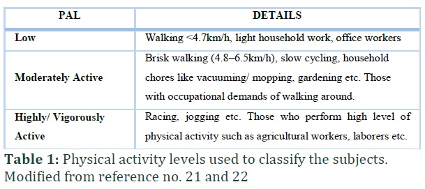
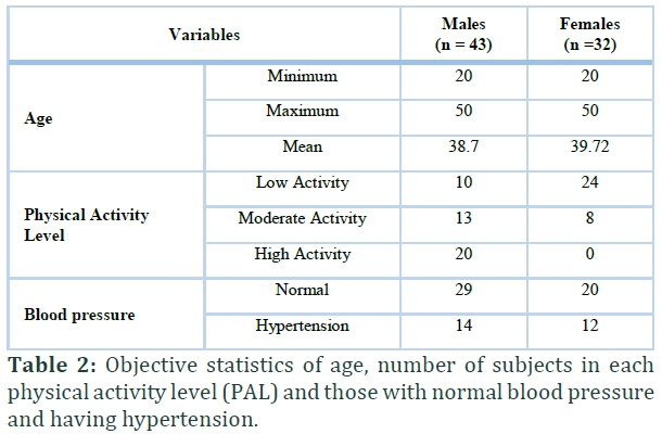
Inquiry based, detailed investigation about physical activity was carried out and physical activity levels (PAL) were assigned to the patients according to Table 1 [21, 22]. The number of patients in each category of PAL and normal and hypertensive categories are given in the Table 2.
Imaging
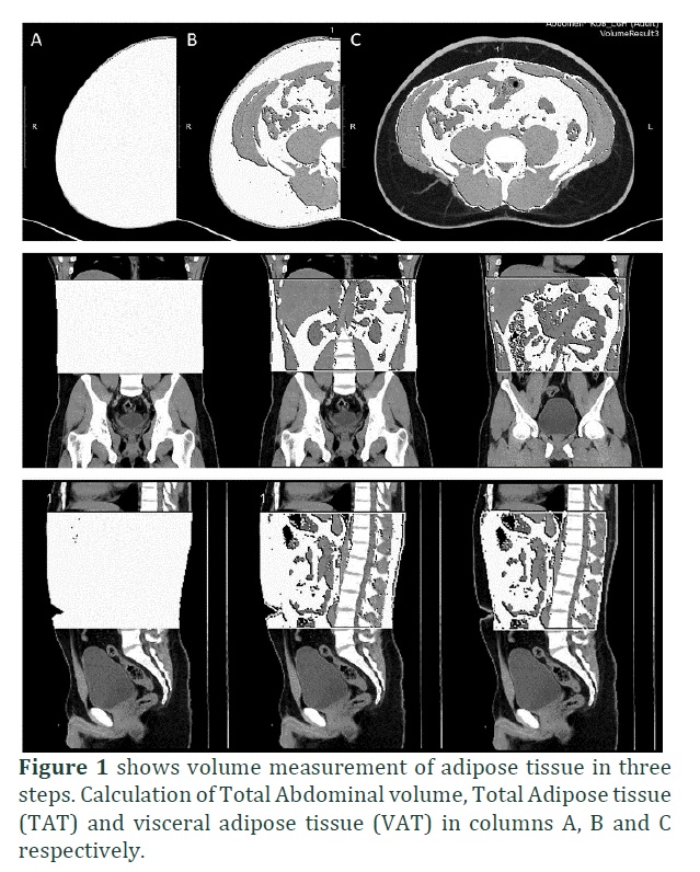
128-Somatom Definition Edge multi slice CT scanner was used to scan and post- processing was then done with slice thickness and gap of 1mm each. Scan area included from xiphoid process superiorly down to superior iliac crest. Care was taken to include lateral, anterior and posterior abdominal walls in the scan. Volumetric measurements were performed in the following steps using Volume application of the CT scanner (Figure 1):
- Total volume of the all the tissues in the scanned area was obtained by setting the image display window between -1024 HU to +3000 HU. This measurement included all the tissues including muscles, bones, air, subcutaneous, visceral fat etc.
- Total abdominal fat volume was then measured by using the Hounsfield Units range between -150 to -30. This measurement included both VAT and SAT volumes.
- The abdominal muscular wall separating the visceral from the subcutaneous adipose compartment was then traced manually to measure VAT and excluding SAT from the measurement.
- SAT volume was then calculated by subtracting VAT from the total abdominal fat volume
Statistical Analysis
The collected data were analyzed using the MEDCALC. Volumes of adipose tissue in different compartments were compared in men and women using Kruskal-Wallis test. Pearson’s correlation coefficient (R) and P values were found out between age and calculated volumes of TAT, SAT and VAT. Kruskal Wallis test was performed to compare the adipose tissue in various compartments in both genders with different PALs and in normal and hypertensive individuals. P-value of 0.05 or less was taken as significant.
Results![]()
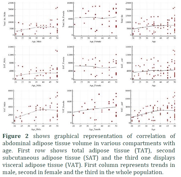
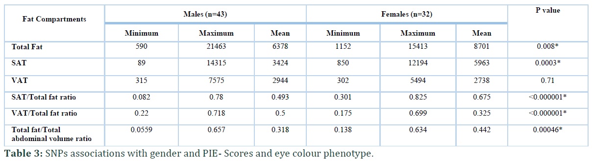
The distribution of fat in both the genders in each compartment is shown in the Table 3. Women showed significantly higher volumes of total fat as well as SAT compared to men. Men had greater volume of VAT but the difference wasn’t significant.
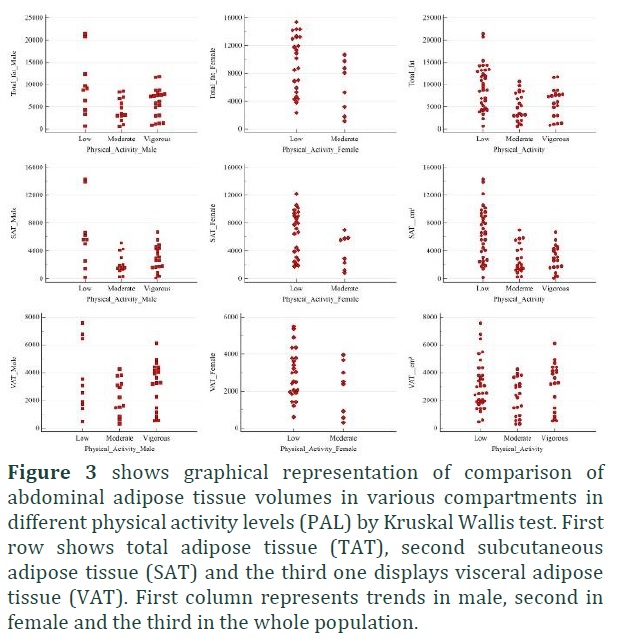
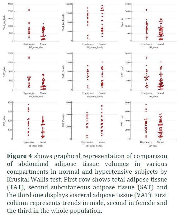
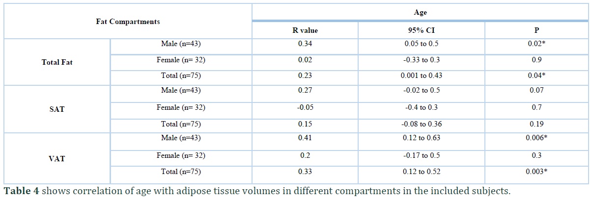
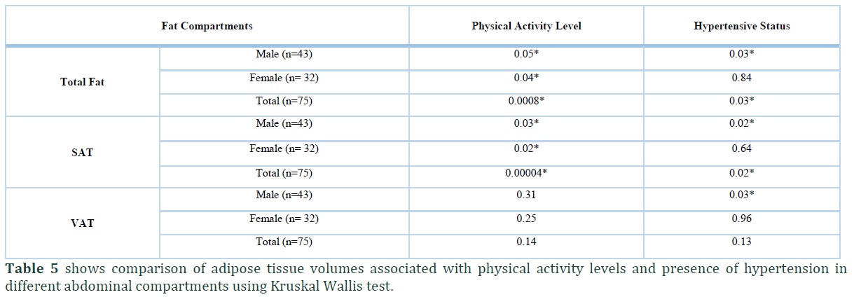
A statistically significant correlation was noted between volume of TAT and advancing age in the total population and in men. The volume of VAT also showed a significantly greater positive correlation with age in men but no such trend was seen in the volumes of SAT (Table 4, Figure 2). SAT volumes were noted to be significantly different in three PALs in the whole population as well as in men and women. VAT on the other hand showed no significant difference in these groups (Figure 3). Men showed higher responsiveness of fat deposition to the grades of hypertension, volumes of each of the SAT, VAT and TAT showed significant association with presence of hypertension. No such relationship was seen in women. (Table 5, Figure 4)
Figures & Tables
Discussion![]()
Factors such as ethnicities, obesity, diabetes, PAL, presence of hypertension, age and gender are all known to be associated with the likelihood of not only deposition but site -specific deposition of adipose tissue [2, 4–11, 20,23].
Irrespective of ethnicities, women have higher TAT and SAT deposits but men have tendency to accumulate VAT [24]. We also found such trend of fat deposition in female population of our study. Men on the other hand, although, showed more volumes of VAT compared to women but this difference wasn’t statistically significant. The reason might be the fact that out of 32 female subjects in our study 15(47%) and 8(25%) were above 45 and 50 years of age respectively. Possible menopause in some of these subjects might have caused increased deposition in the visceral compartment too resulting in the loss of statistical difference between the two genders. A higher propensity of VAT deposition is seen in the post-menopausal women compared to those who are still menstruating [25]. The lack of information about the menopausal status of women is a major limitation in our study.
Increased amount of fat deposition, especially visceral, is associated with essential hypertension [23]. Very interesting finding of our study was that the abdominal adiposity in men showed significant association with presence of hypertension in both subcutaneous and visceral compartments but no such association was seen in the women. This finding of ours is in accordance with a study conducted by Guo et. al on 168 overweight or obese subjects. They found a positive association between reduction in visceral fat and improvements in both systolic and diastolic blood pressures in men but not in women [20]. We didn’t however specifically recorded the antihypertensive medication used by these patients. This knowledge might also be important because of the known side effects of some of the drugs like beta blockers to affect the weight gain and fat redistribution. These may even selectively promote the accumulation of abdominal fat.
A study by Murabito et al., suggested the negative correlation of both SAT and VAT with PAL [26]. This correlation was however stronger in women than in men. In our study, we also noted significant negative correlation of PAL with TAT and SAT which was stronger in women than in men. We however found no association of VAT with activity level in both men and women. The difference might be due to the fact that the mentioned article reports the effect of a planned moderate to vigorous physical activity with accelerometer performed by the participants over a period of 5-7days. In our population on the other hand a questionnaire based grading of physical activity level was performed by inquiring them about their profession, workout etc. Secondly only middle aged men and women were included in their study while our study population comprised of subjects under 50years of age. In another study, Fischer K et. al reported significant negative association of physical activity with VAT volumes in males and with SAT in females in subjects above 62 years and BMI [11]. This study also differs with ours in the age of the studied subjects.
This study of ours might serve as a baseline study for the volumes of fat deposits in various compartments in normal BMI population of this country. The knowledge is very crucial because ethnicity, genetic factors, life style, socioeconomic status, level of awareness about physical fitness, and even type of cuisine in that specific region have roles to play in body fat distribution.
In conclusion, factors such as gender, age, level of physical activity and hypertension affect the site specific deposition of fat even in those individuals who aren’t over-weight or obese.
Hafiz Muhammad Rizwan: Data Collection, Literature search
Zareen Fatima: Study Designing, Statistical Analysis and Manuscript preparation.
Shoaib Rasool: Literature search, data collection
Iqra Ilyas: Literature search, data collection
Khalid Idrees: Data collection, manuscript preparation
Taiba Zulfiqar: Literature search, data collection
Maria Mohsan: Manuscript preparation
Arshad Jamal: Manuscript preparation
Ahmed Bilal Waqar: Study designing, data analysis and manuscript preparation
All the authors declare that they have no competing interest that can negatively affect the current study.
References![]()
- Fox CS, Massaro JM, Hoffmann U, Pou KM, Maurovich-Horvat P, et al. Abdominal visceral and subcutaneous adipose tissue compartments: association with metabolic risk factors in the Framingham Heart Study. Circulation, 2007; 116: 39–48.
- Chandra A, Neeland IJ, Berry JD, Ayers CR, Rohatgi A, et al. The relationship of body mass and fat distribution with incident hypertension: Observations from the dallas heart study. Journal of the American College of Cardiology, 2014; 64: 997–1002.
- Snijder MB, Visser M, Dekker JM, Seidell JC, Fuerst T, et al. The prediction of visceral fat by dual-energy X-ray absorptiometry in the elderly: a comparison with computed tomography and anthropometry. International journal of obesity, 2002; 26(7): 984–93.
- William MJ, Hunter GR, Kekes-Szabo T, Synder S, Treuth MS. Regional fat distribution in women and risk of cardiovascular disease. The American journal of clinical nutrition, 1997; 65: 855-860.
- Chaston TB, Dixon JB. Factors associated with percent change in visceral versus subcutaneous abdominal fat during weight loss: findings from a systemic review. International journal of obesity, 2008; 32: 619-628.
- Britton KA, Massaro JM, Murabito JM, Kreger BE, Hoffman U, Fox CS. Body fat distribution and incident cardiovascular disease, cancer and all-cause mortality. Journal of the American College of Cardiology, 2013; 62: 921-925.
- Staiano AE, Broyles ST, Gupta AK, Katzmarzyk PT. Ethnic and sex differences in visceral, subcutaneous and total body fat in children and adolescents. Obesity (Silver Spring), 2013; 21: 1251–1255.
- Bidulescu A, Liu J, Hickson DA, Hairston KG, Fox ER, et al. Gender differences in the association of visceral and subcutaneous adiposity with adiponectin in African Americans: the Jackson Heart Study. BMC cardiovascular disorders, 2013; 13(1): 9.
- Zahn K, Linseisen J, Heier M, Peters A, Thorand B, et al. Body fat distribution and risk of incident ischemic stroke in men and women aged 50 to 74 years from the general population. The KORA Augsburg cohort study. PLoS One, 2018; 13(2): e0191630.
- Meisinger C, Doring A, Thorand B, Heier M, Lowel H. Body fat distribution and risk of type 2 diabetes in the general population: are there differences between men and women? The MONICA/KORA Augsburg cohort study. The American journal of clinical nutrition, 2006, 84 (3): 483-489.
- Fischer K, Rüttgers D, Müller HP, Jacobs G, Kassubek J, et al. Association of habitual patterns and types of physical activity and inactivity with MRI-determined total volumes of visceral and subcutaneous abdominal adipose tissue in a general white population. PLoS One, 2015; 10: 1–22.
- Micklesfield LK, Goedecke JH, Punyanitya M, Wilson KE, Kelly TL. Dual-Energy X-Ray Performs as Well as Clinical Computed Tomography for the Measurement of Visceral Fat. Obesity, 2012; 20: 1109–1114.
- Mauad FM, Chagas-Neto FA, Benedeti, Augusto César Garcia Saab, Nogueira-Barbosa MH, et al. Reproducibility of abdominal fat assessment by ultrasound and computed tomography. Radiologia brasileira, 2017; 50 (3): 141-147.
- Irlbeck T, Massaro JM, Bamberg F, O'Donnell CJ, Hoffmann U, Fox CS. Association between single-slice measurements of visceral and abdominal subcutaneous adipose tissue with volumetric measurements: the Framingham Heart Study. International journal of obesity, 2010; 34(4): 781–787.
- Tong Y, Udupa JK, Torigian DA. Optimization of abdominal fat quantification on CT imaging through use of standardized anatomic space: a novel approach. Medical physics, 2014; 41(6): 063501.
- Gong W, Ren H, Tong H, Shen X, Luo J, et al. A comparison of ultrasound and magnetic resonance imaging to assess visceral fat in the metabolic syndrome. Asia Pacific journal of clinical nutrition, 2007;16: 339-345.
- Klopfenstein BJ, Kim MS, Krisky CM, Szumowski J, Rooney WD, Purnell JQ. Comparison of 3 T MRI and CT for the measurement of visceral and subcutaneous adipose tissue in humans. The British journal of radiology, 2012; 85(1018): e826–e830.
- Ryckman EM, Summers RM, Liu J, Munoz del Rio A, Pickhardt PJ. Visceral fat quantification in asymptomatic adults using abdominal CT: Is it predictive of future cardiac events? Abdominal imaging, 2015; 40(1): 222–226.
- Neeland IJ, Grundy SM, Li X, Adams-Huet B, Vega GL. Comparison of visceral fat mass measurement by dual-X-ray absorptiometry and magnetic resonance imaging in a multiethnic cohort: the Dallas Heart Study. Nutrition & diabetes, 2016; 6(7): e221.
- Guo X, Xu Y, He H, Cai H, Zhang J, et al. Visceral fat reduction is positively associated with blood pressure reduction in overweight or obese males but not females: an observational study. Nutrition & metabolism, 2019; 16: 44.
- Piepoli MF, Hoes AW, Agewall S, Albus C, Brotons C, et al. 2016 European Guidelines on cardiovascular disease prevention in clinical practice: The Sixth Joint Task Force of the European Society of Cardiology and Other Societies on Cardiovascular Disease Prevention in Clinical Practice (constituted by representatives of 10 societies and by invited experts) Developed with the special contribution of the European Association for Cardiovascular Prevention & Rehabilitation (EACPR). European heart journal, 2016 1; 37(29): 2315-81.
- "Human energy requirements: Energy Requirement of Adults". Report of a Joint FAO/WHO/UNU Expert Consultation. Food and Agriculture Organization of the United Nations. 2004.
- Hall JE, do Carmo JM, da Silva AA, Wang Z, Hall ME. Obesity-induced hypertension: interaction of neurohumoral and renal mechanisms. Circulation research, 2015; 116(6): 991–1006.
- Shen W, Punyanitya M, Silva AM, Chen J, Gallagher D, et al. Sexual dimorphism of adipose tissue distribution across the lifespan: a cross-sectional whole-body magnetic resonance imaging study. Nutrition & metabolism, 2009; 6: 17.
- Janssen I, Powell LH, Kazlauskaite R, Dugan SA. Testosterone and visceral fat in midlife women: the Study of Women's Health across the Nation (SWAN) fat patterning study. Obesity (Silver Spring), 2010; 18 (3): 604–610.
- Murabito JM, Pedley A, Massaro JM, Vasan RS, Esliger D, et al. Moderate-to-vigorous physical activity with accelerometry is associated with visceral adipose tissue in adults. Journal of the American Heart Association, 2015; 4(3): e001379.
This work is licensed under a Creative Commons Attribution-Non Commercial 4.0 International License. To read the copy of this license please visit: https://creativecommons.org/licenses/by-nc/4.0