

Full Length Research Article
Development of a diagnostic scar marker for Vibrio shilonii caused acute hepatopancreatic necrosis disease in whiteleg shrimp
Hoang Tan Quang, Pham Thi Diem Thi, Tran Thuy Lan, Nguyen Duc Huy, Nguyen Duy Quynh Tram, Nguyen Thi Thu Lien*
Adv. life sci., vol. 7, no. 4, pp. 257-263, August 2020
*- Corresponding Author: Nguyen Thi Thu Lien (Email: nttliencnsh@hueuni.edu.vn)
Authors' Affiliations
Abstract![]()
Introduction
Methods
Results
Discussion
References
Abstract
Background: In a previous report, we showed that Vibrio shilonii was found on whiteleg shrimp (Litopenaeus vannamei) with acute hepatopancreatic necrosis disease in Thua Thien Hue province, Vietnam. This study was performed to develop a diagnostic molecular marker generated by random amplified polymorphic DNA (RAPD) for V. shilonii rapid detection.
Methods: Pathogen Vibrio spp. were isolated from shrimps and fishes, and were identified by 16S rRNA sequencing. Genetic diversity of Vibrio strains was analysis by RAPD technique. Specific PCR product for V. shilonii was cloned and sequenced. SCAR marker was developed from specific PCR product.
Result: Twenty random primers were evaluated for RAPD to identify DNA polymorphisms between Vibrio species. The random primer OPN-06 generated a 468-bp DNA fragment specific for V. shilonii. This was then converted into a sequence-characterized amplified region (SCAR) marker designated N6-441.
Conclusion: Specific primers (Vshi-441F/Vshi-441R) amplified a unique DNA fragment in all V. shilonii isolates but not in the other Vibrio spp. This PCR assay showed significantly sensitive to the target DNA and reliably for the amplification the V. shilonii genome.
Keywords: AHPND; RAPD; SCAR; Vibrio shilonii; Vietnam
Introduction![]()
Whiteleg (Litopenaeus vannamei) is the most popular shrimp species for commercial cultivation due to their survival advantages, fast growth rate and strong adaptability to environment [1]. The shrimp is currently cultivated in various areas in the world including Southeast Asia, China, India, United States, Mexico, and Latin America countries. Shrimp industry produced more than 4 million tons in 2016 [2]. Whiteleg and black tiger (Penaeus monodon) shrimps are the most common farm‐based shrimp species cultured in Vietnam [3]. However, the diseases are highly occurred and directly affected to shrimp farming industry [4].
Thua Thien Hue is a province that is located in Central of Vietnam. It has a biggest lagoon in the Southeast Asia (Tam Giang lagoon) which plays significantly importance role in the aquaculture farming industry. Aquaculture has been developing in Thua Thien Hue province for 50 years ago and became the most important livelihood activity. However, aquaculture productivity decreased continuously since 2009 until now. The main reason is because of the disease outbreak, natural disasters, climate change and the environment issues, especially water pollution, impacting on the health of aquatic animals. Many farmers lost their production due to the bloom of pathogenic Vibrio sp. and viral shrimp diseases related to increasing water temperature [5].
In 2009, acute hepatopancreatic necrosis disease (AHPND) has caused remarkable mortality (up to 100%) in populations of shrimp cultured in South East Asia and Latin America countries. This disease is referred to as early mortality syndrome (EMS). AHPND is caused by several Vibrio species, which carry out the genes encode for toxic proteins similar to Photorhabdus insect-related (Pir) toxins [6,7]. The marine bacterium Vibrio shilonii was firstly identified in the coastal areas of the Mediterranean Sea and has been proposed as the agent factor causing the bleaching disease for Oculina patagonica [8]. It was recently reported that V. shilonii caused the acute hepatopancreatic necrosis disease (AHPND) on shrimp in Ecuador and Vietnam [5,7].
Random amplified polymorphic DNA (RAPD) is rapid and powerful tool for generating molecular markers to differentiate organism population. However, RAPD is limited by inherent low reliability and reproducibility of the PCR reaction. Thus, a RAPD-derived codominant and reproducible marker was developed and called as the sequence-characterized amplified region (SCAR) [9]. Using SCAR markers for identification of pathogens such as Pseudomonas syringae [10], Pseudofabraea citricarpa [11], and Bipolaris sorokiniana [12]. However, SCAR markers to identify Vibrio species have not been previously reported.
In Thua Thien Hue, five strains of V. shilonii were found on whiteleg shrimp with AHPND [5]. However, no more information of these five strains has been published. This study aimed to develop a molecular marker for the rapid V. shilonii identification and to provide scientific data as the basis for the study, and to contribute to the production of rapid diagnostic kits for pathogens Vibrio in the future.
Methods![]()
Vibrio strains
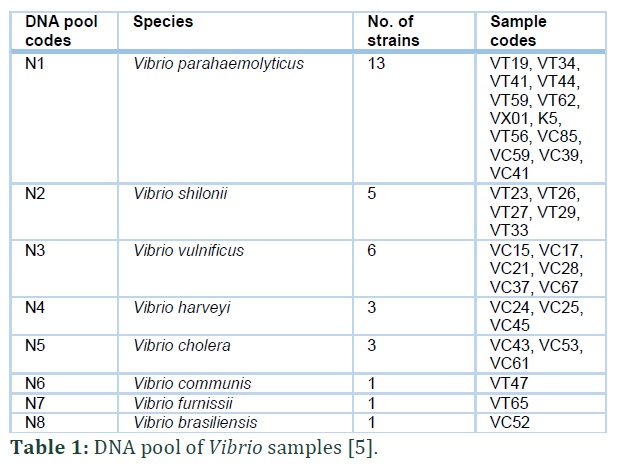
Vibrio strains were provided by the Laboratory of Gene Technology, Institute of Biotechnology, Hue University, Vietnam (Table 1). Characteristics of each strain were previously described [5].
DNA isolation
Vibrio colonies were cultured in alkaline saline peptone water medium with 2% peptone and 2% NaCl, pH 8.6, with shaking at 180 rpm for 18 h at 30°C. Cells were collected using centrifugation with 13,000 rpm at 4°C for 1 min. Total genomic DNA was extracted by the AquaPure Genomic DNA Isolaton Kit (Cat. 732-6340, Bio-rad) according to the manufacturer’s recommendations, and stored at 4°C. Total DNA concentrations were determined using a photo spectrometer at 260/280 nm. Genomic DNA was adjusted to final concentration of 50 ng/µL for PCR amplification.
RAPD analysis
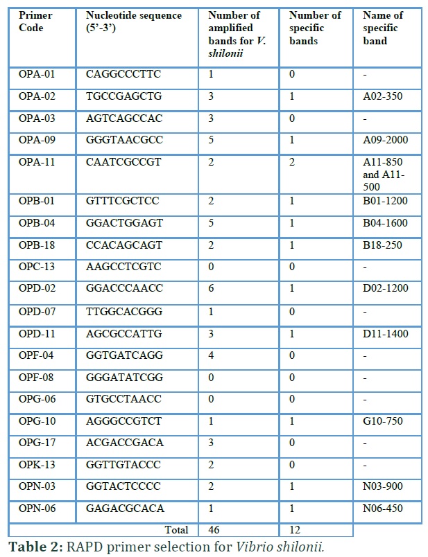
Genomic DNA pools of each species were used as templates for RAPD analysis (Table 1). DNA pool samples were prepared by mixing genomic DNA of all strains from each species into one unique sample with an equal ratio. A total of 20 RAPD primers (Operon Technologies, USA) were assayed for detecting Vibrio shilonii species (Table 2), these primers were used in our previous study [5] of randomly selected. Specific bands (absent in the other Vibrio species) were replicated twice for confirmation used for further studies. PCR-RAPD reactions were carried out according to Quang et al. [5].
DNA fragment cloning and sequencing
The specific amplicon of V. shilonii was extracted, then purified with GeneJET Gel Extraction Kit (Thermo Scientific, Lithuania). The purified PCR product was cloned into pGEM-T Easy vector (Promega, USA) and transferred into Escherichia coli strain TOP10 competent cells heat shock method [13]. Positive clones were confirmed by PCR with M13 primers and sequenced at Firstbase company (Malaysia).
SCAR marker development
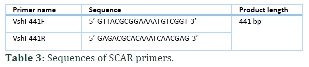
Specific SCAR primers Vshi-441F and Vshi-441R were designed based on the sequenced RAPD amplicon (Table 3). Genomic DNA samples, including those from five V. shilonii strains and other pathogen Vibrio species, were amplified using the Vshi-441F and Vshi-441R primers to verify the SCAR marker specificity. A 12 μL PCR reaction system was developed containing 50 ng of total DNA, 10 pmol of each primer, 6 µL 2× GoTaq® Green Master Mix (Promega, USA), and sterile distilled water. PCR amplification was performed as follows: 95°C for 5 min; 35 cycles at 95°C for 1 min, 55°C for 1 min, and 72°C for 1 min; the final cycle at 72°C for 10 min. To test detection sensitivity, 0.01–10 ng/μL of diluted genomic DNA of V. shilonii were evaluated as DNA templates for PCR amplification.
Results![]()
RAPD analysis
To reduce the number of test samples, we used a DNA pool instead of individual genomic DNA for specific RAPD amplicons. The DNA pool in this study was a mixture of genomic DNA of strains of each Vibrio species at an equal ratio: eight DNA pool samples for V. parahaemolyticus, V. vulnificus, V. shilonii, V. cholera, V. communis, V. harveyi, V. furnissii, and V. brasiliensis (Table 1).
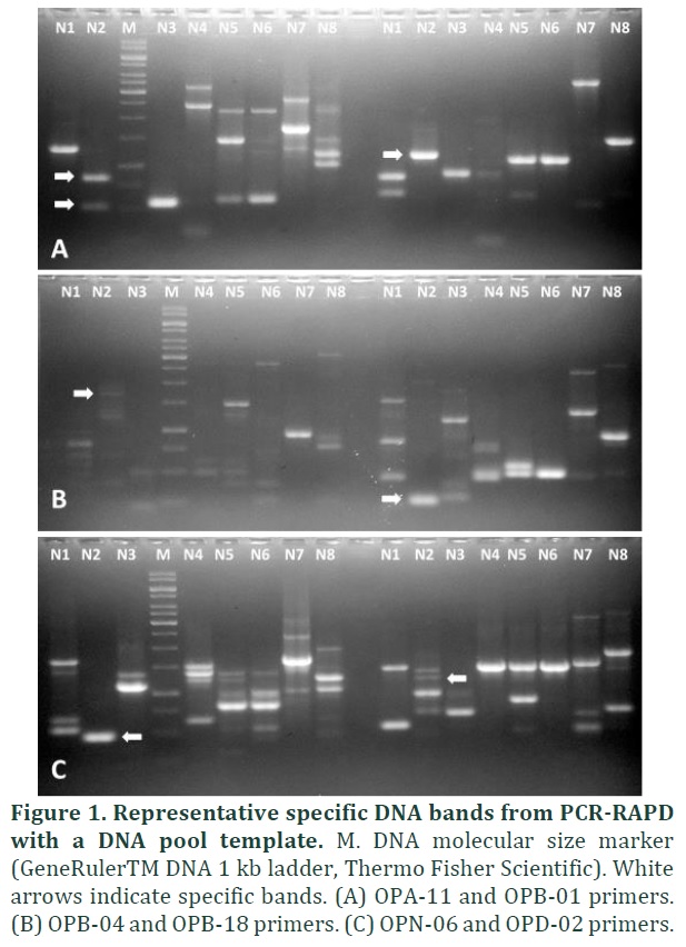
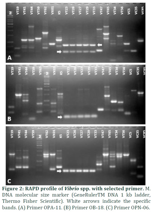
From 20 tested random primers, we obtained 46 DNA fragments from Vibrio shilonii (0–6 DNA bands/primer); most of these were polymorphic bands (Figure 1). Twelve specific bands for V. shilonii were generated from 11 primers (Table 2), and these primers were used for amplification of all samples. Specific PCR products generated from these 11 primers were used to screen specific markers for V. shilonii. Results showed that the 12 specific bands were present in five strains of V. shilonii and absent in other Vibrio species (Figure 2). Of 12 PCR products, N06-450 fragment (designated as the primer name and expected size of the band) was chosen to develop the SCAR marker because it produced a clear band of the suitable size (~450 bp) and was sufficiently different from RAPD patterns of other pathogens. However, the OPN-06 primer amplified only one PCR product, so further research was easily performed with this band.
SCAR marker development
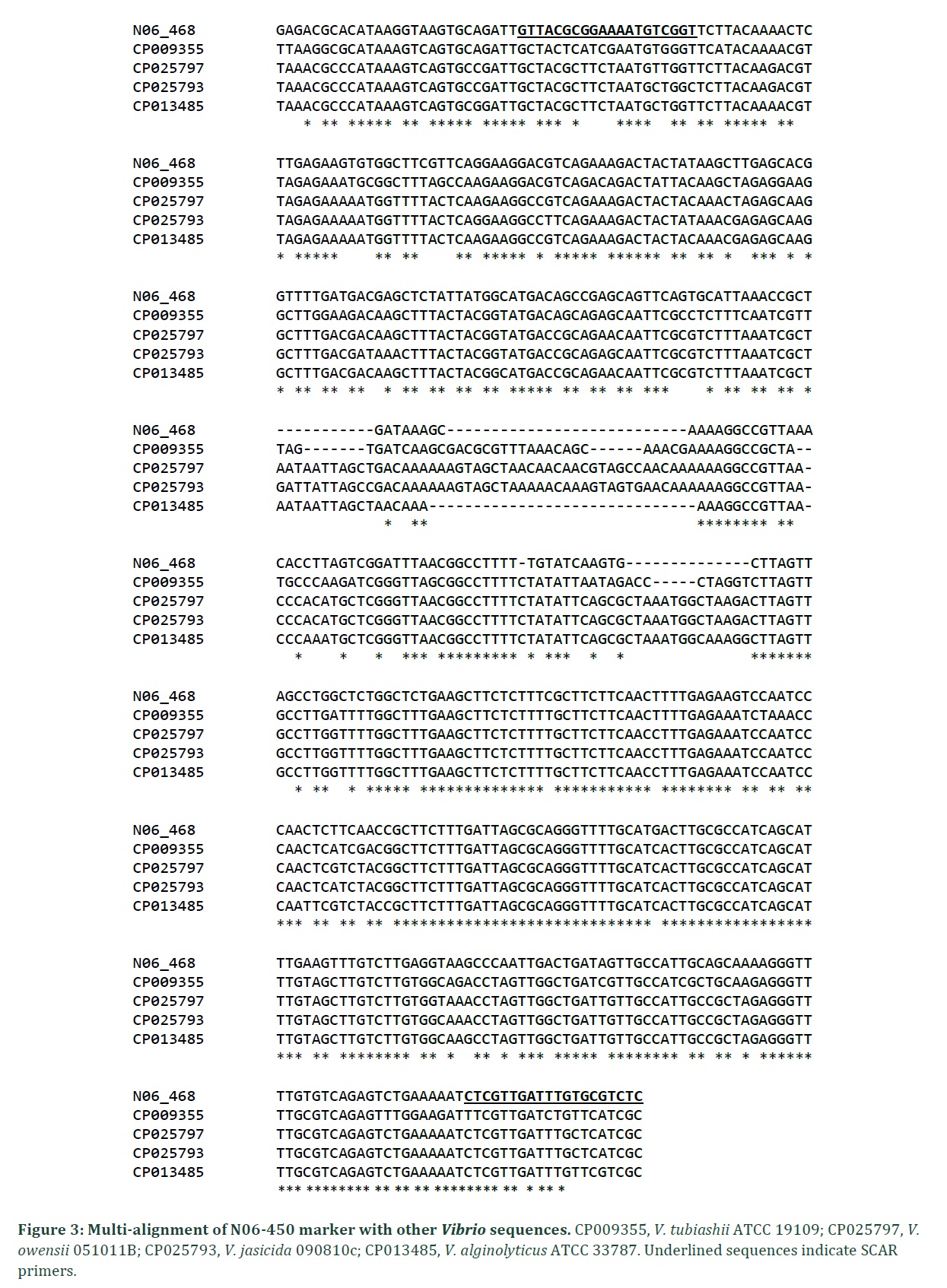
The N06-450 band was chosen to develop the SCAR marker. Thus, the PCR band was cloned into pGEM-T Easy vector and performed DNA sequencing. The results indicated that the length of the amplicon was 468 bp with 44.4% GC content (A = 119, T = 141, G = 111, and C = 97) (Figure 3).
BLAST results showed no significant similar sequence on Genbank database. The similarity coefficient of the N06-450 sequence with other Vibrio species ranged from 74%–78%: V. alginolyticus ATCC 33787 (78.19%), V. hyugaensis 090810a (74.13%), V. tubiashii ATCC 19109 (73.52%), V. owensii 051011B (73.53%), or V. jasicida 090810c (72.75%) (Figure 3). No similar sequences were found from popular Vibrio pathogens such as V. parahaemolyticus, V. brasiliensis, V. communis, V. furnissii, and V. harveyi.
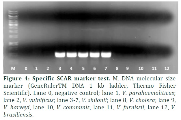
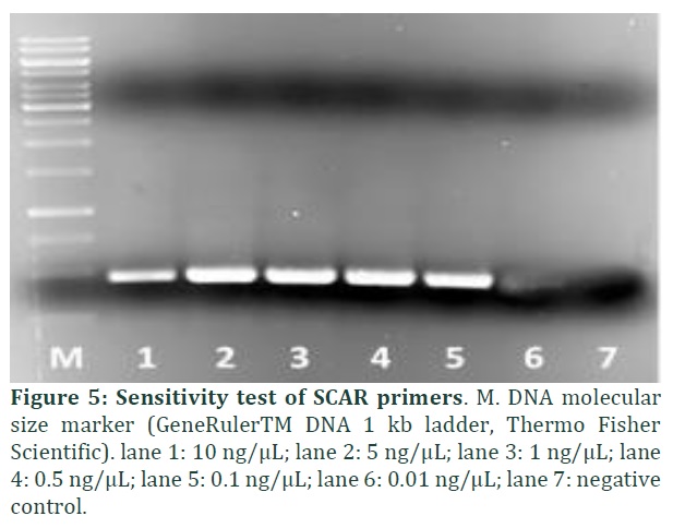
The primer pair Vshi-441F/Vshi-441R (Table 3) was designed based on the comparison of N06-450 sequences and others from Genbank (Figure 3 and Table 3). The Vshi-441F/Vshi-441R primers were selected to amplify on genomic DNA of pathogenic Vibrio isolates. A unique band with the size approximately of 441 bp was observed in V. shilonii isolates only (Figure 4), indicating that the designed SCAR marker was specific for this species, (this SCAR marker was named N6-441). To evaluate the SCAR marker sensitivity for V. shilonii identification, serial diluted V. shilonii genomic DNA were used as templates for the PCR assay using Vshi-441F/Vshi-441R primers. The results revealed that this SCAR marker detected V. shilonii DNA at concentration of 100 pg/μL or higher (Figure 5).
Figures & Tables
Discussion![]()
Vibrio spp. have been traditionally identified by culture the isolate on selective blood agar medium in integration with biochemical and serological testing. These methods are simple but require a lot of time and technicians. Meanwhile, PCR method has high advantages for rapid identification of Vibrio spp. in laboratory targeting the defined DNA sequences on genomic DNA. Furthermore, this method is often selected to amplify the specific targeted DNA sequence of the heterogeneous collection [14].
RAPD is a PCR-based technique to amplify the target or random DNA fragment using random primers. RAPDs are simple assay with fast results. This technique does not require sequence data as well as randomly genome distribution, and nature dominant. RAPD marker disadvantage is that this method is not able to identify the organism species due to their low reproducibility and nature dominant. Thus, RAPD marker recently is being developed into a codominant and reproducible marker (SCAR), which is able for species identification [15]. Using SCAR markers for identification of animal pathogens has been reported for such pathogens as Eimeria species [9] and Actinobacillus pleuropneumoniae [16].
Molecular diagnostic methods for Vibrio spp. detection based on PCR have been reported including loop-mediated isothermal amplification assay for V. parahaemolyticus [17], multiplex PCR for Vibrio spp. [18], or V. vulnificus [19]. Conventional PCR for specific Vibrio spp. genes also have been reported [20]. However, to our knowledge, no SCAR marker methods for diagnosing Vibrio spp. have been published heretofore.
In this study, we developed a rapid and highly specific SCAR marker for V. shilonii identification and detection. The method was developed based on specific primers designed for conventional PCR which is able to replace the traditional microbiology method. Our newly developed method is applicable for epidemiological studies as well as contribution to the protection programs for aquaculture.
Using the RAPD technique, we found a high diversity of Vibrio species except for V. shilonii, which is crucial for specific DNA fragments selection of the species. The designed primers for V. shilonii was evaluated the specificity by sequencing the amplified regions, followed by BLAST analysis because these sequences did not show significant similarities within the NCBI database (Figure 3).
The results in this study therefore show the SCAR primers can be applied to directly detect V. shilonii strains. The specificity of these primers was evaluated based on genomic DNA from eight Vibrio spp. demonstrated the positive amplification only observed on DNA of the targeted species.
The results of this study show a high level of polymorphism in the RAPD pattern of the different Vibrio species, and a SCAR marker (N6-441) was developed based on the 468-bp sequence specific to V. shilonii. Specific primers (Vshi-441F/Vshi-441R) amplified an unique DNA fragment for all V. shilonii isolates but not in the other Vibrio spp. This PCR method was highly sensitive to the V. shilonii genome and thus may be useful in rapid detection of this pathogenic bacterium.
Authors' Contribution
Hoang Tan Quang: experimental design, data analysis, paper preparation
Pham Thi Diem Thi: experimental performant
Tran Thuy Lan: experimental performant
Nguyen Duc Huy: paper preparation
Nguyen Duy Quynh Tram: experimental performant
Nguyen Thi Thu Lien: project leader, experimental design, paper preparation
The authors declare that they have no competing interests.
Acknowledgement
This study was financial supported from Vietnam Ministry of Education and Training (Project No. CT-2018-DHH-02).
References![]()
- Li J, Jiang H, Li L, Zhang X, Chen J. The Effect of Disease and Season to Hepatopancreas and Intestinal Mycobiota of Litopenaeus vannamei. Frontiers in Microbiology, (2019); 10889-889.
- FAO (2020) Cultured aquatic species information programme: Penaeus vannamei (Boone, 1931). http://www.fao.org/fishery/culturedspecies/Penaeus_vannamei/en. Date accessed: 08/20/2020.
- Thakur K, Patanasatienkul T, Laurin E, Vanderstichel R, Corsin F, et al. Production characteristics of intensive whiteleg shrimp (Litopenaeus vannamei) farming in four Vietnam Provinces. Aquaculture Research, (2018); 49(8): 2625-2632.
- Huy ND, Ngoc LMT, Loc NH, Lan TT, Quang HT, et al. Isolation of Weissella cibaria from Pacific white shrimp (Litopenaeus vannamei) gastrointestinal tract and evaluation of its pathogenic bacterial inhibition. Indian Journal of Science & Technology, (2020); 13(10): 1200-1212.
- Quang HT, Lan TT, Hai TTH, Yen PTH, Van TQK, et al. Genetic diversity and toxic genes analysis of Vibrio spp. isolated from white leg shrimp and marine fishes cultured in Tam Giang lagoon in Thua Thien Hue province, Vietnam. Indian Journal of Science & Technology, (2020); 13(13): 1412-1422.
- Han JE, Tang KFJ, Piamsomboon P, Pantoja CR. Evaluation of a reliable non-invasive molecular test for the diagnosis of the causative agent of acute hepatopancreatic necrosis disease of shrimp. Aquaculture Reports, (2017); 5: 58-61.
- Han JE, Tang KFJ, Tran LH, Lightner DV. Photorhabdus insect-related (Pir) toxin-like genes in a plasmid of Vibrio parahaemolyticus, the causative agent of acute hepatopancreatic necrosis disease (AHPND) of shrimp. Diseases of Aquatic Organisms, (2015); 113(1): 33-40.
- González Y, Venegas D, Mendoza-Hernandez G, Camarena L, Dreyfus G. Na+- and H+-dependent motility in the coral pathogen Vibrio shilonii. FEMS Microbiology Letters, (2010); 312(2): 142-150.
- Fernandez S, Katsuyama A, Kashiwabara AY, Madeira A, Durham A, Gruber A. Characterization of SCAR markers of Eimeria spp. of domestic fowl and construction of a public relational database (The Eimeria SCARdb). FEMS Microbiology Letters, (2004); 238(1): 183-188.
- Kałużna M, Albuquerque P, Tavares F, Sobiczewski P, Puławska J. Development of SCAR markers for rapid and specific detection of Pseudomonas syringae pv. morsprunorum races 1 and 2, using conventional and real-time PCR. Applied microbiology and biotechnology, (2016); 100(8): 3693-3711.
- Yang Y, Hu J, Chen F, Ding D, Zhou C. Development of a SCAR marker-based diagnostic method for the detection of the Citrus target spot pathogen Pseudofabraea citricarpa. BioMed Research International, (2018); article ID 7128903.
- Aggarwal R, Gupta S, Banerjee S, Singh V. Development of a SCAR marker for detection of Bipolaris sorokiniana causing spot blotch of wheat. Canadian journal of microbiology, (2011); 57934-942.
- Reid GA. Molecular cloning: A laboratory manual, 2nd edn: by J. Sambrook, E. F. Fritsch and T. Maniatis, Cold Spring Harbor Laboratory Press, 1989. $115.00 (3 vols; 1659 pages) ISBN 0 87969 309 6. Trends in Biotechnology, (1991); 9(1): 213-214.
- Ramazanzadeh R, Rouhi S, Shakib P, Shahbazi B, Bidarpour F, et al. Molecular characterization of Vibrio cholerae Isolated from clinical samples in Kurdistan province, Iran. Jundishapur journal of microbiology, (2015); 8(5): e18119-e18119.
- Sairkar PK, Sharma A, Shukla NP. SCAR marker for identification and discrimination of Commiphora wightii and C. myrrha. Molecular Biology International, (2016); 20161482796-1482796.
- Rossi CC, Pereira MF, Langford PR, Bazzolli DMS. A BOX-SCAR fragment for the identification of Actinobacillus pleuropneumoniae. FEMS Microbiology Letters, (2014); 352(1): 32-37.
- Anupama KP, Chakraborty A, Karunasagar I, Maiti B. Loop-mediated isothermal amplification assay as a point-of-care diagnostic tool for Vibrio parahaemolyticus: recent developments and improvements. Expert Review of Molecular Diagnostics, (2019); 19(3): 229-239.
- Kim HJ, Ryu JO, Lee SY, Kim ES, Kim HY. Multiplex PCR for detection of the Vibrio genus and five pathogenic Vibrio species with primer sets designed using comparative genomics. BMC Microbiology, (2015); 15(1): 239-250.
- Tsai YH, Chen PH, Yu PA, Chen CL, Kuo LT, Kuo CH. A multiplex PCR assay for detection of Vibrio vulnificus, Aeromonas hydrophila, methicillin-resistant Staphylococcus aureus, Streptococcus pyogenes, and Streptococcus agalactiae from the isolates of patients with necrotizing fasciitis. International Journal of Infectious Diseases, (2019); 81: 73-80.
- Alramahy SK. Molecular diagnostics for Vibrio cholera based on recA gene isolated from human in Diwaniyah city. Journal of Pharmaceutical Sciences and Research, (2018); 10(5): 1125-1127.
- Hänninen J, Takala J, Keinänen-Kiukaanniemi S. Quality of life in NIDDM patients assessed with the SF-20 questionnaire. Diabetes Research and Clinical Practice, (1998); 42(1): 17-27.
- Senez B, Felicioli P, Moreau A, Goaziou M-FL. Quality of life assessment of type 2 diabetic patients in general medicin. Presse Médicale, (2004); 33(3): 161-166.
- Kazemi-Galougahi MH, Ghaziani HN, Ardebili HE, Mahmoudi M. Quality of life in type 2 diabetic patients and related effective factors. Indian Journal of Medical Sciences (2012); 66(9-10): 230-237.
- Shaheen F, Basit A, Riaz M, Fawwad A, Hakeem R, et al. Assessing health related quality of life in diabetic subjects by SF 36 questionnaire in a tertiary care diabetes unit of Karachi, Pakistan. . International Journal of Advanced Research, (2014); 6(6): 13-17.
- Moreau A, Senez B, Felicioli P, Goaziou M-FL. Évaluation de la qualité de vie des patients diabétiques de type 2 en médecine générale. Presse Médicale, (2003); 17(608): 1-5.
- Finkelstein MM. Body mass index and quality of life in a survey of primary care patients. Journal of Family Practice, (2000); 49(8): 734-737.
- Eckert K. Impact of physical activity and bodyweight on health-related quality of life in people with type 2 diabetes. Diabetes, Metabolic Syndrome and Obesity, (2012); 5303-311.
- Woodcock AJ, Julious SA, Kinmonth AL, Campbell MJ. Problems with the performance of the SF-36 among people with type 2 diabetes in general practice. Quality of Life Research, (2001); 10(8): 661-670.
- Gulliford MC, Mahabir D. Relationship of health-related quality of life to symptom severity in diabetes mellitus: a study in Trinidad and Tobago. Journal of Clinical Epidemiology, (1999); 52(8): 773-780.
- Larsson D, Lager I, Nilsson PM. Socio-economic characteristics and quality of life in diabetes mellitus–relation to metabolic control. Scandinavian Journal of Public Health, (1999); 27(2): 101-105.
- Clouet F, Excler-Cavailher G, Christophe B, Masson F, Fasquel D. [Type 2 Diabetes and Short Form 36-items Health Survey]. Diabetes & Metabolism, (2001); 27(6): 711-717.
- Ikeda A, Iso H, Toyoshima H, Fujino Y, Mizoue T, et al. Marital status and mortality among Japanese men and women: the Japan Collaborative Cohort Study. BMC Public Health, (2007); 773.Liu H. Till Death Do Us Part: Marital Status and U.S. Mortality Trends, 1986 – 2000. Journal of Marriage and Family, (2009); 71(5): 1158-1173.
- Wee HL, Cheung YB, Li SC, Fong KY, Thumboo J. The impact of diabetes mellitus and other chronic medical conditions on health-related Quality of Life: is the whole greater than the sum of its parts? Health Qual Life Outcomes, (2005); 32.
- Poljicanin T, Ajduković D, Sekerija M, Pibernik-Okanović M, Metelko Z, et al. Diabetes mellitus and hypertension have comparable adverse effects on health-related quality of life. BMC Public Health, (2010); 1012.
- Lloyd A, Sawyer W, Hopkinson P. Impact of long-term complications on quality of life in patients with type 2 diabetes not using insulin. Value Health, (2001); 4(5): 392-400.
This work is licensed under a Creative Commons Attribution-Non Commercial 4.0 International License. To read the copy of this license please visit: https://creativecommons.org/licenses/by-nc/4.0

