

Full Length Research Article
Exposure to low concentrations of heavy metals alone and in combination induces histopathological and genotoxic effects in fish (Labeo rohita)
Asma Yamin1, Saima Naz1, Riaz Hussain2*, Tuba Rehman1, Ansar Shaheen1, Ahmad Manan Mustafa Chatha3, Abdul Ghaffar4, Muhammad Ismail Abbas5, Muhammad Sajjad Moazzam6
Adv. life sci., vol. 7, no. 4, pp. 240-246, August 2020
*– Corresponding Author: Riaz Hussain (Email: dr.riaz.hussain@iub.edu.pk)
Authors' Affiliations
2. Department of Pathology, Faculty of Veterinary and Animal Sciences, The Islamia University of Bahawalpur – Pakistan
3. Department of Entomology, University College of Agriculture and Environmental Sciences, The Islamia University of Bahawalpur – Pakistan
4. Department of Life Sciences (Zoology) Pathology, Faculty of Veterinary and Animal Sciences, The Islamia University of Bahawalpur – Pakistan
5. Poultry Research Institute, Livestock and Dairy Department Punjab – Pakistan
6. Bahawal Victoria Hospital, Quaid e Azam Medical College Bahawalpur – Pakistan
Abstract![]()
Introduction
Methods
Results
Discussion
References
Abstract
Background: The terrestrial and agro-aquatic ecosystems are continuously at the pity of human's negative impacts. Extensive and indiscriminate application of agrochemicals like heavy metals, industrial wastes, germicides, drug residues and different feed additives has become serious threats to public health. The current experimental trial was executed to investigate genotoxic potential of different heavy metals in fresh water fish (Labeo rohita).
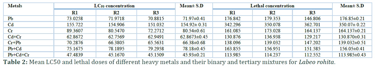
Methods: A total of 120 active, healthy, free from disease and internal parasites fish having three months of age were procured from local fish breeding center and were kept in glass aquarium having 100 liter water. After two weeks of acclimatization, fish were arbitrarily distributed and placed in different eight groups (A-H). Prior to start of experiment, acute toxicity of various heavy metals alone such as lead, cadmium, chromium and in combination (Pb +Cr), (Cr+Cd), (Cd+Pb) and (Pb+Cr+Cd) mixture were determined in-term of 96-h LC50 and lethal doses for Labeo rohita. After assessment of acute toxicity, all the fish were exposed to three sub-lethal concentrations (5, 7 and 9%) of heavy metals alone and in combinations for a period of 30 days in triplicate experiments.
Results: Different clinical ailments like, increased surface breathing, loss of coordination, rapid opercular movement, erratic swimming, air gulping, jerking movement and tremors were noted in fish. Histopathological observations of gills of various fish exhibited severe microscopic alterations. Results on micronucleus assay exhibited increased frequency of formation of micronuclei in red blood cells, while comet assays showed significantly increased DNA damage in peripheral erythrocytes in-term of arbitrary units of comets, average percentile rate of damaged cell and genomic damage index.
Conclusion: From the results of our experimental study, it can be concluded that fresh water fish are useful and reliable bio-indicators of heavy metal toxicity. Micronucleus and comet assays showed that heavy metals alone and in combination induced DNA damage in fish.
Keywords: Labeo rohita; Heavy metals; Histopathology; Erythrocytes; DNA damage
Introduction![]()
Chemical contamination due to industrial wastes, pesticides and heavy metals has been linked as a major factor to decline the life expectancy of variety of target and non-target exposed organisms both inhabiting in terrestrial and aquatic ecosystems [1 – 3]. Due to advancement in technology, development of industries and extensive use of synthetic chemicals, many developing countries in the world are facing contamination of natural ecosystems [4]. Non-target aquatic species including fish thriving in fresh water are commonly exposed to such chemicals and thus become susceptible to various chemicals toxicity because of direct contact of their gills tissues. Aquatic environmental contamination with heavy metals and other toxicants is of great concern due to extreme toxic effects such as carcinogenic, mutagenic and cytotoxicity [1, 5]. Studies have reported that among variety of soil, air, surface water and groundwater contaminants, heavy metal toxicity is the main abiotic stress leading to induce countless abnormalities and disruption of normal physiological functions of the exposed organisms [1, 6- 8]. It is reported that the heavy metal toxicity is mainly induced by deposition and bioaccumulation in different body tissues depending upon the mechanisms of accumulation, water habitats and other pathways of exposure including dietary or waterborne exposure [5, 9]. Previous different toxicological studies have shown exposure to different heavy metals causes mitochondrial damage, DNA abnormalities, oxidative stress and programmed death in different visceral tissues of the organisms [1, 10, 11]. In addition it has been estimated that exposure to water containing heavy metals causes infertility and DNA fragmentation [5]. Furthermore, heavy metal pollution induces harsh effects on aquatic organisms and alters ecological balance between biodiversity and ecosystems [12]. Fish are the useful aquatic bio-indicators and are frequently used as sentinel organisms because of part of the food chain and their sensitivity to low concentrations of mutagenic toxicant compounds [3, 13, 14]. Different levels of water pollution effluents containing, environmental toxins and industrial heavy metals induces DNA damage in various aquatic species including fish [15, 16]. The contaminated water severely affects the aquatic organisms and reduces the life span of target and non-target species, especially fish [17, 18].
Previously several published studies have proved that the micronucleus assay is reliable, important and useful technique to investigate the mutagenic changes induced by drugs and different environmental pollutants in red blood cells of various organisms [3, 19, 20]. DNA damage assessment using comet assay is regarded the most unique technique to determine the genotoxic potential of toxicants [21, 22]. Therefore, monitoring of deleterious effects of xenobiotics/pollutants in local species is crucial which can assist in the estimation of quality of the environment and to lower the exposure of environmental contaminants in human [5]. In Pakistan the fresh water fish are mainly affected by various contaminants present in untreated water [23, 24]. Acute and chronic toxic effects of various heavy metals may influence development of target and non-target species owing to metal accumulation in their bodies. Therefore, this work was aimed to investigate the deleterious effects of different heavy metals alone, in binary and their tertiary mixtures on fresh water fish (Labeo rohita).
Methods![]()
The current experimental trial was executed at laboratory of department of Zoology, Government Sadiq College Women University Bahawalpur. All the experimental research work was performed according to the guidelines regarding use of laboratory animals for research devised by ethical Committee of the Government Sadiq College Women University Bahawalpur.
Fish management and Chemicals
A total of 120 fresh water fish (Labeo rohita) having three months of age and about 200-225 body weight (gm) were collected from the local fish breeding center Bahawalpur. All the fish were transported in plastic bags supplemented with sufficient amount of oxygen to the experimental laboratory and were placed in glass aquariums having 100 liter water. All the fish were acclimatized prior to start of the trial. Feed (34% DP and 3.0Kcal/gm DE) was provided to experimental fish daily. During acclimatization period, water of the entire aquarium was changed on daily basis. For sufficient amount of oxygen supplementation to fish, all the aquariums were equipped with oxygenators with constant air pumps. Analytical grade of different heavy metals such as lead (98% pure; CAS-Nr: 7558-95-4), cadmium (99% pure; CAS-Nr: 7440-43-9) and chromium (98% pure; CAS-Nr: 10060-12-5) were selected to test their effects on fish. For acute toxicity tests the stock solutions were diluted to make required concentrations of metallic ions. All the chemicals used in this experimental research were obtained from Sigma-Aldrich (St. Louis Missouri, USA) and Merck (Germany). Physico-chemical features of water were measured prior to start and at the end of experiment (Table 1).
Experimental design and treatment
After 15 days of acclimatization period all the test specimens (active fish) free from clinical ailments, disease, behavioral changes and parasites were haphazardly picked and placed in eight groups (A-H). Each experimental group contained 15 fish. The fish of group A served as control group and were kept separately on normal feed in parallel to other groups. LC50 and lethal doses of different heavy metals and their binary and tertiary mixtures for Labeo rohita was determined prior to start of experiments (table 2). Heavy metals alone and in combinations were mixed in clean distilled water and fish kept in all groups (B-H) except A were exposed to various concentrations (5, 7 and 9% of LC50) for a period of 30 days. All the fish were carefully monitored twice a day to observe any clinical and behavioral signs.
Histopathological and Genotoxic studies
After 30 days of exposure all the experimental fish were killed by a sharp blow of head for tissue collection. About 2 ml blood sample was collected with the help of 26 gauge hypodermic needle in EDTA vacutainers separately from each test specimen. For micronucleus assay thin smear were prepared from fresh blood collected without anticoagulant. All the smears were air dried, fixed in ethanol and stained by using Giemsa stain [25]. All the smears were examined with the help of light microscope using oil immersion lens. For histopathological changes gills tissues were obtained from all the fish separately, preserved in 10% neutral buffered formaldehyde solution. After few days of fixation all the gills tissues were processed using standard procedures [13, 26]. Briefly, about 4-5 μm thick microscopic slices from gills were obtained using semi-automatic microtome, embedded in paraffin wax, mounted in DPX and stained with hematoxylin and eosin [27].
Single cell preparation and comet assay
Comet assay technique was performed under alkaline conditions on peripheral red blood cells [19, 28]. Briefly, thin smear of melted normal (1%) and low melting point agarose (0.75%) was made on frosted glass slide from each fish. The prepared slides containing cells were solidified and immediately kept in fresh lysing solution. After that the slides were placed in electrophoresis tank. The electrophoresis was conducted for 25 min at 20V. All the slides were neutralized and stained with ethidium bromide stain and were observed under fluorescent microscope. A total of 1500 cells were observed for DNA damage.
Statistical analysis
Physical observations
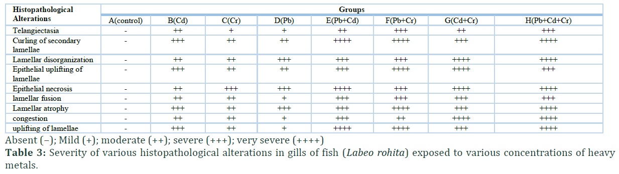
In present study, no mortality, physical and behavioral alterations were observed in the fish of the control group. All the fish kept in this group remain active, healthy and did not show any abnormal signs while the fish treated with different levels of heavy metals in different groups exhibited various physical changes. Due to heavy metals treatments variety of behavioral and clinical disorders like trembling of fins, increased opercular movement, loss of balance, irregular swimming, swallowing of air, increased mucous secretion, loss of coordination and increased surface breathing. Results on light microscopic observation of histological sections from gill tissues of various fish of control group showed normal structures while various microscopic alterations were observed in gills of various fish due to heavy metals treatments. Severity of occurrence of various histopathological abnormalities in gills of treated fish (Labeo rohita) is presented in table 3. Fish exposed to individual metals showed minute changes like curling/twisting of gills and disorganized primary and secondary lamellae. Severe abnormalities in gills sections of heavy metal treated fish like lamellar fusion, necrosis of primary and secondary lamellar epithelial cells, uplifting of lamellae and congestion were observed during the experimental trial. The lamellar disorganization, atrophy, curling of secondary lamellae and necrosis of lamellar epithelial cells were observed in fish exposed to different metals (Figure 1). Disruption and disorganization of primary lamellae, disruption of cartilaginous core, congestion, necrosis of lamellar epithelial cells and infiltration of leukocytes in gills of fish exposed to heavy metals were observed.
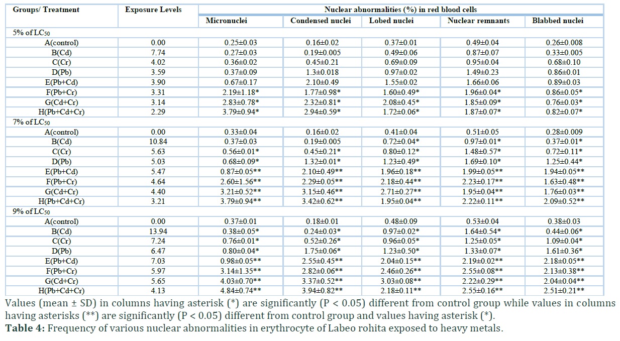
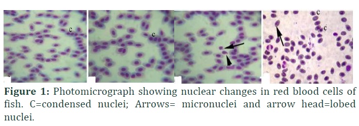
Different nuclear abnormalities in red blood cells of fish exposed to various concentrations of heavy metals alone and in combinations are presented in table 4. The frequency of formation of micronuclei, condensed nuclei, lobed nuclei, nuclear remnants and blabbed nuclei was significantly increased in fish exposed to various concentrations of heavy metals alone and in different combinations (Figure 2). The results showed significantly increased nuclear abnormalities in red blood cells of fish exposed to individual metals, binary and tertiary metal mixtures. Results also revealed that the nuclear abnormalities in red blood cells of fish exposed to binary and tertiary metal mixtures were significantly higher than the fish exposed to individual metals.
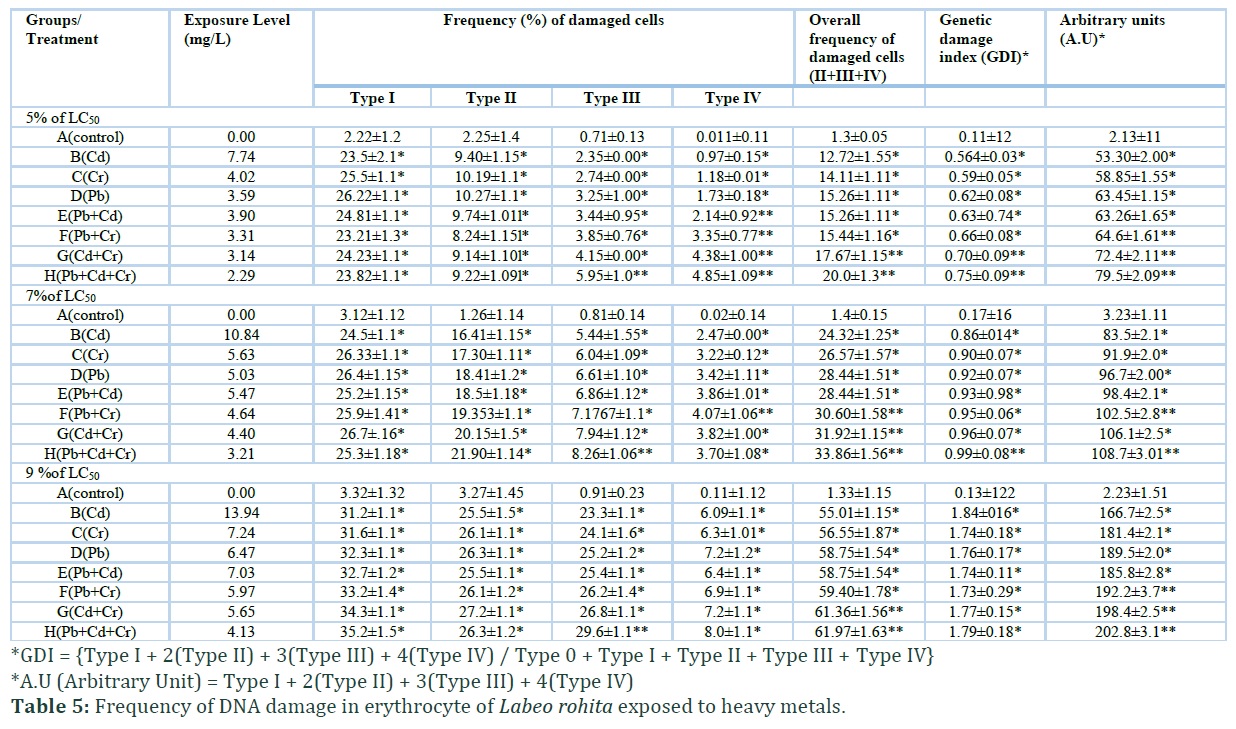
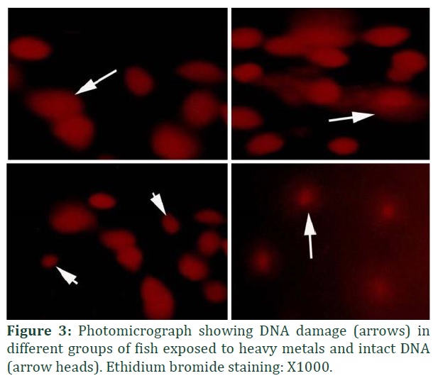
The results on comet assay (Figure 3) showed increased percentile rate of DNA damage in erythrocyte of treated fish, higher frequency of genomic damage index (GDI) and arbitrary units of comets in comparison to untreated control group (Table 5). The results showed increased frequency of DNA damage in fish due to various metals (alone) and their mixtures (binary and tertiary). Results showed that the percentile rate of DNA damage in erythrocyte of treated fish with tertiary mixture was higher as compared to other treatments and control group. The results showed non-significant GDI in erythrocytes of fish due to lead, chromium and binary mixtures treatments.
Figures & Tables


Discussion![]()
Excessive release of industrial effluents (pesticides, fungicides, herbicides and heavy metals) causes a serious threat to environment, domestic animals and public health [30, 31, 32]. It is interesting to know that variety of chemicals including industrial effluents and heavy metals may not show obvious adverse effects when they are present in lower concentrations in the ecosystem but their long-term and continuous exposure cause variety of abnormalities in exposed organisms [33, 34, 35]. Monitoring and assessment of toxic effects of industrial and environmental pollutants is of vital importance to reduce the release of such type of chemicals into the environment and to enhance the life expectancy of target and non-target species of life including human being. Different physical, nervous and behavioral signs including fin tremors, mucus secretion, erratic swimming, and operculum movements observed in present study might be due to due disruption CNS activities and obstruction of chloride channels in brain.
Previous similar clinical and behavioral changes due to various toxicants have also been observed in various fresh water fish [33, 36 – 38]. Blood and gill tissues in fish are sensitive and are known as useful markers for toxicity assessment [39, 40]. Histologically, gills sections from treated fish exhibited atrophy of secondary lamellae, pyknosis of lamellar epithelial pillar cells, disruption and disorganization of primary and secondary lamellae, lamellar degeneration, congestion, aneurysm and curling of secondary lamellae. Previously, due to metal poisoning similar histopathological disorders in gills of fish including
atrophid lamellae, lamellar fusion, uplifting of lamellae and disruption of primary and secondary lamellae have also been observed in gills of J. multidentata [41] Jenynsia multidentata [42] and tilapia [13]. In contrast to our findings, interstitial edema and increased oxidative stress has also been observed in gills of the fish exposed to heavy [8, 43]. The histological changes in gills of exposed fish could be due to increased generation of reactive oxygen species and inflammatory responses.
In present study increased frequency of nuclear abnormalities in erythrocyte of treated fish was observed. The significantly increased frequency of formation of micronuclei, condensed nuclei, blabbed nuclei, nuclear remnants and lobed nuclei in erythrocyte of fish exposed to heavy metals could be due to activation of DNase caspases resulting in degradation of gelsolin, tubulin, fodrin and vimentin proteins [44-46]. The nuclear alterations in red blood cells of fish might also be due to oxidative stress [47, 48]. Previously in published literature, higher frequency of formation of micronuclei, increased DNA damage and increased percentage of tail DNA due to heavy metals in gills and red blood cells of exposed Labeo rohita has also been observed [1].
It is reported that increased oxidative stress may lead to the DNA damage in erythrocytes of exposed fish. In addition, studies have shown that oxidative stress causes genomic instability and is responsible for inability of fish to repair its DNA [19, 25]. Moreover, the increased percentile rate of erythrocyte with nuclear abnormalities, arbitrary units of comets and GDI in exposed Labeo rohita can also be due to long term exposure to individual and mixture metals resulting increased generation of free radicals and oxidative stress [47]. The increased arbitrary units of comets, GDI and percentage of damaged cells in fish exposed to metals (mixture) might be due to interactions of heavy metals leading to toxic effects [49] in fish. In present study increased comet tail was observed in fish exposed to metals has also been determined [45, 48]. Moreover, increased frequency of DNA damage might also be related to oxygen free radical scavenging actions on metal binding, metal-chelating agent due to oxidative stress and poor detoxification mechanisms of metals in exposed fish [50].
The results of current experimental research suggested that exposure to heavy metals even at low level (alone) and in combination (mixtures) induced physical, histopathological and genomic abnormalities in fresh water fish (Labeo rohita).
Authors' Contribution
Asma Yamin, Ansar Shaheen and Tuba Rehman conducted the experiments. Saima Naz planned the experiment. Riaz Hussain analyzed the data, wrote manuscript and interpreted the results. Ahmad Manan Mustafa Chatha, Abdul Ghaffar, Muhammad Ismail Abbas and Muhammad Sajjad Moazzam involved in manuscript preparation.
The authors declare that they have no competing interests.
References![]()
- Ghaffar A, Hussain R, Aslam M, Abbas G, Khan A. Arsenic and urea in combination alters the hematology, biochemistry and protoplasm in exposed rohufish (Labeorohita) (Hamilton, 1822). Turkish Journal of Fisheries and Aquatic Sciences, (2016); 16 (2):289–296.
- Gul ST, Khan A, Saleemi MK, Ahmad M, Zahoor A, Naseem MN, Hussain R. Immuno-toxicological effects of allethrin (Active ingredient in mosquito repellent coils) in BALB/c mice following oral administration. Pakistan Veterinary Journal, (2019); 39 (2): 256-260.
- Ghaffar A, Hussain R, Abbas G, Khan R, Akram K,Latif H, Ali S, Baig S, Du X Khan A Assessment of genotoxic andpathologic potentials of fipronil insecticide in Labeorohita(Hamilton, 1822), Toxin Reviews, (2019);
- Razali NSM, Amin NM, Omar WBW, Ikhwanuddin M, Kadir NHA. Proteomic analysis and assestment of heavy metals in hepatopancreas of mud crabs from Setiu and Kuala Sepetang. Asian Journal of Agriculture and Biology, (2019); (Special issue): 17-24.
- Kousar S, Javed M. Diagnosis of metals induced DNA damage in fish using comet assay. Pakistan Veterinary Journal, (2015); 35 (2): 168-172.
- Javed M. Chronic dual exposure (waterborneþdietary) effects of cadmium, zinc and copper on growth and their bioaccumulation in Cirrhina mrigala. Pakistan Veterinary Journal, (2015); 35(2):143–146.
- Sattar A, Khan A, Hussain HI, He C, Hussain R, Zhiqiang S, Saleemi MK, Gul ST. Immunosuppressive effects of arsenic in broiler chicks exposed to Newcastle disease virus, Journal of Immunotoxicology, (2016); 13(6):861-869
- Bashir MA, Javed M, Latif F, Ambreen F. Effects of various doses of copper sulphate on peroxidase activity in the liver, gills, kidney and brain of Cirrhina mrigala. Asian Journal of Agriculture and biology, (2018); 6(3):367-372
- Nkwunonwo UC, Odika PO, Onyia NI. A Review of the Health Implications of Heavy Metals in Food Chain in Nigeria. The Scientific World Journal Volume, (2020); Article ID 6594109, 11 pages https://doi.org/10.1155/2020/6594109.
- Shahzad M., Javed MT, Shabir S, Irfan M. Hussain R. Effects of feeding urea and copper sulphate in different combinations on live body weight, carcass weight, percent weight to body weight of different organs and histopathological tissue changes in broilers. Experimental Toxicologic Pathology, (2012); 64(3): 141- 147
- Li S, Jiang X, Luo Y, Zhou B, Shi M, Liu F, Sha A, Sodium/calcium overload and Sirt1/Nrf2/OH-1 pathway are critical events in mercuric chloride-induced nephrotoxicity. Chemosphere, (2019); 234 (1): 579-588.
- Noor N, Zutshi B. Bioaccumulation of trace metals in tissues of Rohu fish for environmental risk assessment. Journal of Water Resource and Protection, (2016); 8(4): 472-481.
- Rahman URA, Ismail SNS, Abidin EZ, Praveena SM. Heavy metals accumulation in gills and muscles of Mozambique Tilapia (Oreochromis mossambicus) exposed to crude leachate. Asian Journal of Agriculture and Biology, (2019); (Special Issue): 111-115
- Lopes PA, Pinheiro T, Santos MC, da Luz Mathias M, Collares-Pereira MJ, Viegas-Crespo AM. Response of antioxidant enzymes in freshwater fish populations (Leuciscus alburnoides complex) to inorganic pollutants exposure. Science of the total environment, (2001); 280 (1-3): 153-163.
- Ergene S, Çavaş T, Çelik A, Köleli N, Kaya F, Karahan A. Monitoring of nuclear abnormalities in peripheral erythrocytes of three fish species from the Goksu Delta (Turkey): genotoxic damage in relation to water pollution. Ecotoxicology, (2007); 16(4): 385-391.
- Ghaffar A, Hussain R, Khan A, Abbas RZ. Hemato-biochemical and genetic damage caused by triazophos in fresh Water fish, Labeorohita. International Journalof Agriculture and Biology, (2015a); 17(3): 637-642.
- Greig S, Sear D A, Carling P A. The impact of fine sediment accumulation on the survival of incubating salmon progeny: implications for sediment management. Science of the total environment, (2005); 344(1-3): 241-258.
- Varsha G, Malik D S, Dinesh K. Risk assessment of heavy metal pollution in middle stretch of river Ganga: an introspection. International Research Journal of Environmental Sciences, (2017); 6(2): 62-71.
- Hussain R, Ali F, Rafique A, Ghaffar A, Jabeen G, Rafay M, Liaqat S, Khan I, Malik R, Khan MK, Niaz M, Akram K, Masood A. Exposure to sub-acute concentrations of glyphosate induce clinico-hematological, serum biochemical and genotoxic damage in adult cockerels. Pakistan Veterinary Journal, (2019): 39(2): 181-186.
- Ghaffar A, Hussain R, Noreen S, Chodhary IR, Abbas G, Khan A, Ahmed Z, Khan MK, Akram K, Ulhaq M, Ahmad N, Ali F, Niaz M. Dose and time-related pathological and genotoxic studies on thiamethaxam in fresh water fish (Labeorohita) in Pakistan. Pakistan Veterinary Journal, (2020); 40(2):151-156.
- Miller WR, Anderson TJ, Dixon JM. Anti-tumor effects of letrozole. Cancer investigation, (2002); 20(2): 15-21.
- Nagarani N, Devi V J, Kumaraguru AK.Mercuric chloride induced proteotoxicity and structural destabilization in marine fish (Therapon jarbua). Toxicology and Environmental Chemistry, (2011); 93(2): 296-306.
- Jabeen G, Javed M, Azmat H. Assessment of heavy metals in the fish collected from the river Ravi, Pakistan. Pakistan Veterinary Journal, (2012); 32(1): 107-111.
- Rauf A, Javed M, Ubaidullah M, Abdullah S. Assessment of heavy metals in sediments of the river Ravi, Pakistan. International Journal of Agriculture and Biology, (2009); 11(2): 197-200.
- Hussain R, Ghaffar Al, Ali HM, Abbas RZ, Khan JA, Khan IA, Ahmad I, Iqbal Z. Analysis of different toxic impacts of Fipronil on growth, hemato-biochemistry, protoplasm and reproduction in adult cockerels, Toxin Reviews, (2018); 37(4): 294-303.
- Ciaputa R, Szymerowski A, Janus I, Prządka P, Kandefer-Gola M, Nowak M. Immunohistochemical and histological features of a spontaneous Leydig cell tumour in a rat. Pakistan Veterinary Journal, (2019); 39(4): 603-605.
- AL-Samawy ERM, Jarad AS, Al-Saffar FJ, Kadhim DMH. Histological and histochemical study on the large intestine of one-humped camel in Iraq. Asian Journal of Agriculture and Biology, (2019); 7(3):373-380.
- Hussain R, Mahmood F, Khan MZ, Khan A, Muhammad F. Pathological and genotoxic effects of atrazine in male Japanese quail (Coturnix japonica), Ecotoxicology, (2011); 20 (1): 1–8.
- Steel, RGD, Torrie JH and Dicky DA. Principles and Procedures of Statistics, A Biometrical Approach. 3rd Edition, McGraw Hill, Inc. Book Co., New York, (1997); 352-358.
- Riaz A, Ulhaq M, Khan IA, Khan A, Hussain R, Yousaf A, Muhammad F. Chlorpyrifos induced dermal toxicity in albino rabbits. Pakistan Veterinary Journal, (2018); 38(1): 91-95
- Hussain R, Mahmood F, Khan A, Javed MT, Rehan S, Mehdi T. Cellular and biochemical effects induced by atrazine on blood of male Japanese quail (Coturnix Japonica). Pesticide Biochemistry and Physiology, (2012); 103(1):38–42.
- Ghaffar A, Hussain R, Khan A, Abbas RZ, Asad M. Butachlor induced clinico-Hematological and cellular changes in fresh water fish Labeorohita (Rohu). Pakistan Veterinary Journal, (2015b); 35(2): 201-206.
- Qureshi IZ, Bibi A, Shahid S, Ghazanfar M. Exposure to sub-acute doses of fipronil and buprofezin in combinationor alone induces biochemical, hematological, histopathological andgenotoxic damage in common carp (Cyprinuscarpio L.). Aquatic Toxicology, (2016); 179(4): 103–114.
- Ghaffar A, Hussain R, Abbas G, Ali MH, Saleem M, Khan T, Malik R, Ahmed H. Cumulative effects of sodium arsenate and diammonium phosphate on growth performance, hemato-biochemistry and protoplasm in commercial layer. Pakistan Veterinary Journal, (2017); 37(3): 257-262.
- Ghazanfar M, Shahid S, Qureshi IZ. Vitamin C attenuates biochemical and genotoxicdamage in common carp (Cyprinuscarpio) upon joint exposure to combined toxic doses of fipronil and buprofezin insecticides Aquatic Toxicology, (2018); 196 (4): 43–52.
- Zheng N, Cheng J, Zhang W, Li W, Shao X, et al. Binding difference of fipronil with GABAARs in fruit fly and zebra fish: insights from homology modeling, docking, and molecular dynamics simulation studies. Journal of Agriculture and Food Chemistry, (2014); 62(44): 10646–10653
- El-Murr A, Imam TS, Hakim Y, Ghonimi WAM. Histopathological, immunological, hematological and biochemical effects of fipronilon Nile tilapia (Oreochromisniloticus). Journal of Veterinary Science and Technology, (2015); 6(5):1-9.
- EI-Serafy SS, Abdel-Hameid NAH, EI-Daly A. A Histological and histochemical alterations induced by phenol exposure in Oreochromis aureus (Steindachner, 1864) juveniles, Egyptian Journal of Aquatic Biology and Fishries, (2009); 13(2):151-172.
- Au DWT. The application of histocytopathological biomarkers in marine pollution monitoring: a review, Marine Pollution Bulletin, (2004); 48(9-10): 817-834.
- Sanchez J, Varela Jr, Corcini C, Da Silva J, Primel E, Caldas S, Klein R, Martins C, Effects of Roundup formulations on biochemical biomarkers and male sperm quality of the live bearing Jenynsia multidentata. Chemosphere, (2017); 177: 200-210. doi:10.1016/j.chemosphere.2017.02.147
- Sanchez JAA, da Costa Klosterhoff M, Romano LA, De Martinez Gaspar Martins C. Histological evaluation of vital organs of the livebearer Jenynsia multidentata (Jenyns, 1842) exposed to glyphosate: A comparative analysis of Roundup® formulations. Chemosphere, (2019); 217: 914-924; https://doi.org/10.1016/j.chemosphere.2018.11.020
- Rossman TG. Mechanism of arsenic carcinogenesis: an integrated approach. Mutation Research/Fundamental and Molecular Mechanisms of Mutagenesis, (2003); 533(1-2): 37-65.
- García-Medina S, Razo-Estrada AC, Gómez-Oliván L M, Amaya-Chávez A, Madrigal-Bujaidar E, Galar-Martínez M. Aluminum-induced oxidative stress in lymphocytes of common carp (Cyprinus carpio). Fish physiology and biochemistry, (2010); 36(4): 875-882.
- Ternjej I, Mihaljević Z, Stanković I, Kerovec M, Sipos L, Želježić D, Kopjar N. Estimation of DNA integrity in blood cells of eastern mosquitofish (Gambusia holbrooki) inhabiting an aluminium-polluted water environment: an alkaline comet assay study. Archives of environmental contamination and toxicology, (2010); 59: 182-193.
- Abdulqadir SZ, Aziz FM. Nickel nanoparticles induced nephrotoxicity in rats: influence of particle size. Pakistan Veterinary Journal, (2019); 39(4): 548-552.
- Ghaffar A, Hussain R, Abbas G, Ahmad MN, Abbas A, Rahim Y, Younus M, Shahid M, Mohiuddin M. Sodium arsenate and/or urea differently affect clinical attributes, hemato-biochemistry and DNA damage in intoxicated commercial layer birds, Toxin Reviews, (2018); 37(4): 206-215
- Nagpure NS, Srivastava R, Kumar R, Kushwaha B, Srivastava SK, Kumar P, Dabas A. Assessment of genotoxic and mutagenic potential of hexavalent chromium in the freshwater fish Labeo rohita (Hamilton, 1822). Drug and Chemical Toxicology, (2015); 38(1):9-15.
- Frenzilli G, Nigro M, Lyons B. The Comet assay for the evaluation of genotoxic impact in aquatic environments. Mutation research/reviews in mutation research, (2009) 681(1): 80-92.
- Kalpaxis D L, Theos C, Xaplanteri M A, Dinos G P, Catsiki A V, Leotsinidis M. Biomonitoring of Gulf of Patras, N. Peloponnesus, Greece. Application of a biomarker suite including evaluation of translation efficiency in Mytilus galloprovincialis cells. Environmental Research, (2004); 94(2): 211-220.
- Hussain R, Khan A, Mahmood F, Rehan S, Ali F. Clinico-hematological and tissue changes induced by butachlor in male Japanese quail (Coturnix japonica), Pesticide Biochemistry and Physiology, (2014); 109:58–63.
This work is licensed under a Creative Commons Attribution-Non Commercial 4.0 International License. To read the copy of this license please visit: https://creativecommons.org/licenses/by-nc/4.0