

Full Length Research Article
Anticancer, antimicrobial and antioxidant compounds of quinoa inflorescence
Iqra Haider Khan*, Arshad Javaid
Adv. life sci., vol. 8, no. 1, pp. 68-72, November 2020
*– Corresponding Author: Iqra Haider Khan (Email: iqrahaider_khan@yahoo.com)
Authors' Affiliations
Abstract![]()
Introduction
Methods
Results
Discussion
References
Abstract
Background: Chenopodium quinoa is a newly introduced drought resistant crop in Pakistan. Studies regarding the efficacy of bioactive compounds present in this plant are scarce. Therefore, the current investigation was carried out to identify the antimicrobial, antioxidant and anticancer compounds present in ethyl acetate fraction of methanolic extract of inflorescence of C. quinoa.
Methods: Dry powdered inflorescence of the test plant was macerated with methanol and partitioned through different organic solvents on the basis of increase in polarities beginning with n-hexane followed by chloroform and ethyl acetate. GC-MS analysis was performed for the identification of bioactive constituents present in ethyl acetate fraction.
Results: The GC-MS analysis revealed the presence of 15 different phytochemicals. Among these, 1,2-benzedicarboxylic acid, diisooctyl ester (15); 9,12-octadecadienoic_acid-(Z,Z) (13); 8,11-octadecadienoic_acid, methyl ester (12); hexacosanoic acid, methyl-ester (11); hexadecenoic acid,2-hydroxy-1-(hydroxymethyl) ethyl ester (14); n-hexadecanoic acid (10); hexadecenoic-acid, methyl ester (8); 2-propenoic acid,3-[4-(acetyloxy)-3-methoxyphenyl]-, methyl ester (7); 1,6,10,14,18,22-tetracosahexaen-3-ol,2,6,10,15,19,23-hexamethyl-,(all-E)-(9) and undecane (1) were present in moderate to abundant concentrations. Biological activities of the identified compounds were searched in the previous literature.
Conclusion: The present study concluded that ethyl acetate fraction of methanolic extract of the inflorescence of C. quinoa contains a diverse range of potent bioactive constituents with antimicrobial, antifungal, antibacterial, cancer preventive, anti-inflammatory and cytotoxic properties.
Keywords: Chenopodium quinoa; Antimicrobial and antioxidant compounds
Introduction![]()
Plants are considered as a natural source of certain bioactive molecules that are used as raw material for pharmaceutical, medicine and agrochemical industry [1]. The use of herbal medicines to cure infectious diseases is an old age practice [2]. Numerous studies have shown antimicrobial and antioxidant potential [3, 4]. Synthetic antimicrobial drugs have toxic side effects on the host cells, which make grounds for the search and development of novel antibiotics isolated from plant origin [5]. Plant products are considered as a potential source for the discovery of organic compounds with beneficial medicinal effects [6]. These compounds are mostly secondary metabolites such as flavonoids, ketones, alcohols, steroids, tannins, alkaloids, terpenes, diterpenes, sesquiterpene lactones, triterpenes, phytoestrogens, carotenoids, curcumin and curcuminoids with the ability to produce definite physiological effect on hosts [7]. World Health Organization (WHO) is also working to screen antimicrobial agents from medicinal plants for the exploitation of traditional medicine system [8].
Chenopodium quinoa Willd., family Amaranthaceae (previously Chenopodiaceae), is native to the South America and is being used as a food crop for more than 5000 years [9]. It is known as a pseudo-cereal recently introduced in Asia, Southeast Asia, Australia and European countries. It is an annual herbaceous drought, cold resistant plant easily grown on acidic soils [10]. It is a rich source of amino acids, carbohydrates, fibers, proteins, vitamins, iron, calcium, minerals and magnesium [11]. It is also low in fat contents and naturally gluten free plant, making it a wonderful choice for the people [12]. In addition, quinoa seeds contain essential phenolics, saponins, flavonol glycosides, betaines, tannins, triterpene, ecdysteroids, and terpenoids that are rich in anti-inflammatory and antimicrobial properties [13]. Knowledge of the biological activities of phytochemical compounds present in quinoa inflorescence is limited. Therefore, the present study was carried out to analyze ethyl acetate fraction of methanolic inflorescence extract of C. quinoa through GC-MS to search compounds with antimicrobial, antioxidant and anticancer properties.
Methods![]()
Seeds of quinoa var. 2WANT (origin was New Mexico, USA) were obtained from Prof. Dr. Shahzad Ahmed Basra, Department of Agronomy, University of Agriculture, Faisalabad. Seeds were sown in the field at Experimental Station, Institute of Agricultural Sciences, University of the Punjab, Lahore, Pakistan during quinoa growing season of 2017-18. Inflorescence was collected from mature quinoa plants before drying. Quinoa inflorescence (10 g) was washed carefully under running tap water for the removal of associated debris. The material was shade dried first and then completely dried at 40 °C in an electric oven. The obtained dried biomass was passed through a mechanical grinder to pulverize it into a fine powder. Next, the powder was dipped in methanol (200 mL) for two weeks at room temperature followed by a filtration process using double layer of Whatman No. 1 filter paper and the filtrates were evaporated on a rotary evaporator at 45 °C to obtain a concentrated crude methanolic extract. The traces of methanol were evaporated by putting the crude extract in an electric oven at 45 ºC. The obtained extract was then mixed well in autoclaved distilled water (50 mL) and partitioned with n-hexane (5 × 100 mL) using a separating funnel. The remaining aqueous material was then extracted with chloroform (100 mL) followed by ethyl acetate (100 mL) [14].
Thereafter, ethyl acetate fraction was subjected to analysis of different volatile organic compounds using GC-MS. Analysis was performed by using a Shimadzu GC-2010 plus system coupled with an auto injector AOC-20i, an auto sampler AOC-20s and a gas chromatograph. Helium was used as a carrier gas, sample volume 1.0 µL was injected through setting injector at a temperature of 250 °C and interface temperature was calibrated at 320 °C. After injection of sample, the initial column temperature was 100 °C for 60 s that was enhanced from 100 to 200 °C at 20 °C min-1 and hold for 2.0 min, finally from 200 °C to 300 °C at 40 °C min-1. The total run time was 10.9 min [15]. A thorough literature survey was conducted for the evaluation of possible bioactive constituents.
Results![]()
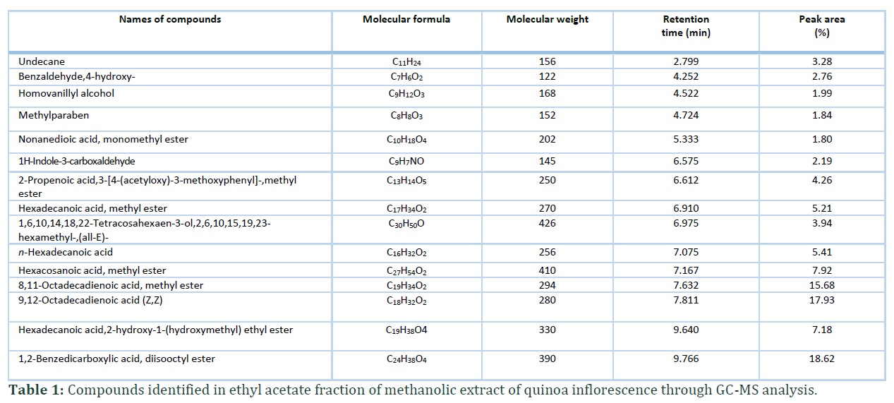
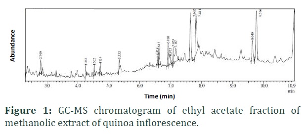
GC-MS analysis was performed to identify possible bioactive phytoconstituents present in the ethyl acetate fraction of methanolic extract of inflorescence of quinoa plant. GC-MS chromatogram indicates the presence of 15 major peaks (Figure 1).
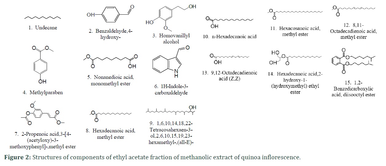
Details of the retention times, peak area percentages and molecular weights of the isolated compounds are provided in Table 1 whereas their structures are illustrated in Figure 2. The most abundant compounds were 1,2-benzedicarboxylic acid, diisooctyl ester (15); 9,12- octadecadienoic acid (Z,Z) (13) and 8,11-octadecadienoic acid, methyl ester (12) with peak areas of 18.62%, 17.93% and 15.68%, respectively. The compounds namely hexacosanoic acid, methyl ester (11); hexadecenoic acid, 2-hydroxy-1-(hydroxymethyl)ethyl ester (14); n-hexadecanoic acid (10); hexadecanoic acid, methyl ester (8); 2-propenoic acid, 3-[4-(acetyloxy)-3-methoxyphenyl]-, methyl ester (7); 1,6,10,14,18,22-tetracosahexaen-3-ol, 2,6,10,15,19,23-hexamethyl-,(all-E)- (9) and undecane (1) were present in moderate concentrations with 7.92%, 7.18%, 5.41%, 5.21%, 4.26%, 3.94% and 3.28% peak areas, respectively. On the other hand, the least abundant compounds were benzaldehyde, 4-hydroxy- (2); 1h-indole-3-carboxaldehyde (6); homovanillyl alcohol (3); methylparaben (4) and nonanedioic acid, monomethyl ester (5) with peak areas ranging from 1.80 to 2.76%.
Figures & Tables
Discussion![]()
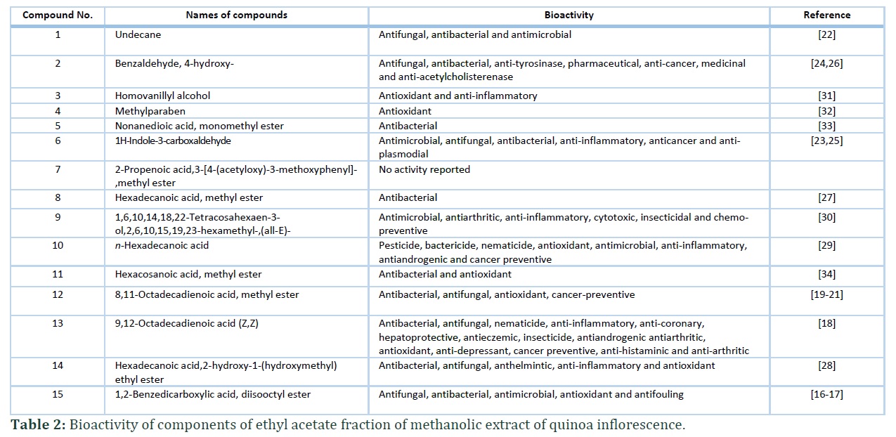
Among identified phytoconstituents, compound 15 was previously isolated from a medicinal plant Artemisia princeps leaves with an excellent antimicrobial efficacy against the pathogenic microbes including Candida albicans, Bacillus subtilis, Staphylococcus epidermis, S. aureus and Aspergillus niger [16]. It has also been reported from a medicinal plant Saccharum spontaneum with potent antimicrobial properties [17]. Similarly, compound 13 also known as linoleic acid has been previously identified from the leaf, root and stem extract of Cenchrus biflorus with potent antibacterial, antifungal, nematicide, anti-inflammatory, anti-coronary, hepatoprotective, antieczemic, insecticide, antiandrogenic, antiarthritic, antioxidant, anti-depressant, cancer preventive, anti-histaminic and anti-arthritic properties [18]. In previous studies, compound 12 was isolated from methanolic extracts of Melastoma beccarianum and M. malabathricum. The compound was investigated to evaluate its antibacterial activity against Bacillus anthracis, the pathogenic bacterial strain responsible for infections in humans [19,20]. Moreover, it was also tested against phytopathogenic fungal strains such as Penicillium digitatum and Aspergillus niger and showed profound antifungal activity against these fungi [21]. Likewise, compound 1 was previously isolated from Zingiber officinale and tested against a number of fungal and bacterial species namely Pseudomonas aeruginosa, Bacillus subtilis, Penicillium spp., A. niger, Candida albicans and Saccharomyces cerevisiae by disc diffusion method. It was noted that the compound had marked inhibitory activity towards C. albicans, Penicillium spp. and P. aeruginosa [22]. Similarly, compound 2 and 6 were tested against B. subtilis, S. aureus, A. niger, A. fumigatus, A. clavatus, S. pyogenes and C. albicans. The compound showed an excellent potential against all the tested microbes but the results were more promising in reducing the population of C. albicans, B. anthracis and A. fumigatus in comparison to the reference drugs namely amphotericin B, gentamicin and ampicillin [23,24]. Both the compounds also possessed strong medicinal, pharmaceutical, anti-plasmodial, antimicrobial, antifungal, antibacterial, anti-tryosinase, anti-cancer and anti-acetylcholisterenase properties [25,26]. Compounds 8, 9, 10 and 14 are well known due to their remarkable anti-inflammatory, cytotoxic, antibacterial, insecticidal, antifungal, anthelmintic, cancer preventive, antimicrobial and antioxidant activities [27-30]. Similarly, compound 3, 4, 5 and 11 were reported to possess strong antioxidant, anti-inflammatory and antibacterial properties [31-34]. This study concludes that ethyl acetate fraction is a potent source of antimicrobial, antioxidant and medicinally important compounds.
Author Contributions
Iqra Haider Khan did experimental work and wrote a part of paper. Arshad Javaid supervised the research and contributed in paper writing.
The author declares that there is no conflict of interest regarding the publication of this paper.
References![]()
- Sbhatu DB, Abraha HB. Preliminary antimicrobial profile of Solanum incanum L.: A common medicinal plant. Evidence-Based Complementary and Alternative Medicine, (2020); 20: Article ID 3647065.
- Aswal S, Kumar A, Semwal RB, Chauhan A, Kumar A, Lehmann J, Semwal DK. Drimia indica: a plant used in traditional medicine and its potential for clinical uses. Medicina, (2019); 55(6): 255-271.
- Bakal SN, Bereswill S, Heimesaat MM. Finding novel antibiotic substances from medicinal plants—antimicrobial properties of Nigella sativa directed against multidrug resistant bacteria. European Journal of Microbiology and Immunology, (2017); 7(1): 92-98.
- Chander PM, Vijayachari P. Antimicrobial properties of ethnomedicinal plants against selected human pathogens. Journal of Traditional Medicine and Clinical Naturopathy, (2018); 7: Article ID 10000253.
- Teixeira MC, Sanchez-Lopez E, Espina M, Calpena AC, Silva AM, Veiga FJ, Souto EB. Advances in antibiotic nanotherapy: Overcoming antimicrobial resistance. In: Emerging Nanotechnologies in Immunology, Elsevier. (2018); pp. 233-259.
- Mehmood A, Ishaq M, Zhao L, Safdar B, Rehman AU, Munir M, Wang C. Natural compounds with xanthine oxidase inhibitory activity: A review. Chemical Biology and Drug Design, (2019); 93(4): 387-418.
- Beran F, Kollner TG, Gershenzon J, Tholl D. Chemical convergence between plants and insects: biosynthetic origins and functions of common secondary metabolites. New Phytologist, (2019); 223(1): 52-67.
- World Health Organization. Essential medicines and health products information portal, (2019); https://apps.who.int/medicinedocs/en/d/Jh2945e/2.2.html. Accessed on February 10, 2020.
- Rodrigues DB, Tunes LVM, Villela FA, Gadotti GI, Costa CJ, Rosa TD, Nunes C. Production potential and quality of Chenopodium quinoa Willd. seed cultivated in different seeding seasons. Journal of Agricultural Science, (2019); 11(1): 251-260.
- Hinojosa L, Matanguihan JB, Murphy KM. Effect of high temperature on pollen morphology, plant growth and seed yield in quinoa (Chenopodium quinoa Willd.). Journal of Agronomy and Crop Science, (2019); 205(1): 33-45.
- Pathan S, Eivazi F, Valliyodan B, Paul K, Ndunguru G, Clark K. Nutritional composition of the green leaves of quinoa (Chenopodium quinoa Willd.). Journal of Food Research, (2019); 8: 55-65.
- Zelada E, Rene C. Nutritional composition of quinoa (Chenopodium quinoa Willd.) and its use in the elaboration of gluten-free bread of optimized quality. (2019); Ph. D. Thesis, University of Minho Library.
- Sobota A, Swieca M, Gęsinski K, Wirkijowska A, Bochnak J. Yellow‐coated quinoa (Chenopodium quinoa Willd)–physicochemical, nutritional, and antioxidant properties. Journal of the Science of Food and Agriculture, (2020); 100(5): 2035-2042.
- Akhtar R, Javaid A. Biological management of basal rot of onion by Trichoderma harzianum and Withania somnifera. Planta Daninha, (2018); 35: Article ID e017164713.
- Khan IH, Javaid A. Comparative antifungal potential of stem extracts of four quinoa varieties against Macrophomina phaseolina. International Journal of Agriculture and Biology, (2020); 24(3): 441-446.
- Mamun MIR, El-Aty AA, Rahman MM, Choi JH, Yun KW, Shin HC, Shim JH. Characterization of secondary metabolite compounds correlated with the seasons in Artemisia princeps var. orientalis (Pamp.) H. hara leaves using direct sample injection and gas chromatography–mass spectrometry: contribution to phytotoxicity. Journal of the Korean Society for Applied Biological Chemistry, (2015); 58(1): 173-183.
- Devi J, Muthu AK. Gas chromatography-mass spectrometry analysis of bioactive constituents in the ethanolic extract of Saccharum spontaneum Linn. International Journal of Pharmacy and Pharmaceutical Sciences, (2014); 6(2): 755-759.
- Arora S, Kumar G. Phytochemical screening of root, stem and leaves of Cenchrus biflorus Roxb. Journal of Pharmacognosy and Phytochemistry, (2018); 7(1): 1445-1450.
- Diris MN, Basri AM, Metali F, Ahmad N, Taha H. Phytochemicals and antimicrobial activities of Melastoma malabathricum and Melastoma beccarianum leaf crude extracts. Research Journal of Phytochemistry, (2017); 11(1): 35-41.
- Yuan W, Lee HW, Yuk HG. Antimicrobial efficacy of Cinnamomum javanicum plant extract against Listeria monocytogenes and its application potential with smoked salmon. International Journal of Food Microbiology, (2017); 260(2): 42-50.
- Kianinia S, Farjam MH. Chemical and biological evolution of essential oil of Arum maculatum. Iranian Journal of Science and Technology, Transactions A: Science, (2018); 42(2): 395-399.
- Sasidharan I, Menon AN. Comparative chemical composition and antimicrobial activity fresh & dry ginger oils (Zingiber officinale Roscoe). International Journal of Current Pharmaceutical Research, (2010); 2(4): 40-43.
- Salman AS, Mahmoud NA, Abdel-Aziem A, Mohamed MA, Elsisi DM. Synthesis, reactions and antimicrobial activity of some new 3-substituted indole derivatives. International Journal of Organic Chemistry, (2015); 5(2): 81-99.
- Kortiwala N, Patel J, Desai VA. Novel imidazolinones derivatives with diverse biological activities. World Journal of Pharmaceutical Research, (2016); 5(4): 1371-1379.
- El-Sawy ER, Abdelwahab AB, Kirsch G. Utilization of 1H–indole-3-carboxyaldehyde as a precursor for the synthesis of bioactive indole alkaloids. Synthesis, (2018); 50(23): 4525-4538.
- Kumar BR, Anupam A, Manchikanti P, Rameshbabu AP, Dasgupta S, Dhara S. Identification and characterization of bioactive phenolic constituents, anti-proliferative, and anti-angiogenic activity of stem extracts of Basella alba and rubra. Journal of Food Science and Technology, (2018); 55(5): 1675-1684.
- Kumar V, Bhatnagar AK, Srivastava JN. Antibacterial activity of crude extracts of Spirulina platensis and its structural elucidation of bioactive compounds. Journal of Medicinal Plants Research, (2011); 5(32): 7043-7048.
- Al-Marzoqi AH, Hameed IH, Idan SA. Analysis of bioactive chemical components of two medicinal plants (Coriandrum sativum and Melia azedarach) leaves using gas chromatography-mass spectrometry (GC-MS). African Journal of Biotechnology, (2015); 14(40): 2812-2830.
- Vats S, Gupta T. Evaluation of bioactive compounds and antioxidant potential of hydroethanolic extract of Moringa oleifera Lam. from Rajasthan, India. Physiology and Molecular Biology of Plants, (2017); 23(1): 239-248.
- Jenecius A, Uthayakumaria F, Mohan VR. GC-MS determination of bioactive components of Sauropus bacciformis blume (Euphorbiaceae). Journal of Current Chemical and Pharmaceutical Sciences, (2012); 2(4): 347-358.
- Garcia-Perez ME, Royer M, Herbette G, Desjardins Y, Pouliot R, Stevanovic T. Picea mariana bark: A new source of trans-resveratrol and other bioactive polyphenols. Food Chemistry, (2012); 135(3): 1173-1182.
- Manorenjitha MS, Norita AK, Norhisham S, Asmawi MZ. GC-MS analysis of bioactive components of Ficus religiosa Linn. stem. International Journal of Pharm and Bio Sciences, (2013); 4(2): 99-103.
- Chaudhery R, Ahmed D, Liaqat I, Dar P, Shaban M. Study of bioactivities of lipid content of fresh Lagenaria siceraria seeds pulp and identification of its chemical constituents. Journal of Medicinal Plant Research, (2014); 8(31): 1014-1020.
- Hagr TE, Adam IA, Almain AA, Mohammed MM. Phytochemical screening, gc-ms analysis, antibacterial and antioxidant activity of seeds oil of Annona squmosa L. Sudanese medicinal plant. Journal of Pharmacy and Pharmacology, (2019); 7(1): 1-6.
This work is licensed under a Creative Commons Attribution-Non Commercial 4.0 International License. To read the copy of this license please visit: https://creativecommons.org/licenses/by-nc/4.0