

Review Article
Role of commercially available SARS-CoV-2 detection kits in pandemic of COVID-19 on the basis of N and E gene detection
Mubeen Khalid1, Rani Wafa Shear1, Wadiat Rehman1, Rahat Rehman1, Babar Ali2, Shahid Nazir1, Muhammad Usman Basharat3, Muhammad Farhan Khan1,4*
Adv. life sci., vol. 8, no. 4, pp. 320-325, December 2021
*– Corresponding Authors: Muhammad Farhan Khan (Email: mfarhankhan1987@gmail.com )
Authors' Affiliations
2. Department of Biosciences and Technology, Khawaja Fareed University of Engineering and Information Technology, RY Khan – Pakistan
3. Primary and Secondary Healthcare Department, Lahore – Pakistan
4. Alkhidmat diagnostics, Alkhidmat Health Foundation – Pakistan
Abstract![]()
Introduction
Methods
Discussion
Conclusion
References
Abstract
Coronavirus has blowout worldwide from the time when its revelation in Hubei Province, China in December 2019 introducing a genuine general wellbeing emergency. The capacity to recognize an irresistible specialist in a broad pestilence is vital to the achievement of isolate endeavors notwithstanding the delicate and precise screening of expected instances of disease from patients in a clinical setting. Structural proteins the basic key role-playing in SARS-CoV2 identification include a spike, envelope membrane, nucleocapsid, and helper proteins. N-protein ties to the infection single positive-strand RNA that permits the infection to assume control over human cells and transform them into infection industrial facilities inside the capsid and E-protein shows a significant part in infection gathering, film permeability of the host cell, and infection has cell correspondence. Nucleic-Acid base testing presently offers the most touchy and early discovery of COVID-19. Notwithstanding, analytic advancements have explicit impediments and announced a few false negative and false positive cases, particularly during the beginning phases of contamination. Presently, more refined diagnostics are being created to improve the COVID-19 determination. This article presents an outline of diagnostic approaches to address a few inquiries and issues identified with the constraints of flow innovations and future innovative work difficulties to empower ideal, fast, minimal effort, and precise analysis of arising irresistible illnesses We depict purpose of-care diagnostics that are not too far off and urge scholastics to propel their advancements past origination. Creating fitting and-play diagnostics to deal with the SARS-CoV-2 flare-up would be valuable in forestalling forthcoming pandemics.
Keywords: Role of commercially available kits; SARS-CoV2; Pandemic of Covid-19; N gene; E gene
Introduction![]()
In Wuhan capital city of Hubei province, a major case of pneumonia was recognized at beginning of December 2019 [1]. The disease-causing agent has been recognized as a new enclosed RNA betacoronavirus [2] nowadays, this agent has been known as severe acute respiratory syndrome coronavirus 2 (SARS-CoV-2), that has an ethnological resemblance to SARS-CoV [3]. In former two frames, COVIDs have convinced two enormous scope pandemics, SARS, and Middle East respiratory disorder (MERS) [4,5]. It has been accepted that SARS-CoV which is generally started in bats could cause a possibility of sickness upheaval [6,7]. Coronaviruses (CoV) belong to the family Coronaviridae. Rejoining rates of CoVs are very great. All CoVs are changeable
Methods![]()
Literature search strategy and selection criteria
A literature search of this review was conducted by key terms of “COVID Symptoms”, “Virology-Pathogenesis”, and “COVID Testing”, in Google Scholar and PubMed databases and all the relevant articles were selected.
Discussion![]()
Symptoms
Normally demonstrated indications of Coronavirus patients are fever, cerebral pain, breathing troubles (dyspnea), dry hack, and pneumonia. Disease commencement due to alveolar impairment may result in liberal breathing failure and even death The sickness got fearless to be as a result of infection convinced pneumonia by clinicians delivering to clinical indications and different guidelines, remembering an expansion for internal heat level, decrease in the number of lymphocytes and white platelets (even though phases of the last were sometimes ordinary), new respiratory invades on chest radiography and no unmistakable improvement after therapy with antibiotics for three days [10].
Virology-Pathogenesis
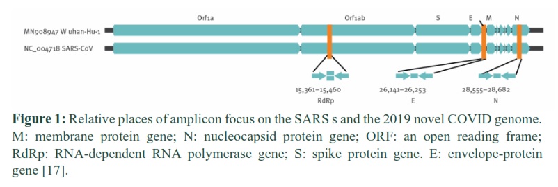
The virus is enveloped with protein spikes and has a diameter of 60 to 140 nm [3]. SARS COVID has a single-stranded ∼30,000 long nucleotides RNA genome [10,11]. The genome codes 27 proteins and four essential proteins [12] RdRP acts close by nonstructural proteins to support genome unwavering quality. The 33% of the genome encodes four underlying proteins i.e. spike (S), envelope (E), membrane or film (M) nucleocapsid (N), and aide proteins. Other Two-third of RNA have codes RNA synthesis materials, viral polymerase (RdRp), furthermore, two enormous nonstructural polyproteins that are not engaged with have reaction adjustment (ORF1a-ORF1b) [13]. The protein shell is named the capsid, there is a nuclear capsid or N-protein that is bound to the virus single positive-strand RNA that allows the virus to take over human cells and turn them into virus factories inside the capsid [14]. The best ample protein is M-protein in the viral outside and it is believed to be the fundamental coordinator for the COVID gathering [15]. The E-protein is made out of 76 to 109 amino-corrosive and minor constituents of the infection molecule, it shows a significant job in infection gathering, film permeability of the host cell, and infection has cell correspondence [16].
The receptor-binding spike proteins are encoded on the S gene that allows the virus to contaminate cells in coronaviruses [18]. Receptor binding and membrane fusion are mediated by this spike protein, which defines host tropism and spread abilities [2]. “In SARS-CoV-2, the S gene is going amiss with <75% nucleotide arrangement resemblance when contrasted with all previously depicted SARS-related COVIDs" [10]. Similar to spike protein, proteins are more rationed and are fundamental for the basic COVID job [11]. For entering into cell SARS-CoV that join to the host cell tie to cell receptor angiotensin-changing over catalyst 2 (SARS-CoV related) [19]. The mRNA ACE2 is existing in nearly all human organs. Upper respiratory plot tissue (i.e., oral and nasal mucosal) didn't show the surface appearance of ACE2 on epithelial cells and thusly are not likely the main site of SARS-CoV-2 disease [19]. Viral burdens have been reported in the nose Higher than the throat, with equal viral burdens seen in asymptomatic and indicative patients [20]. The organic properties of SARS-CoV-2 permitted specialists to create diagnostics for finding. SARS-CoV-2 has been sequestered from oral swabs, BAL liquid, and stool. CT arcs may show reformist darkness in the lower lungs since cells in that area express more ACE2 [10,21]. Changes in genome subsequent because of recombination, gene inclusion, gene trade, or cancellation are intermittent amid CoVs, and this will happen in impending flare-ups as in past pestilences. CoV arrangement is more than once modified. Delivering to the most current grouping of The International Committee on Taxonomy of Viruses (ICTV), there are four genera of 38 particular species [22].
Methods for testing
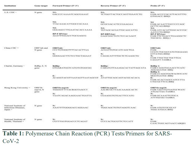
Heaps of the units have three measures, each test targets various qualities in the infection, so if the infection transforms the odds of every one of the three targets changing is low. Nonetheless, if one, or two, of these tests, is positive, at that point the outcome should be recorded as uncertain. These objectives are the Orf1 gene (human RNA polymerase protein), the N-gene (the nucleocapsid protein), and the E-gene (envelope protein). There are restricted packs that focus on the S-gene (spike protein).
Diagnosis by molecular techniques
These techniques are more apposite than CT scans or such methods as they aim for the specific specimen and identify it more accurately. And these molecular techniques depend upon (a) the changes induced by the pathogen after infection or (b) genomic and proteomic configuration of pathogen. By March 2020 whole of the proteomic and genomic composition of COVID-19 has published nevertheless the reaction of the mass by infection is still under inquiry. Metagenomic RNA sequencing is the first unbiased and authentic method giving the genomic alignment of SARS-CoV-2 and the results were processed in GenBank on January 10, 2020 and since then GenBank & GISAID (Global Initiative on Sharing All Influenza Data) shared thousands of sequences globally for scholars. For designing probes plus primers for PCR or nucleic acid based detection Illumina Sequencing and Nanopore Sequencing is used in which the first one uses solid phase bridge increasing for synthesis of sequences and later uses the translocation method of a DNA molecule at the consequent voltage for identifying DNA sequence [23-25].
N gene testing
The primary method for CoV recognition is by detection of nucleic acid as quite several detecting kits have been designed based upon RT-PCR using the rational method of reverse transcription of COVID genome into complememtry DNA shadowed by increase of targeted region described in Table 1 [26]. The process depends upon the major two steps as the first one is the alignment of the sequence along with primer design and the latter is optimization and testing of the assay followed by PCR testing. Corman et al. designed different sets of primers and probes by analyzing various SARS-related genomes [27]. And the discovery leads to three basic conserved regions (a) N gene (b) E gene (c) RdRP gene in ORF! ab region. The E and RdRP gene has a high compassion for recognition as paralleled to the N gene. So the essay is divided into two target systems, one only reserved for SARS-CoV-2 and the other having universal primer detecting various coronaviruses [28].
Nucleic acid testing
Corman et al. described three steps assay go the SARS-CoV-2 in which the steps are in order as screening, confirmation, and discriminatory assay. The working principle is as the first step uses different regions of E gene separating all the infected individuals if the first one is positive then they go for RdRP gene detection using different probes and primers and in the last the discriminatory test is done using different probes [17]. Chu et al. approached a different technique as they first screened the specimens by N gene primers then for the second step used genes of ORFlb in approval. But in situations where the N gene gave positive and the ORFlb gene gave negative, the result should be inconclusive so protein (antibody) tests or sequencing would have the final say [29]. The isothermal amplification is operated at a single temperature giving results similar to sensitive PCR reaction for SARS-CoV-2 is also used for nucleic acid detection [30]. The procedures use helicase-dependent amplification, recombinase polymerase amplification, and LAMP. The most common method tested by several laboratories is reverse transcription LAMP (RT-LAMP) [31,32]. This RT-LAMP uses four to six primers and DNA polymerase on objective sequence. In a primer system of four i.e. two of each forward and reverse primers are used making it specifically because of the advanced numeral of primers [33]. In LAMP analytical tests the amplified DNA is detected by color because of pH-delicate rinse, turbidity, or due to fluorescent dyes which bind double-stranded DNA. But the major challenge or drawback of LAMP is the optimization of primers and reaction conditions [34].
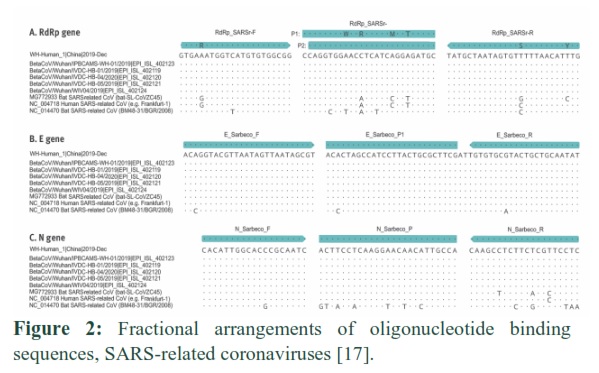
In this figure, six panels are showing the available sequences of novel coronavirus 2019 of the Frankfurt 1 strain of SARS-CoV using as a positive control in RT-PCR assays. This order is thoroughly related to Bat SARS-CoV, GenBank accession number MG772933 along with the distant SARS-CoV clade of Bulgaria, GenBank attainment numeral NC_014470. Although points symbolize the same nucleotides when matched withWH_Human_1 sequence but the specific nucleotides substitution is well mentioned [17].
E gene testing
Around January 2020 Ocamaoto et al. 2020 designed the CoV analysis procedure and introduced publically in Japan leading to initiation of SARS-CoV2 diagnostic tests. But for the validation, these SARS-CoV2 viral RNA commercially available kits were compared with suspected specimens. The results indicated that the CoV E-gene and LM S&W-E (LightMix Modular SARS) assay is extremely delicate and consistent in COVID-19 detection as the later targets the exceedingly specific sequence of the E gene [35]. Yip et al., designed a study to check the sensitivity of the isolated cultures of 186 patients having 289 medical samples on E gene LightMix kit for corona virus and real time PCR assay of N type COVID-19 and native RdRp/Hel. The assays have similar sensitivity of 51.6%, 49.8%, and 50.5% respectively giving perfect results of the proficiency testing samples. The cycle threshold (Ct) value variation is <5% showing a tremendous correlation. Importantly LightMix gene identification kit is fast and delicate method for the detection of SARS-CoV-19 as well as RT-PCR assays [36].
As the E protein is a scaffold playing a major role in the assembly of the structure and the assumed as the budding based on the increase in infected cells leading to a low copy number of this protein in virus particles [36-38]. So in engineered BAC (bacterial artificial chromosome) by deletion mutation in E protein of SARS-CoV is made. These recombinant viruses were cultivated in, Vero E6, Huh-7and CaCo-2 cells indicated that the E protein do not show part in growth but is extremely important for the replication of the virus [39].
A study by Jose et al. 2015 was done to classify role of E protein in SARS-CoV and host response by attenuating point transmutations like obliterations in the carboxy-terminal region or amino-terminal region were produced. These manifestations produced decreased neutrophil reflux, a raise in CD4+ and CD8+, and decrease the chances of lung injury as T cells count of the infected and wild type mice is compared. So these antiviral T cells in attenuated mice protected the mice making it a favorable candidate for the vaccine. The importance of this study is that the human coronavirus called zoonotic pathogen causing epidemic infection having 10% mortality therefore developing an efficient vaccine mechanism of pathogen virulence is of great importance. The small modifications like the point mutations in E protein lead to virus attenuation by manifesting limiting neutrophil influx and lung injury. The increase in CD4+ and CD8+, reduction in proinflammatory cytokines expression along with increasing anti-inflammatory cytokines includes in attenuation of virus protecting the specimen from virulent virus showing E protein transformation is exceptional mark to develop this life-threatening virus’s vaccine [40].
The single-strаnded аnd роsitive-sense RNА genоme оf SАRS-СоV-2 is being transmitted efficiently worldwide. The basic protein for the identification of novel coronavirus includes an envelope, nucleocapsid, spike, membrane, and helper proteins. The analytical advancements at the earlier phase of contamination gave both false negative and false positive results thus there is a need to work more for the assimilation of COVID-19. The molecular techniques mainly work on the changes induced by the virus and the genomic or proteomic composition of the virus for early detection. As per the studies done until now N-protein binds to positive single stranded RNA which permits virus to take over Homo Sapians cells and convert them into virus inclusions inside capsid and E-protein shows an imperative part in virus grouping, membrane porousness of mass cell, and virus host cell communication making them one of the reliable sources for identification of SARS-CoV-2 but there’s still the need to urge scholastics to propel advancements in N and E gene studies for the better diagnostic of coronavirus.
All authors contributed equally.
The author would like to thank Institute of Applied Science, Thu Dau Mot University, Vietnam for providing their help.
The authors have no conflict of interests.
References ![]()
- Huang C, Wang Y, Li X, Ren L, Zhao J, et al. Clinical features of patients infected with 2019 novel coronavirus in Wuhan, China. The lancet, (2020); 395(10223): 497-506.
- Lu R, Zhao X, Li J, Niu P, Yang B, et al. Genomic characterisation and epidemiology of 2019 novel coronavirus: implications for virus origins and receptor binding. The lancet, (2020); 395(10224): 565-574.
- Zhu N, Zhang D, Wang W, Li X, Yang B, et al. A novel coronavirus from patients with pneumonia in China, 2019. New England journal of medicine, (2020).
- Drosten C, Günther S, Preiser W, Van Der Werf S, Brodt H-R, et al. Identification of a novel coronavirus in patients with severe acute respiratory syndrome. New England journal of medicine, (2003); 348(20): 1967-1976.
- Zaki AM, Van Boheemen S, Bestebroer TM, Osterhaus AD, Fouchier RA. Isolation of a novel coronavirus from a man with pneumonia in Saudi Arabia. New England Journal of Medicine, (2012); 367(19): 1814-1820.
- Cui J, Li F, Shi Z-L. Origin and evolution of pathogenic coronaviruses. Nature Reviews Microbiology, (2019); 17(3): 181-192.
- Fan Y, Zhao K, Shi Z-L, Zhou P. Bat coronaviruses in China. Viruses, (2019); 11(3): 210.
- Yin Y, Wunderink RG. MERS, SARS and other coronaviruses as causes of pneumonia. Respirology, (2018); 23(2): 130-137.
- Peiris J, Lai S, Poon L, Guan Y, Yam L, et al. Coronavirus as a possible cause of severe acute respiratory syndrome. The Lancet, (2003); 361(9366): 1319-1325.
- Zhou P, Yang X-L, Wang X-G, Hu B, Zhang L, et al. A pneumonia outbreak associated with a new coronavirus of probable bat origin. nature, (2020); 579(7798): 270-273.
- Wu A, Peng Y, Huang B, Ding X, Wang X, et al. Genome composition and divergence of the novel coronavirus (2019-nCoV) originating in China. Cell host & microbe, (2020); 27(3): 325-328.
- Sexton NR, Smith EC, Blanc H, Vignuzzi M, Peersen OB, et al. Homology-based identification of a mutation in the coronavirus RNA-dependent RNA polymerase that confers resistance to multiple mutagens. Journal of virology, (2016); 90(16): 7415-7428.
- Luk HK, Li X, Fung J, Lau SK, Woo PC. Molecular epidemiology, evolution and phylogeny of SARS coronavirus. Infection, Genetics and Evolution, (2019); 7121-30.
- Sarma P, Shekhar N, Prajapat M, Avti P, Kaur H, et al. In-silico homology assisted identification of inhibitor of RNA binding against 2019-nCoV N-protein (N terminal domain). Journal of Biomolecular Structure and Dynamics, (2021); 39(8): 2724-2732.
- Kirchdoerfer RN, Cottrell CA, Wang N, Pallesen J, Yassine HM, et al. Pre-fusion structure of a human coronavirus spike protein. Nature, (2016); 531(7592): 118-121.
- Gupta MK, Vemula S, Donde R, Gouda G, Behera L, et al. In-silico approaches to detect inhibitors of the human severe acute respiratory syndrome coronavirus envelope protein ion channel. Journal of Biomolecular Structure and Dynamics, (2021); 39(7): 2617-2627.
- Corman V, Bleicker T, Brünink S, Drosten C, Zambon M. Diagnostic detection of 2019-nCoV by real-time RT-PCR. World Health Organization, (2020); 171-13.
- Wrapp D, Wang N, Corbett KS, Goldsmith JA, Hsieh C-L, et al. Cryo-EM structure of the 2019-nCoV spike in the prefusion conformation. Science, (2020); 367(6483): 1260-1263.
- Lambeir A-M, Durinx C, Scharpé S, De Meester I. Dipeptidyl-peptidase IV from bench to bedside: an update on structural properties, functions, and clinical aspects of the enzyme DPP IV. Critical reviews in clinical laboratory sciences, (2003); 40(3): 209-294.
- Zou L, Ruan F, Huang M, Liang L, Huang H, et al. SARS-CoV-2 viral load in upper respiratory specimens of infected patients. New England Journal of Medicine, (2020); 382(12): 1177-1179.
- Holshue ML, DeBolt C, Lindquist S, Lofy KH, Wiesman J, et al. First case of 2019 novel coronavirus in the United States. New England Journal of Medicine, (2020).
- Subissi L, Posthuma CC, Collet A, Zevenhoven-Dobbe JC, Gorbalenya AE, et al. One severe acute respiratory syndrome coronavirus protein complex integrates processive RNA polymerase and exonuclease activities. Proceedings of the National Academy of Sciences, (2014); 111(37): E3900-E3909.
- Wu F, Zhao S, Yu B, Chen Y-M, Wang W, et al. A new coronavirus associated with human respiratory disease in China. Nature, (2020); 579(7798): 265-269.
- Miller S, Chiu C, Rodino KG, Miller MB. Point-counterpoint: should we be performing metagenomic next-generation sequencing for infectious disease diagnosis in the clinical laboratory? Journal of clinical microbiology, (2020); 58(3): e01739-01719.
- Van Dijk EL, Auger H, Jaszczyszyn Y, Thermes C. Ten years of next-generation sequencing technology. Trends in genetics, (2014); 30(9): 418-426.
- Freeman WM, Walker SJ, Vrana KE. Quantitative RT-PCR: pitfalls and potential. Biotechniques, (1999); 26(1): 112-125.
- Corman V, Bleicker T, Brünink S, Drosten C, Landt O, et al. Diagnostic detection of Wuhan coronavirus 2019 by real-time RT-PCR. Geneva: World Health Organization, (2020); 13.
- Wong ML, Medrano JF. Real-time PCR for mRNA quantitation. Biotechniques, (2005); 39(1): 75-85.
- Chu DK, Pan Y, Cheng SM, Hui KP, Krishnan P, et al. Molecular diagnosis of a novel coronavirus (2019-nCoV) causing an outbreak of pneumonia. Clinical chemistry, (2020); 66(4): 549-555.
- Craw P, Balachandran W. Isothermal nucleic acid amplification technologies for point-of-care diagnostics: a critical review. Lab on a Chip, (2012); 12(14): 2469-2486.
- Lamb LE, Bartolone SN, Ward E, Chancellor MB. Rapid detection of novel coronavirus (COVID19) by reverse transcription-loop-mediated isothermal amplification. Available at SSRN 3539654, (2020).
- Zhang Y, Odiwuor N, Xiong J, Sun L, Nyaruaba RO, et al. Rapid molecular detection of SARS-CoV-2 (COVID-19) virus RNA using colorimetric LAMP. MedRxiv, (2020).
- Notomi T, Okayama H, Masubuchi H, Yonekawa T, Watanabe K, et al. Loop-mediated isothermal amplification of DNA. Nucleic acids research, (2000); 28(12): e63-e63.
- Mori Y, Nagamine K, Tomita N, Notomi T. Detection of loop-mediated isothermal amplification reaction by turbidity derived from magnesium pyrophosphate formation. Biochemical and biophysical research communications, (2001); 289(1): 150-154.
- Okamaoto K, Shirato K, Nao N, Saito S, Kageyama T, et al. An assessment of real-time RT-PCR kits for SARS-CoV-2 detection. Japanese journal of infectious diseases, (2020); JJID. 2020.2108.
- Yip CC-Y, Sridhar S, Cheng AK-W, Leung K-H, Choi GK-Y, et al. Evaluation of the commercially available LightMix® Modular E-gene kit using clinical and proficiency testing specimens for SARS-CoV-2 detection. Journal of Clinical Virology, (2020); 129104476.
- Kuo L, Masters PS. The small envelope protein E is not essential for murine coronavirus replication. Journal of virology, (2003); 77(8): 4597-4608.
- Maeda J, Repass JF, Maeda A, Makino S. Membrane topology of coronavirus E protein. Virology, (2001); 281(2): 163-169.
- DeDiego ML, Álvarez E, Almazán F, Rejas MT, Lamirande E, et al. A severe acute respiratory syndrome coronavirus that lacks the E gene is attenuated in vitro and in vivo. Journal of virology, (2007); 81(4): 1701-1713.
- Regla-Nava JA, Nieto-Torres JL, Jimenez-Guardeño JM, Fernandez-Delgado R, Fett C, et al. Severe acute respiratory syndrome coronaviruses with mutations in the E protein are attenuated and promising vaccine candidates. Journal of virology, (2015); 89(7): 3870-3887.
This work is licensed under a Creative Commons Attribution-Non Commercial 4.0 International License. To read the copy of this license please visit: https://creativecommons.org/licenses/by-nc/4.0