

Full Length Research Article
Antibacterial Evaluation of The Roots of Moroccan Aristolochia longa Against Referenced Gram-positive and Gram-negative Bacteria
Latifa Doudach1, Samiah H. Al-Mijalli2*, Emad M. Abdallah3, Hanae N. Mrabti4, Fatiha Chibani5, Moulay El Abbes Faouzi4
Adv. life sci., vol. 9, no. 1, pp. 116-121, May 2022
*– Corresponding Author: Samiah H. Al-Mijalli (Email: shalmejale@pnu.edu.sa)
Authors' Affiliations
2. Department of Biology, College of Science, Princess Nourah bint Abdulrahman University, Riyadh – Saudi Arabia.
3. Department of Science Laboratories, College of Science and Arts, Qassim University – Saudi Arabia.
4. Laboratory of Pharmacology and Toxicology, Bio Pharmaceutical and Toxicological Analysis Research Team, Faculty of Medicine and Pharmacy, Mohammed V University in Rabat – Morocco.
5. Laboratory of microbiology, Hospital Ibn Baja, Taza – Morocco
Abstract![]()
Introduction
Methods
Results
Discussion
References
Abstract
Background: Interest in medicinal plants has recently risen as a consequence of its therapeutic capabilities, which require further investigation. Aristolochia longa (A. longa) roots are commonly employed in traditional Moroccan medicine as an anticancer agent and against some cutaneous infections, but little is known about their antibacterial capabilities.
Methods: The disc-diffusion and minimal inhibitory concentration (MIC) methods were carried out to determine the antibacterial activity of aqueous and methanol extracts of A. longa roots against several reference bacterial strains.
Results: Both aqueous and methanolic extracts had antibacterial activity to varying degrees with the disc-diffusion assay, however the methanolic extract shown significant activity at a higher dosage (200 mg/ml). Escherichia coli (22.0 mm) was the most sensitive bacterium tested, followed by Staphylococcus aureus (16.0 mm) and Bacillus subtilis (12.0 mm). The bacteria with the lowest susceptibility were Klebsiella pneumonia, Pseudomonas aeruginosa, and Micrococcus luteus, which all had a 10.0 mm zone of inhibition. The MIC values corroborated these observations, with E. coli MICs of 6.25±1.5 mg/ml, S. aureus MICs of 25±1.02 mg/ml, M. luteus MICs of 12.5±1.25 mg/ml, K. pneumonia MICs of 50.0±0.75 mg/ml, and B. subtilis MICs of 100±0.6 mg/ml, respectively. The findings of the methanolic extract were statistically significant and equivalent to those of the antibiotics studied.
Conclusion: Roots of A. longa have significant broad-spectrum antibacterial agents, notably in the methanolic extract, which validated their applications in Moroccan folkloric medicine to treat soft-tissue and skin infections.
Keywords: Aristolochia longa; Antibacterial activity; Aqueous extract; Methanolic extract; Disc-diffusion; Minimal inhibitory concentration
Introduction![]()
Plants have been utilized as a source of traditional medicine for centuries to cure a variety of ailments; due to their diverse biological and medicinal properties, they contribute significantly to the creation of effective therapeutic agents [1]. Numerous medicinal plants said to be useful in folk medicine need scientific examination to determine their efficacy and toxicity, and subsequently to develop alternative medications and treatment procedures [2]. Since the late nineteenth century, science investigation has shown that some spices, herbs, and their constituents possess antibacterial characteristics [3]. Numerous plants have been shown to have a high and potent antibacterial action on a global scale. In the last few years, Scientific methods for evaluating natural compounds with biological activity in vitro have emerged, including the development of highly automated bioassay screening approaches based on colorimetric methods that measure biological activity [4,5]. These procedures were deemed to be expedient and affordable for determining antibacterial activity [6,7]. However, in developing countries, conventional procedures and a significant number of natural product extracts are still employed, which enables the separation and purification of their biologically active components to be guided easily [8]. Traditional medicine is widely used in Morocco. The ethnobotanical and ethnopharmacological studies undertaken in various parts of Morocco resulted in the development of a 360-species inventory and the recording of over 500 prescriptions [9]. Moroccan medicinal herbs have previously been investigated for their bioactivity against a variety of human ailments [10]. Aristolochia plants have shown intriguing anticancer properties, including cytotoxic and apoptotic activity. However, herbal treatments including Aristolochia species are prohibited in a number of countries due to its nephrotoxic aristolochic acid [11]. They contain aristolochic acids, which may cause so-called “aristolochic acid nephropathy,” which can result in upper urinary tract cancer and renal fibrosis [12].
Aristolochia species are often used in Moroccan traditional medicine to treat a range of ailments, most notably cancer, diabetes, and digestive system issues [13]. Aristolochia longa (A. longa) root powder and salted butter are used topically to treat skin infections [14-16]. Additionally, certain traditional healers utilize a little amount of this powder in combination with honey or salted butter to treat upper respiratory tract infections and stomach pain [17]. To the best of our knowledge, the antibacterial activity of Moroccan A. longa is unknown. As such, the aim of this study was to evaluate the antibacterial activity of A. longa methanolic and aqueous extracts, a plant that is often used in Moroccan traditional medicine.
Methods![]()
Plant material
Aristolochia longa roots was gathered in a wild location in Morocco, 30 kilometers south of Marrakesh. Taxonomists from Rabat's Scientific Institute examined authoritative sources to identify the plant [18,19]. There has been a deposit of a voucher specimen (RAB 6135). The roots were separated and cleaned several times with water before being dried in the shade at room temperature.
Plant extraction
The dried roots of A. longa were crushed to a fine powder using a machine grinder, and 500 g of the powdered powder was extracted sequentially with 1 liter of 100% methanol during a 48-hour maceration period at room temperature (25°C). After that, the extract was filtered through Whatman paper and vacuum-distilled in a rotary evaporator at 65°C. The brownish residues that resulted were used in the screening process. Finally, the methanolic extract was dried for 2 hours at 35°C to eliminate any remaining solvent. The final extract seemed to be a dense mass of dark green material. The methanolic extract had a dry weight percentage of 21.8% and was kept at -20°C until use. On another flask, 500 mL distilled water was added to 100 g powdered plant root and macerated for up to 3 days. After gravity filtering, the extract was concentrated under reduced pressure, and then lyophilized (FreeZone 4.5 Liter Benchtop Freeze Dry System, USA). A. longa extract was recovered at a concentration of 5.27 % (w/w). To maintain stability, the lyophilized material was stored at -20°C.
Microorganisms
Six bacterial strains, namely Pseudomonas aeruginosa (ATCC 15442), Bacillus subtilis (ATCC 6633), Escherichia coli (ATCC 54127), Klebsiella pneumoniae (ATCC 53153), Micrococcus luteus (ATCC 9341) and Staphylococcus aureus (ATCC 6538), were employed for antibacterial testing; The cultures of bacteria were kept in Nutrient agar slants and stored in the fridge at 4° and employed as stock cultures.
Disc-diffusion assay
To evaluate the antibacterial activity of A. longa extracts, the disc-diffusion test was performed. A suspension of the tested microorganism (106 cfu/ml) was prepared by taking samples of bacterial culture at regular intervals, measuring their optical density (at 600 nm), and serial dilutions, and then adding the desired dilution (106 cfu/ml) to the surface of each plate containing 10 ml Mueller Hinton. Individual sterile filter discs (in diameter equals 5 mm, Whatman No. 1) were saturated with 200, 100, 50, and 25 mg/ml of the aqueous and methanolic extracts and then inserted onto previously inoculated agar plates. Plates were incubated for 24 hours at the recommended culture temperature. As a negative control, discs impregnated with sterile distilled water were used. Positive controls included Cefoxitin (30 mcg/disc) and Amikacin (30 mg/disc). After incubation, we assessed the growth inhibition rings by measuring the diameter of the inhibition zone in millimeters [20,21]. All tests were conducted in triplicate, and the mean value was calculated.
Determination of Minimum Inhibitory Concentration (MIC)
Muller Hinton Agar was used to suspend the test strains (MHA). Spectro-photometric adjustments were made to the suspension to meet the 0.5 McFarland turbidity standard (resulting in a final density of 107cfu/ml). The minimal inhibitory concentration was determined using the viability indicator: MTT (3-(4, 5 dimethylthiazol -2-yl)-2, 5 diphenyltetrazolium bromide (Sigma-Aldrich, Ireland) using the fast microplates technique [22,23]. Serial dilutions of extracts in distilled water were prepared in sterile test tubes at concentrations ranging from 25 to 200 mg/ml. Each test tube was seeded with the methanolic and aqueous extracts of A. longa, which were added to 5ml of Muller Hinton Agar in tubes containing 107 cfu/ml live bacterial cells. The tubes were subsequently incubated at 37°C for an additional 18 hours under optimal conditions. Following incubation, each well was treated with 1 mg/ml MTT to determine bacterial growth. Negative controls included a control without plant extracts with MTT and bacterial inoculums after incubation durations ranging from 3 to 5 hours at 37°C. The fluid became blue as the bacteria multiplied. The MIC was established as the highest dilution (lowest concentration) at which no detectable growth occurred. All studies were performed in triplicate, and the MIC was calculated precisely.
Statistical analysis
Data were validated for statistical significance by One-Way ANOVA. At the P < 0.05 level, differences were deemed statistically significant.
Results![]()
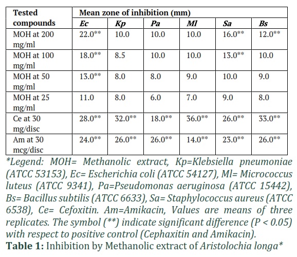
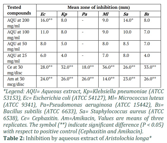
Both of the disc diffusion test and the micro-dilution assay were used to determine the antibacterial activity of methanolic and aqueous crude extracts of A. longa. As demonstrated in (Table 1), Escherichia coli (22 mm) had the largest zone of inhibition (most sensitive bacteria), followed by Staphylococcus aureus (16 mm) and Bacillus subtilis (12 mm). The bacteria with the lowest susceptibility were K. pneumoniae, P. aeruginosa, and M. luteus, which had a 10 mm zone of inhibition at a 200 mg/ml extract concentration. On the other side, the antibacterial potential of A. longa aqueous extract is illustrated in (Table 2). E. coli (16 mm) was the most sensitive bacteria to the aqueous extract, followed by S. aureus (14 mm), M. luteus (9 mm), B. subtilis (8 mm), and K. pneumonia (8 mm). P. aeruginosa, on the other hand, exhibited no sensitivity to the aqueous extract. The optimum bactericidal dosage in the research was 200 mg/ml for both methanolic and aqueous extracts.
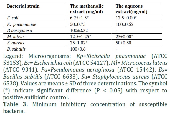
The Results of MIC tests are shown in (Table 3), The MIC values supported the disc-diffusion results, the least MIC values were reported with the methanolic extract compared to the aqueous extract, meaning that the bioactive principles are concentrated in the methanolic extract. The MIC of methanol extract of Aristolochia longa was 6.25±1.5 mg/ml for E. coli, 25±1.02 mg/ml for S. aureus, 12.5±1.25 mg/ml for M. luteus, 50±0.75 mg/ml for K. pneumonia, and 100±0.6 mg/ml for B. subtilis and P. aeruginosa, respectively.
Figures & Tables
Discussion![]()
Generally, our results revealed that the best inhibition was observed with a methanol root extract of Aristolochia longa and range of antibacterial inhibition zone was 12–22 mm at concentration 200 mg/ml of the extract, which was the best concentration and was in particular significant for E. coli and S. aureus compared with the tested antibiotics at (P < 0.05). The MIC values obtained confirmed the previous results with a significant activity against tested microorganisms. Overall, Escherichia coli were the most sensitive towards this extract with MIC ratios of 6.25 mg/mL. Medicinal plants are a valuable part of the local heritage. Herbs provide medicinal properties due to the presence of several biochemical compounds of variable composition found in one or more parts of these plants called secondary plant metabolites [24, 25]. Secondary metabolites contribute to plants' therapeutic capabilities [26]. The pre-experimental ethnobotanical survey of this study (not published) showed that roots of Aristolochia longa are mostly used for treatment of cancer, similar to reports from Algeria cited that A. longa are used traditionally for cancer treatment [27, 28] Aristolochic acid, a major active component of Aristolochiaceae plants, exhibited a dose-dependent cytotoxic effect in HK-2 cells and altered the expression profiles of genes involved in DNA damage response, carbohydrate metabolic process, DNA repair, macromolecule metabolic process, transcription, DNA metabolic process, apoptosis, and cell cycle [29]. The recent investigation established that this plant had antibacterial properties. Among the scarce research on this plant, many papers state that numerous plant extracts from the genus Aristolochia have been proven to have significant in vitro antibacterial activity [30-32]. Additionally, a number of research on antimicrobial screening of medicinal plant extracts have been published. The methanolic extract of A. longa had the best efficacy against S. aureus, E. coli, and M. luteus in these investigations [33, 34].
Aristocholic acid, polyphenols, flavonoids, tannins, c-heterosides, carbohydrates, saponins, phenolic compounds, and flavonoids were detected in A. longa. Because medicinal plants' biological functions are inextricably linked to their chemical components [35], As a result, the A. longa's significant biological activity may be a result of the chemicals discovered. The antibacterial activity of dried extracts (ethyl acetate and methanolic extracts) of the species Aristolochia bracteata against a variety of Gram-negative and Gram-positive microorganisms, as well as the aristolochic acid extracted from both extracts, were previously shown. All crude extracts and extracted aristolochic acid exhibited antibacterial activity throughout a wide range [36, 37], When compared to prior publications, our investigation also revealed significant antibacterial activity.
The inclusion of these key components, as well as the possible antibacterial activity of aristolochic acid against both gram-positive and gram-negative bacteria, may contribute to the extracts' antibacterial activity [38, 39]. Also, it is hypothesized that biologically active components disrupt the permeability of the bacterial cytoplasmic membrane, so facilitating the influx of antibiotics [40, 41], that is confirmed by the MIC values, in fact the aqueous extract of A. Longa L had a low MIC value against Gram-negative and Gram-positive. While the majority of antibacterial medicinal herbs are effective against Gram-positive bacteria, just a minority are effective against Gram-negative bacteria [42-44]. Gram-negative bacteria have been shown to be resistant to extract due to the existence of a hydrophilic outer membrane that prevents hydrophobic extract chemicals from penetrating the target cell membrane. Additionally, gram-negative bacteria' periplasmic region includes enzymes capable of degrading foreign molecules supplied from the outside [45] .
Additionally, A. Longa is responsible for 16% of renal failure [15] and aristolochic acid is a combination of two structurally distinct acids, aristolochic acid-I and aristolochic acid-II [46, 47]. Aristolochic acids are nephrotoxic substances that produce abrupt, atrophy of tubular lesions necrosis, renal failure, and lymphocytic infiltrates characterized by a benign (noncancerous) accumulation of white blood cells in experimental animals and humans [48], This toxicity is a result of both aristolochic acid-II and aristolochic acid-I undergoing nitro group reduction to yield reactive cyclic nitrenium ions capable of building covalent DNA adducts with the exocyclic amino groups of guanine and adenine [49, 50], resulting in cell cycle arrest [45,46]. Aristolochic acid-I was revealed to be more harmful than aristolochic acid-II, while other structural equivalents were shown to be less dangerous overall or to be nontoxic [51, 52]. The fact that cell cycle arrest is responsible for the destruction or injury of renal tissue explains the immunomodulatory features of aristolochic acids and implies their participation in immune responses during toxic lesions [15, 53]. Additionally, Aristolochic acid compounds are commercially generated and utilized as reference standards and research chemicals [54]. Finally, the current study's findings were in accordance with previous international research on A. longa, which demonstrated that this plant possesses antibacterial activity against a variety of microorganisms [55-57], and thus could be used in pharmacological studies to isolate bioactive molecules for pharmaceutical formulation as new antibacterial drugs.
In conclusion, our investigation indicated that extracts of the Aristolochia longa roots, inhibit efficaciously several bacterial strains, supporting the folkloric applications of this plant against diverse bacterial illnesses on skin and alimentary canal. This demonstrates that this plant may be beneficial for the generation of alternative compounds that are potentially more active and less toxic. Further research and development of a suitable and safe self-administered dosage for treating infections caused by these antibiotic resistant microorganisms is required. However, based on the studies, its toxicity profile should be examined to isolate or separate the possible hazardous components.
Authors' Contribution
All authors contributed equally to this study.
The authors declare that there is no conflict of interest in this paper.
References
- Sargın SA, Akçicek E, Selvi S. An ethnobotanical study of medicinal plants used by the local people of Alaşehir (Manisa) in Turkey. Journal of ethnopharmacology, (2013); 150(3): 860-874.
- ATEŞ DA, TURGAY Ö. Antimicrobial activities of various medicinal and commercial plant extracts. Turkish Journal of Biology, (2003); 27(3): 157-162.
- Xavier-Júnior F, Silva K, Farias I, Morais A, Alencar E, et al. Prospective study for the development of emulsion systems containing natural oil products. Journal of drug delivery science and technology, (2012); 22(4): 367-372.
- Yong J, Jie-ying S, Hong-ju F. Studies on Chemical Compositions of Volatile Oil from Seeds of Cuminum cyminum L. Journal of Integrative Plant Biology, (1990); 32(5).
- Mosmann T. Rapid colorimetric assay for cellular growth and survival: application to proliferation and cytotoxicity assays. Journal of immunological methods, (1983); 65(1-2): 55-63.
- Denizot F, Lang R. Rapid colorimetric assay for cell growth and survival: modifications to the tetrazolium dye procedure giving improved sensitivity and reliability. Journal of immunological methods, (1986); 89(2): 271-277.
- Doudach L, Meddah B, Alnamer R, Faouzi M, Chibani F, et al. In vitro antibacterial potential activity of the methanolic and aqueous extracts of corrigiola telephiifolia pourr. and mesembryanthemum nodiflorum. Journal of Biologically Active Products from Nature, (2012); 2(5): 284-291.
- Alnamer R, Alaoui K, Doudach L, Bouidida H, Chibani F, et al. Investigation of methanolic and aqueous extract of Lavandula officinalis for toxicity and antibacterial activity. WJPR, (2012); 5(1): 1-12.
- Cordell GA. Changing strategies in natural products chemistry. Phytochemistry, (1995); 40(6): 1585-1612.
- Bellakhdar J Pharmacopée marocaine traditionnelle. Chapter: Book Name. 1997 of publication; Ibis press.
- González-Tejero M, Casares-Porcel M, Sánchez-Rojas C, Ramiro-Gutiérrez J, Molero-Mesa J, et al. Medicinal plants in the Mediterranean area: synthesis of the results of the project Rubia. Journal of Ethnopharmacology, (2008); 116(2): 341-357.
- Saidi F, Cherif H, Metidji H, Rouibia A, Chaouia C, et al. Studies on the in vitro multiplication by direct organogenesis of a medicinal plant: Aristolochia longa L. Agricultura-Revistă de Scedilla˜ tiintcedilla˜ ă scedilla˜ i Practică Agricolă, (2009); (3/4): 53-65.
- Yamani A, Bunel V, Antoine M-H, Husson C, Stévigny C, et al. Substitution between Aristolochia and Bryonia genus in North-Eastern Morocco: toxicological implications. Journal of Ethnopharmacology, (2015); 166250-260.
- Debelle FD, Nortier JL, De Prez EG, Garbar CH, Vienne AR, et al. Aristolochic acids induce chronic renal failure with interstitial fibrosis in salt-depleted rats. Journal of the American Society of Nephrology, (2002); 13(2): 431-436.
- BENZAKOUR G, BENKIRANE N, AMRANI M, OUDGHIRI M. Immunostimulatory potential of Aristolochia longa L. induced toxicity on liver, intestine and kidney in mice. Journal of Toxicology and Environmental Health Sciences, (2011); 3(8): 214-222.
- Bellakhdar J, Claisse R, Fleurentin J, Younos C. Repertory of standard herbal drugs in the Moroccan pharmacopoea. Journal of ethnopharmacology, (1991); 35(2): 123-143.
- Merzouki A, Ed-Derfoufi F, El Aallali A, Molero-Mesa J. Wild medicinal plants used by local Bouhmed population (Morocco). Fitoterapia, (1997); 68(5): 444-460.
- Benchaâbane A, Abbad A Les plantes médicinales commercialisées à Marrakech. Chapter: Book Name. 1997 of publication; Traces du présent.
- Maire R. Flora of North of Africa. Biol Encyclopedia, Lechevalier, Paris, (1961); 7216-230.
- Fennane M. Contribution à l’étude phytosociologique des tetraclinaies marocaines. Bulletin de l’Institut Scientifique, Rabat, (1986); (10): 57-78.
- Zampini IC, Vattuone MA, Isla MI. Antibacterial activity of Zuccagnia punctata Cav. ethanolic extracts. Journal of Ethnopharmacology, (2005); 102(3): 450-456.
- Singh G, Kumar P. Antibacterial Potential of Alkaloids of Withania somnifera L. and Euphorbia hirta L. Int J Pharmacy Pharm Sci, (2012); 478-81.
- Grare M, Fontanay S, Cornil C, Finance C, Duval RE. Tetrazolium salts for MIC determination in microplates: Why? Which salt to select? How? Journal of microbiological methods, (2008); 75(1): 156-159.
- Andrew W. Manual of Food Quality Control 4. Rev. 1. Microbiological analysis. FAO of the United Nations. Rome. 1992. FAO food and nutrition paper, (1992); 14(4).
- Abdallah EM. Plants: An alternative source for antimicrobials. Journal of Applied Pharmaceutical Science, (2011); 1(6): 16-20.
- Patel SB, Naikwade NS, Magdum CS. Review on Phytochemistry and Pharmacological Aspects of Euphorbia hirta Linn. Asian Journal of Pharmaceutical Research and Health Care, (2009); 1(1): 113-133.
- Cherif H, Saidi F, Boutoumi H, Rouibi A, Chaouia C. Identification and characterization of some chemical compounds in Aristolochia longa L. Agricultura-Revistă de Scedilla˜ tiintcedilla˜ ă scedilla˜ i Practică Agricolă, (2009); (3/4): 76-82.
- Cherif HS, Saidi F, Lazouri H, Aid K, Rouibi A, et al. Determination of the lipid compounds of Aristolochia Longa L. from Algeria. Bulletin UASMV Agriculture, (2009); 661.
- Chen Y-y, Chiang S-y, Wu H-c, Kao S-t, Hsiang C-y, et al. Microarray analysis reveals the inhibition of nuclear factor-kappa B signaling by aristolochic acid in normal human kidney (HK-2) cells. Acta Pharmacologica Sinica, (2010); 31(2): 227-236.
- Navarro-García VM, Luna-Herrera J, Rojas-Bribiesca M, Álvarez-Fitz P, Ríos MY. Antibacterial activity of Aristolochia brevipes against multidrug-resistant Mycobacterium tuberculosis. Molecules, (2011); 16(9): 7357-7364.
- Alviano WS, Alviano DS, Diniz CG, Antoniolli AR, Alviano CS, et al. In vitro antioxidant potential of medicinal plant extracts and their activities against oral bacteria based on Brazilian folk medicine. Archives of oral biology, (2008); 53(6): 545-552.
- Camporese A, Balick M, Arvigo R, Esposito R, Morsellino N, et al. Screening of anti-bacterial activity of medicinal plants from Belize (Central America). Journal of Ethnopharmacology, (2003); 87(1): 103-107.
- Alnamer R, Alaoui K, Doudach L, Bouidida E, Chibani F, et al. In vitro antibacterial activity of Rosmarinus officinalis methanolic and aqueous extracts. International Journal of Pharmaceutics, (2013); 3(1): 1-6.
- Angalaparameswari S, Mohamed Saleem T, Alagusundaram M, Ramkanth S, Thiruvengadarajan V, et al. Anti-microbial activity of aristolochic acid from root of Aristolochia bracteata Retz. World Acad Sci Eng Technol, (2011); 5(9).
- Mekkaoui M, Assaggaf H, Qasem A, El-Shemi A, Abdallah EM, et al. Ethnopharmacological Survey and Comparative Study of the Healing Activity of Moroccan Thyme Honey and Its Mixture with Selected Essential Oils on Two Types of Wounds on Albino Rabbits. Foods, (2022); 11(1): 28.
- Zhao W-H, Hu Z-Q, Okubo S, Hara Y, Shimamura T. Mechanism of synergy between epigallocatechin gallate and β-lactams against methicillin-resistant Staphylococcus aureus. Antimicrobial agents and chemotherapy, (2001); 45(6): 1737-1742.
- Bartha GS, Tóth G, Horváth P, Kiss E, Papp N, et al. Analysis of aristolochlic acids and evaluation of antibacterial activity of Aristolochia clematitis L. Biologia Futura, (2019); 70(4): 323-329.
- Benarba B, Meddah B. Ethnobotanical study, antifungal activity, phytochemical screening and total phenolic content of Algerian Aristolochia longa. Journal of Intercultural Ethnopharmacology, (2014); 3(4): 150.
- El Omari N, Akkaoui S, El Blidi O, Ghchime R, Bouyahya A, et al. HPLC-DAD/TOF-MS chemical compounds analysis and evaluation of antibacterial activity of Aristolochia longa root extracts. Natural Product Communications, (2020); 15(8): 1934578X20932753.
- Abdallah EM, Mujawah AA, Al Mijalli SH. GC-MS and Antibacterial Potential of Methanolic Extract Hyphaene Thebaica L Fruit Pulp against Antibiotics-resistant Pathogens. Journal of Pure and Applied Microbiology, (2021); 15(3): 1655-1665.
- Meng J, Zhu Q, Tan R. New antimicrobial mono-and sesquiterpenes from Soroseris hookeriana subsp. erysimoides. Planta Medica, (2000); 66(06): 541-544.
- Abdallah EM. Antibacterial efficiency of the Sudanese Roselle (Hibiscus sabdariffa L.), a famous beverage from Sudanese folk medicine. Journal of intercultural ethnopharmacology, (2016); 5(2): 186.
- Srinivasan D, Nathan S, Suresh T, Perumalsamy PL. Antimicrobial activity of certain Indian medicinal plants used in folkloric medicine. Journal of ethnopharmacology, (2001); 74(3): 217-220.
- Tomás-Barberán F, Iniesta-Sanmartín E, Tomás-Lorente F, Rumbero A. Antimicrobial phenolic compounds from three Spanish Helichrysum species. Phytochemistry, (1990); 29(4): 1093-1095.
- Cosyns J-P. Aristolochic acid and ‘Chinese herbs nephropathy’. Drug safety, (2003); 26(1): 33-48.
- Lee T-Y, Wu M-L, Deng J-F, Hwang D-F. High-performance liquid chromatographic determination for aristolochic acid in medicinal plants and slimming products. Journal of Chromatography B, (2002); 766(1): 169-174.
- Schmeiser H, Schoepe K-B, Wiessler M. DNA adduct formation of aristolochic acid I and II in vitro and in vivo. Carcinogenesis, (1988); 9(2): 297-303.
- Pfau W, Schmeiser HH, Wiessler M. Aristolochic acid binds covalently to the exocyclic amino group of purine nucleotides in DNA. Carcinogenesis, (1990); 11(2): 313-319.
- Pfau W, Schmeiser HH, Wiessler M. N6-adenyl arylation of DNA by aristolochic acid II and a synthetic model for the putative proximate carcinogen. Chemical research in toxicology, (1991); 4(5): 581-586.
- Li Y, Liu Z, Guo X, Shu J, Chen Z, et al. Aristolochic acid I-induced DNA damage and cell cycle arrest in renal tubular epithelial cells in vitro. Archives of toxicology, (2006); 80(8): 524-532.
- Dey A, Hazra AK, Mukherjee A, Nandy S, Pandey DK. Chemotaxonomy of the ethnic antidote Aristolochia indica for aristolochic acid content: Implications of anti-phospholipase activity and genotoxicity study. Journal of Ethnopharmacology, (2021); 266113416.
- Balachandran P, Wei F, Lin R-C, Khan IA, Pasco DS. Structure activity relationships of aristolochic acid analogues: toxicity in cultured renal epithelial cells. Kidney international, (2005); 67(5): 1797-1805.
- Xian-Hua C, Yu-Zhe Z, Eun-Jeon P, Dong-Hwan S. Tanshinone II A Induces S Phase Cell-cycle Arrest and Apoptosis in Activation of R at Hepatic Stellate Cells in vitro. 춘계총회 및 학술대회, (2008); 8324-324.
- Cheung TP, Xue C, Leung K, Chan K, Li CG. Aristolochic acids detected in some raw Chinese medicinal herbs and manufactured herbal products–a consequence of inappropriate nomenclature and imprecise labelling? Clinical Toxicology, (2006); 44(4): 371-378.
- M’hamed Aneb AT, Bouyahya A, Boury HE, Amzazi S, Benjouad A, et al. In vitro cytotoxic effects and antibacterial activity of Moroccan medicinal plants Aristolochia longa and Lavandula multifida. (2016).
- Dhouioui M, Boulila A, Chaabane H, Zina MS, Casabianca H. Seasonal changes in essential oil composition of Aristolochia longa L. ssp. paucinervis Batt.(Aristolochiaceae) roots and its antimicrobial activity. Industrial Crops and Products, (2016); 83301-306.
- Latha S, Selvamani P, Dhivya P, Benaseer Begam R. A review on pharmacological activities of Aristolochia species. European journal of biomedical and pharmaceutical sciences, (2015); 2(5): 160-167.
This work is licensed under a Creative Commons Attribution-Non Commercial 4.0 International License. To read the copy of this license please visit: https://creativecommons.org/licenses/by-nc/4.0