

Metadata Analysis and Survey Report
Prevalence of Nemathelminthes in Cart Pulling Camels
Muhammad Ahmad*, Majid Hussain Soomro, Fayaz Hussain, Ghulam Jelani
Adv. life sci., vol. 9, no. 3, pp. 235-238, October 2022
*- Corresponding Author: Muhammad Ahmad (Email: mahmad118@yahoo.com)
Authors' Affiliations
Abstract![]()
Introduction
Methods
Results
Discussion
Conclusion
References
Abstract
Background: Camels are multipurpose animals, raised for the source of animal protein and transportation. Pakistan is also a major camel raising country and its population is one million. Parasitic disease cause impaired camel production, although the camels are less affected by the parasites, but some helminths affect them.
Methods: The present study aimed to determine camels’ gastrointestinal helminths (nemathelminthes) in Sakrand, Sindh. The study was carried out in a total 100 dromedaries. The samples were collected and processed through the direct smear and floatation techniques.
Results: The overall data showed a high infestation of nemathelminthes (62%) with the presence of following parasites; Trichostrongylus, Moniezia, Ostertagia, Haemonchus, Marshallagia, Trichuris, Toxocara, Ascaria, Escaria.
Conclusion: To conclude nemathelminthes are major problem in camels under traditional husbandry. Regarding the high prevalence of infection use of parasitic control programmes are essential to improve camel health and productivity because camels play an important role in human lives by helping in transportation, work and provide production.
Keywords: Camelidae; Coproscopy; Dromedary; Nematodes; Parasite; Strongyloides
Introduction![]()
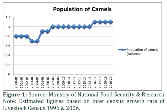
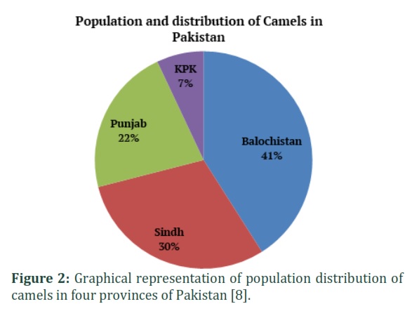
Camel (Camelus dromedaries) is an ancient animal that is well known in the history of human civilization. It belongs to the order Artiodactyla and the family Camelidae [1]. The Camel is a multipurpose animal that plays an important economical role in the dry and semi-arid regions of the globe [2]. The camels not only help the folks who live in the desert with transportation, but it is also a great source of animal protein (Milk & Meat) [3]. Camel milk contains low cholesterol and sugar levels and a higher amount of vitamin C which makes camel milk a healthier food for people living in arid and semi-arid zones as compared to other kinds of milk [4]. The total population of camels is 35 million heads in the globe in this world the major camel raising country is Pakistan [5]. The total camel population in Pakistan is 1.1 million number of heads which is increasing day by day as shown in Fig.1 and the population distribution of camels in different provinces of Pakistan is 41%, 30%, 22% and 7% in Baluchistan, Sindh, Punjab and KPK respectively as shown in Fig.2 [6–8]. Camels are less susceptible to helminthic ailments due to their general browsing practice; however, numerous parasites infect camels. The gastrointestinal nematodes that usually affect the camels are Trichostrongylus, Nematodirella, Haemonchus, Trichuris, Strongyloides, Nematodirus, Coopera, Camelostrongylus and Ostertagia Marshallagia, while camel trematodes are D. dendriticum, Fasciola hepatica, F. gigantica, Paramphistomum and major cestodes reported from camels are Stilesia vittata, Hydatid cyst, Moniezia expansa, Cysticercus tenuicollis and Cysticercus dromedarii [6]. The parasitic diseases are very harmful in livestock production [9]. Damages are due to mortality, pitiable nutrition lowers the development and reproduction rate, loss of weight and rising cost of management and prevention. There is no sufficient published or research data on the incidence of various helminths of camels. Therefore, this study is planned to determine the prevalence rate of nemathelminthes (gastrointestinal parasites) of cart pulling camels in the Sakrand region of province Sindh, Pakistan, to arrange baseline data for advance effort on gastrointestinal parasites in camels and effective control methods can be well proposed.
Methods![]()
Study Site
This study focuses on the camels of the Sakrand District Nawabshah (Shaheed Benazir bad), Sindh Pakistan. The total area of Shaheed Benazirabad (Nawabshah) is 4502 km2. Nawabshah is located at 26.25 latitude and 68.41 longitudes, and it is situated at an elevation of 34 meters above sea level. Sakrand is about 18 kilometres from Nawabshah city and it is situated at an elevation of 25 meters above sea level. From the Sakrand area, a cross-sectional study was conducted on local breeds. Information on age, sex, vaccination, deworming, and herd management was recorded during fecal sample collection for the identification of internal parasites.
Fecal samples collection and material method:
The indigenous camels were 1-18 years ancient, followed traditional husbandry practices and not any anthelmintic drug has been used in previous years. After clinical examination, 100 fecal samples were collected from the rectum and freshly dropped feces by using disposable plastic gloves and placed in a fecal container or polythene bags containing 10% formalin. Total one hundred male camels of Sindhi and Thari breed fecal samples were collected only once from each animal. The collected samples were labeled with appropriate information such as date, locality, and kept cool before reaching the veterinary parasitology analysis research laboratory “Shaheed Benazir Bhutto University of Veterinary and Animal Sciences” where they were immediately examined. Samples were processed on the day of collection.
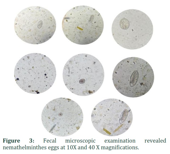
The coproscopy was performed by direct smear method, direct and centrifugal floatation techniques and MacMaster Egg counting techniques were used for the detection of the presence of gastro-intestinal eggs in the fecal samples. The 5 grams of feces were mixed with 70ml of NaCl and sugar dense solution with a density of 1.2 and 1.5 respectively. The mixture sieve via the strainer, fill a dry tube with the mixture and then put cover slip (lamella). After 15 minutes cover slip was placed on the microscopic slide and examined at 10X and 40 X magnifications under a compound microscope. Concentration flotation measures were taken using concentrated salt and sugar solutions for the diagnosis of various eggs and oocysts. The identification of the parasitic genus was based on morphological evidence [10,11]. All the experiments were carried out according to the guidelines of the Institutional Animal Care Committee..
![]()
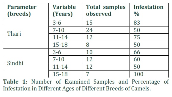
This study shows the prevalence of different types of nemathelminthes that infect the camel and causes a huge loss. Common clinical manifestations of infested camels with gastrointestinal helminths are loss of body weight, soft and bad smell of fecal matter and ill-thriftiness. A total of one hundred male camels of Sindhi and Thari breeds of different ages herd are physically examined in the pen. The fecal sample was macroscopically and microscopically examined in a laboratory.
According to different ages, we divide the total of hundred camels into four different groups according to their age. The macroscopic examination of fecal samples revealed the presence of 2 larvae (2%) from 100 animals. After microscopic examination, 62% of samples were shown positive results with different types of nematode eggs that are shown in table 1.
These all-infested camels were protecting more than one pest. The gastrointestinal parasite's eggs analyzed from the illustrations belong to Trichostrongylus, Moniezia, T. equorum, Ostertagia, H. contortus, Marshallagia, Trichuris, Toxocara, Ascaria, Escaria, C. ovina as shown in Fig.3 presents detected eggs in the samples.
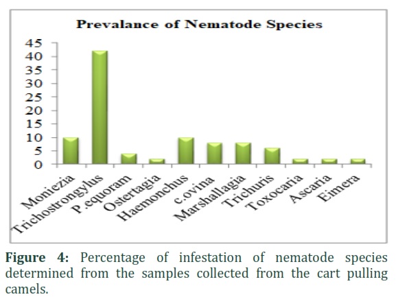
The percentage of identified nematode species Trichostrongylus 42%, Moniezia 10%, T. equorum 4%, Ostertagia 2%, Haemonchus 10%, Marshallagia 8%, Trichuris 6%, Toxocara 2%, Ascaria 2%, Escaria 2%, C. ovina 8% as shown in Fig.4. Parasitic infestation was higher in young animals as compared to other age groups. Animals older than 15 years had the lowest infestation rate. This study was conducted only on males (cart-pulling camels) which shows a high rate of infestation as compared to females.
Tables & Figures
In arid and semi-arid zones camels are adopted better than other domestic animals. Nevertheless, it suffers from various internal and external parasites infection that causes economic losses due to a decrease in productivity, growth and working capacity. In current research illustrated the gastrointestinal nematode prevalence among camels in Tehsil Sakrand District Shaheed Benazir Abad in Sindh region because to our knowledge, there is no previous study conducted in this region with this objective [12]. The microscopic examined 100 camel’s feces samples, and more than half (62%) of camels are infected with gastrointestinal nemathelminthes infection. The microscopic fecal examination revealed that helminthiasis is an important health problem in the study area. This finding agrees with the findings of other researchers that helminthiasis is one of the major problems in camels globally [13,14].
The current study results were in agreement with Birhanu et al. reported 55.5% in 2015 in Ethiopia [15]. This result is incongruent compared with the results of Al- Megrin which found the prevalence was 42.2% in 2015 [16]. Moreover, the prevalence reported by Ijaz et al. was 26.30% in Cholistan desert Pakistan during 2018 [17]. This finding has a lower prevalence rate as compared to Borji et al. which found 75.1% [18], Bekele in Ethiopia reported 75% [19], and Ukashatu et al. reported 77.8% in Nigeria [20]. The high prevalence of nemathelminthes was reported because this study was carried out during the rainy season. The present work indicates that the nematode infestation in camels needs effective anthelminthic treatment. The deworming of camels using broad spectrum anthelminthic can prevent outbreak of parasites in dry and rainy seasons. Further investigations are required to conduct epidemiological study, effects on different body parameters, and to formulate appropriate and cost-effective methods to prevent camels from nematode infestations.
From this study, we concluded that parasitism is a major health problem for camels because parasites get food and shelter from the host and cause disease. This research shows the high prevalence rate of Nemathelminthes of different species. In this research area, people use camels for meat and draught purpose. This study shows the high attack of gastrointestinal Nemathelminthes on camels and their production is used as food by people. For this purpose, it is suggested that deworming should be done properly after regular intervals with safe hand low cost effective anthelmintic drugs (Albendazole, levamizole and Ivermectin). Farmers should be educated through the trained team of the Camel Center to assess the aspects of camel health, management and breeding. Due to the high cost of anthelmintic drugs and checkup fees of veterinarians poor owners do not connect with them and destroy the health of animals which causes a huge earning loss.
Author Contributions
MA wrote the manuscript. GJ & FH collected samples and help MA to perform procedures. MH was convinced of the idea and designs the manuscript.
All listed authors declare no conflict of interests in any capacity including competing and financial.
References
- Al haj OA, Al Kanhal HA. Compositional, technological and nutritional aspects of dromedary camel milk. International Dairy Journal. (2010); 20 (12): 811–21.
- Korlepara RG, Adarsh MK, et al. Camel Milk An White Gold Of Dessert-A Review. International Archive of Applied Sciences and Technology. (2017); 8(3): 74-83
- Hussain R, Khan A, Abbas RZ, Ghaffar A, Abbas G, ur Rahman T, et al. Clinico-Hematological and Biochemical Studies on Naturally Infected Camels with Trypanosomiasis. Pakistan Journal of Zoology. (2016); 48(2) :311–6.
- Mullaicharam A. A review on medicinal properties of camel milk. World Journal of Pharmaceutical Sciences. (2014); 2(3): 237–42.
- FAOSTAT. (2020) Available from: http://www.fao.org/faostat/en/#home
- Parsani H, Singh V, Momin R. Common Parasitic Diseases of Camel. Vet World. (2008);1(10):317. Available from: www.veterinaryworld.org
- Pakistan Economic Survey 2019-20. (2020). Available from: http://www.finance.gov.pk/survey/chapter_20/PES_2019_20.pdf
- Pakistan Livestock Census 2006 | Pakistan Bureau of Statistics. (2006). Available from: http://www.pbs.gov.pk/content/pakistan-livestock-census-2006
- Schwartz HJ, Dioli M. The one-humped camel in Eastern Africa. A pictorial guide to diseases, health care and management. Margraf; 1992.
- Zajac AM, Conboy GA. Veterinary Clinical Parasitology. John Wiley & Sons.; (2012); (20)7; 4–113.
- Khelifi-Ouchene NA, Ouchene N, Dahmani A, Kaaboub EA, Ouchetati I, Haif A. Investigation of internal and external parasites of the camels (Camelus dromedarius) in Algeria. Annals of Parasitology. (2020); 66(3): 331–7.
- Saidi R, Mimoune N, Chaibi R, Baazizi R, Abdelouahed K, Khelef D, et al. Camel gastrointestinal parasites in southern Algeria. Veterinarska stanica. (2022); 53 (3):283–94.
- Kaufmann J. Parasitic infections of domestic animals: a diagnostic manual. ILRI (aka ILCA and ILRAD); (2013).
- Fadhil AI, Abed HH, RFadel S, Al-Zubaidi MtS. Molecular Diagnosis of Nematode Worms Parabronema Skrjabini in Camels (Camelus dromedaries) in Iraq. IRAQI Journal of Agriculture Sciences. (2022); 53 (3): 584–8.
- Birhanu T, Alebie A, Giro B, Chanie M. Prevalence of gastro intestinal nematodes of camel slaughtered at Akaki abattoir, Addis Ababa, Ethiopia. Acta Parasitologica Globalis, (2014); 5(3): 177-182.
- Al-Megrin WA. Prevalence rate of intestinal parasites in camels in Riyadh, Saudi Arabia. International Journal of Zoology Research, (2015); 11(2): 65.
- Ijaz M, Zaman MA, Mariam F, Farooqi SH, Aqib AI, Saleem S, et al. Prevalence, Hematology and Chemotherapy of Gastrointestinal Helminths in Camels. Pakistan Veterinary Journal. (2018); 38(1): 81–5.
- Borji H, Razmi GR, Movassaghi AR, Naghibi A, Maleki M. A study on gastrointestinal helminths of camels in Mashhad abattoir, Iran. Iranian Journal of Veterinary Research. (2010); 11(9): 118-127.
- Bekele T. Epidemiological studies on gastrointestinal helminths of dromedary (Camelus dromedarius) in semi-arid lands of eastern Ethiopia. Veterinary Parasitology. (2002); 105(2): 139–52.
- Ukashatu S, Saulawa MA, Magaji AA. Epidemiology of gastrointestinal parasites of one-humped camel (Camelus dromedarius) slaughtered in Sokoto central abattoir, Sokoto state, Nigeria. Scientific Journal of Veterinary Advances. (2012); 1(4) 789-793.
This work is licensed under a Creative Commons Attribution-Non Commercial 4.0 International License. To read the copy of this license please visit: https://creativecommons.org/licenses/by-nc/4.0

