

Full Length Research Article
Antiparasitic Activity of Chemically Synthesized Magnesium Oxide Nanoparticles against Small Ruminant Haemonchosis
Muhammad Imran1, Abdullah F Alsayeqh2*
Adv. life sci., vol. 9, no. 3, pp. 356-362, October 2022
*– Corresponding Author: Abdullah F Alsayeqh (Email: a.alsayeqh@qu.edu.sa)
Authors' Affiliations
2. Department of Veterinary Medicine, College of Agriculture and Veterinary Medicine, Qassim University, Buraidah – Kingdom of Saudi Arabia
[Date Received: 29/09/2022; Date Revised: 14/10/2022; Date Published: 31/10/2022]
Abstract![]()
Introduction
Methods
Results
Discussion
References
Abstract
Background: Haemonchosis, a debilitating parasitic disease of small ruminants, is a major threat to food security globally. The development of alternative antiparasitic interventions is an important measure toward reducing the progress of veterinary drugs resistance in food animals. Hence, reducing the risks related to chemical foodborne hazards exposure. This study was designed to investigate the anti-parasitic activity of magnesium oxide nanoparticles (MgO-NPs) against different stages of Haemonchus contortus.
Methods: Preparation of magnesium oxide nanoparticles was conducted by sol-gel method at room temperature. For anti-parasitic activity of nanoparticles and commercially available anthelmintics, the adult worms and eggs (collected from gravid female worms) were subjected to adult motility assay and egg hatch assay, respectively. The relative efficacy of both nanoparticles and anthelmintics was classified by identifying the lethal dose LC50.
Results: Wormicidal effects of MgO-NPs were found to depend on both the concentration and the amount of time, they were exposed to. The lower concentrations of the nanoparticle (1 to 5 µg/mL) have not shown any mortality during the first four hours of the incubation. However, the higher concentrations of the nanoparticle (10, 15, 20, and 25 µg/mL) harmed the parasite during this time. The highest dose (25 µg/mL) was capable of killing almost all the worms during the first 16 hours. The highest concentration of nanoparticle induced 90±2.89% egg hatch inhibition and 91±3.3% adulticidal activity for egg hatch assay and adult motility assay, respectively.
Conclusion: Magnesium oxide nanoparticles have shown potent anti-parasitic properties. The study may provide potential alternative anthelmintic to control gastrointestinal parasites particularly haemonchosis in the scenario of emerging anthelmintic resistance.
Keywords: Haemonchosis; Magnesium Oxide Nanoparticles; In-vitro Assays; Anthelmintic; Small Ruminants
Introduction![]()
Parasitism represents one of the key factors threatening global food security and safety. Gastrointestinal (GI) parasitic nematodes are a major health concern for small ruminants in developing countries. Among various GI parasites, Haemonchus (H.) contortus, is a primary, economically authorized and very pathogenic abomasal nematode parasite, and is the main culprit associated with limited performance and production of small ruminants in the subtropical and tropical regions [1,2]. The disease caused by this nasty parasite is hemonchosis. The parasite affects an exceedingly large number of small ruminants ensuing in considerable economic losses in terms of declined meat, milk and wool production, reduced body weight, and cost of anthelmintic drugs [3]. Adult H. contortus invade abomasal walls, attach themselves, and feed on blood. Muscle thickening, cell hyperplasia, necrosis of mucosal layers, and fibrosis of glandular and inflammatory cells are the chief histopathological changes associated with haemonchosis [4].
In developing countries like Pakistan, the economic losses related to H. contortus in small ruminants have been reported more than 8800.09 million rupees in different areas due to condemnation of abomasa. It is also reported that about 29% reduction and 27% reduction in milk yield and weight occurred, respectively due to haemonchosis so, the annual loss in Pakistani rupees is 13406.39 million and 40 million, respectively [5]. The control of GI parasites is mainly centered on the use of anthelmintic drugs (benzimidazoles, levamisoles, and macrocyclic lactones) and plant extracts [6,7]. However, the control of these parasites with current anthelmintic drugs is becoming exceedingly difficult due to the emerging issue of drug resistance [7]. Developing drug resistance is a threat to global food safety and security. Resistance of sheep nematodes to levamisole, oxfendazole, and ivermectin has been reported in some studies as 59%, 88%, and 41%, respectively. Therefore, it necessitates the development of an alternate but eco-friendly and cost-effective product by using nanotechnology.
Nanoparticles (NPs) are widely used for the control of parasites. They cause the death of parasites as they can easily cross the cell membrane due to their smaller size and produce reactive oxygen which results in reactivity and death [8,9]. Magnesium oxide (MgO-NPs) are diversely used in vitro and in vivo biological applications. It may have good anthelmintic properties, as the antiprotozoal activity of MgO-NPs has been reported in Saudi Arabia [10]. But no studies have been reported to determine the in vitro efficacy of chemically synthesized MgO-NPs against nematodes parasites, particularly H. contortus. Thus, the present study was designed to analyze the comparative anthelmintic efficacy of chemically synthesized MgO-NPs and economically available anthelmintics.
Methods![]()
Collection and processing of samples
Abomasa of small ruminants, immediately after slaughtering regardless of sex and age of animals were collected and transported to the Molecular Parasitology Laboratory, Department of Parasitology, Faculty of Veterinary Science, University of Agriculture, Faisalabad. The adult female and male worms were collected from the abomasum. The collected worms were washed and finally placed in a petri dish comprising phosphate buffer saline (1x PBS) at pH 7.4 and macerated to obtain eggs which were repeatedly washed with distilled water.
Egg hatch assay
The efficacy of nanoparticles and anthelmintics was determined through egg hatch assay [11]. For collection of eggs, gravid female worms were reduced by using mortar and pestle. The suspension was made using PBS and constrained through a tea strainer to remove all kinds of debris from the suspension. In the end, 200 eggs/mL were maintained through the McMaster technique [12]. Among three experimental groups formed, group one received chemically synthesized MgO-NPs while group two was treated with commercially available anthelmintic (Albendazole) as the positive control. The third group having egg solution and diluent was considered as the negative control. In the assay, approximately 200 eggs in 0.5mL of suspension were placed in each test tube (5mL). Serial dilutions of MgO-NPs consisting of (0.00001, 0.0001, 0.001, 0.01, 0.1 and 1μg/mL) serve as experimental group while Albendazole dissolved in DMSO at different concentrations 0.03125, 0.0625, 0.125, 0.250 and 0.5 μg/mL as positive control group. Test tubes were covered and left at room temperature for 48 hours. The hatching of eggs ceased by adding 2 drops of Lugol’s iodine to each tube. Total eggs (dead and embryonated) and the number of hatched larvae were counted in every tube under the microscope at 40X magnification.
Worm in-vitro assay
Worm in-vitro assay was performed to check the efficacy of nanoparticles and anthelmintic as described by [13]. 10 adult worms were selected and kept in each tube containing chemically synthesized MgO-NPs at different concentrations. Worms were placed in PBS and then transferred in petri plate, separately. Each petri plate was provided with serial dilutions of MgO-NPs consisting of 1, 5, 10, 15, 20, and 25 μg/mL. Albendazole dissolved in DMSO at different concentrations of 16, 32, 64, 128, and 256 μg/mL for positive control while phosphate buffer saline as a negative control group. The anthelmintic efficacy of MgO-NPs was assessed by restricted movement of the worm. After 24 hours duration, the test solution was washed thoroughly, and the worm was suspended in PBS for 30 minutes to recover the remaining movement of the worm. The number of actively moving worms was recorded under a microscope. The mortality percentage was calculated by using the following formulae.
Statistical Analysis
Probit analysis was performed to calculate LC50 values for egg hatch assay and worm in-vitro assay.
Results![]()
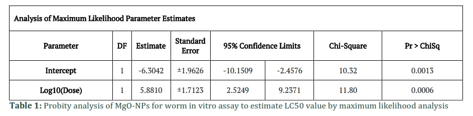
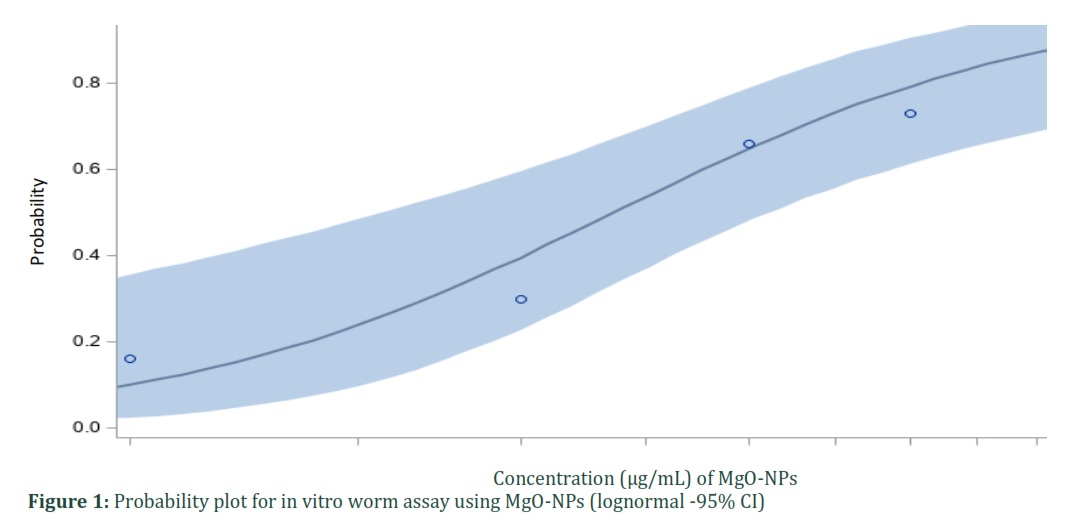
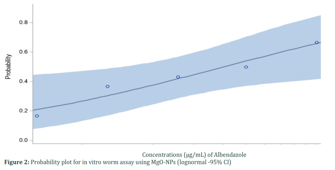
For in vitro worm assay, the variation in adult worm motility was depicted both in the experimental and positive control groups. As the incubation duration and concentrations were increased, there was a significant decrease in the amount of movement presented by the worms. With an increase in the concentration of the NPs, the motility was significantly reduced and the mortality percentage increased. In comparison, the mortality was higher even at lower doses of the nanoparticles while the mortality was lower even at much higher concentrations of Albendazole. Figure 1 and 2 show probability plots for antiparasitic effects of MgO-NPs and albendazole on adult worm mortality, respectively. Table 1 and 2 depict probit analysis of MgO-NPs and albendazole to estimate LC50 value by maximum likelihood analysis.
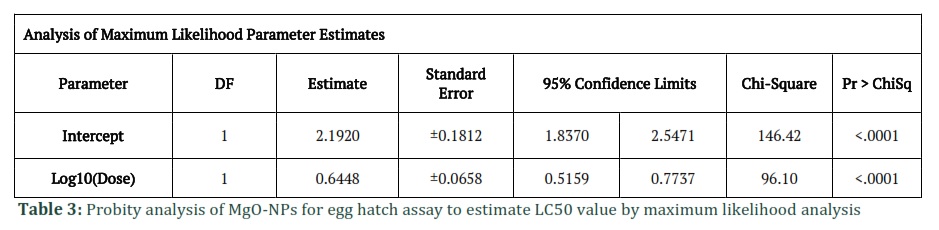
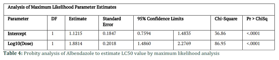
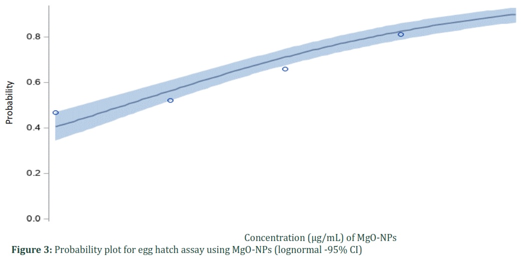
The results of the egg hatch assay were recorded after 48 hours under the microscope at 40X. The hatched larvae and unhatched eggs were counted. The MgO-NPs had a greater impact on inhibition of egg hatching as compared to albendazole, which is less effective even at higher doses. The data unambiguously demonstrate that MgO-NPs deliver stronger antiparasitic effects at the same dose and time point. Figures 3 and 4 indicate probability plots for antiparasitic effects of MgO-NPs and albendazole on the hatching of eggs, respectively. Table 3 and 4 show probit analysis of MgO-NPs and albendazole to estimate LC50 value by maximum likelihood analysis.
Figures & Tables

Haemonchus contortus is one of the leading causes of reduced animal production due to high morbidity and mortality rates [14]. Infection with this nasty parasite causes a decrease in protein absorption, incorrect protein metabolism, and mineral consumption [15]. Although the mortality percentage may not be high during infection, its indirect consequences on animal output are more significant [16]. Decreases in growth, diarrhea, anorexia, loss of appetite, and even anaemia are the indirect losses caused by H. contortus [17]. Moreover, inappropriate and misuse of anthelmintics, made the parasite capable of developing resistance against different anthelmintic groups [18,19,20,21]. Hence, to combat the increasing anthelmintic resistance there is a need to adapt alternate ways. In this regard, nanoparticles are one of the emerging alternatives against resistant parasites. Their small size, extraordinary surface reactivity, and wide range of biological applications make them a suitable candidate for parasite control [22]. They can permeate membranes, and produce reactive oxygen species (ROS), which cause intense reactivity and ultimately results in the death of pathogenic agents [23]. The anthelmintic potential of nanoparticles is currently being continuously assessed for the management of parasitic illnesses [9].
The current study investigates the ovicidal and adulticidal efficacy of chemically produced MgO-NPs against H. contortus using adult mortality and egg hatch assays. MgO-NPs with dimensions of 6 nm in diameter, and 461 m2 in the surface area produced potent antiparasitic effects. After 48 hours, MgO-NPs had egg hatch inhibition of 94.66%, with the highest concentration. Albendazole, a synthetic substance, caused 65.33 % egg hatch inhibition at a dosage of 0.5 g/mL. The MgO-NPs treatment's LC50 value for the egg hatch assay was calculated to be 0.0003984g/mL. According to the study, MgO-NPs were particularly successful at preventing egg hatching at very low concentrations, and nano-sized MgO-NPs must have caused hindrance in molting and other physiological functions necessary for H. contortus eggs to hatch [24]. Therefore, it is evident from the literature review that the findings of present study demonstrated the NPs to be more effective [10,25,26,27]. The greatest concentration of MgO-NPs (25ug/mL) caused 96% of adult mortality in the current study. The LC50 value for the MgO-NPs treatment was 11.80238 g/mL. In the current investigation, morphological changes i.e. coil formation by the worms to prevent stress, caused by the test compounds have been utilized as prospective anthelmintic agents. Remarkable anthelmintic activity of MgO-NPs has been found during the current study. The mode of action of MgO-NPs is still debatable.
Transcuticular diffusion has been demonstrated to be a typical route of entry for non-electrolytic compounds in worms in earlier research [28]. Additionally, research has indicated that, as compared to oral intake, this is the preferred method for the absorption of powerful broad-spectrum anthelmintics [29]. Lipophilic anthelmintics, such as albendazole, are better able to penetrate the helminths’ outer surface than hydrophilic substances [30]. However, the extremely small size of the nanoparticles, i.e., 15–25 nm, which likely allowed for transcuticular absorption of MgO-NPs by the parasite, may have contributed to a higher mortality rate of adult
parasites following MgO-NPs treatment in the current study as compared to treatment with the anthelmintics.
The anthelmintic effects of MgO-NPs against the GI nematode, H. contortus, have been studied for the first time in this study. However, various scientists have studied the antibacterial properties of MgO-NPs and reported that the antibacterial efficacy was size dependent, with the smallest sizes having an impact on both Gram-positive and -negative bacteria while bigger sizes exclusively have an impact on Gram-negative ones [31,32,33,34,35,36,37,38]. Additionally, investigations have shown that MgO-NPs with a particle size of less than 50 nm partially suppressed bacterial growth while having a substantial bactericidal impact at that size [39,24,20,21].
The production of superoxide anions O2 on the MgO surface is one explanation for the strong anthelmintic effects of increasing MgO NP concentrations. The concentration of MgO and OH- on the target's surface is increased as a result of MgO being hydrated and generating Mg (OH)2, which covers the surface. As a result, the target is more effectively destroyed as the O2 concentration rises [40-42]. Although NPs size, dosages, and the characteristics of the microbe, including cell wall structure and cellular enzymes, play an essential role as an antibacterial agent, other parameters such as NPs structure, surface qualities, and the temperature of synthesis may also be contributing to this role. The effects of MgO nanoparticles on Leishmania parasites, which exhibited the best antiparasitic activity (anti-leishmanial) among all evaluated NPs, were in line with the findings of the current study on treating worms and eggs [24].
Comparing MgO-NPs to other anthelmintics previously utilized against H. contortus, treated worms in the current study showed that they were more effective with MgO-NPs. According to the study's findings, H. contortus is very resistant to anthelmintics. It has been determined that among gastrointestinal nematodes, H. contortus is the most prevalent species affecting the production of small ruminants. It was discovered that nanoparticles were more efficient than commercially available anthelmintics.
The resistance to commercially available chemical anthelmintics is growing daily as a result of the widespread usage of chemical substances. The findings of this study showed that MgO-NPs are more effective against H. contortus. However, haemonchosis and other worm-related disorders can be controlled with the help of nanoparticle-based anthelmintics, which are also effective against H. contortus. Animals that have already developed resistance to chemical anthelmintics, i.e. sheep and goats are unable to eradicate GI nematodes even after treatment with the required amount of anthelmintics, benefit more from nanoparticle-based anthelmintics. Commercially available anthelmintics have less efficacy even at higher concentrations as compared to nanoparticles. These agents are needed in higher doses to have desired effects. Nanoparticles have good anthelmintic activity even at very low concentrations The result of the study shows that the anthelmintic effect of nanoparticles is in a dose-dependent manner for both parameters, which suggests the use of these NPs for control of GI nematodes.
Author Contributions
Muhammad Imran planned, executed, and conducted the research in the laboratory. Abdullah F Alsayeqh helped in troubleshooting, statistical analysis, and drafted the manuscript.
![]()
The author declares that there is no conflict of interest regarding the publication of this paper.
![]()
References
- Imran M, Khan MN, Sajid MS, Saqib M. Comparative Evaluation of Natural Resistance of Dera Din Panah and Nachi Goat Breeds Towards Artificial Infection with Haemonchus contortus. Pakistan Veterinary Journal, (2018); 38(4).
- Strbac F, Bosco A, Amadesi A, Rinaldi L, Stojanović D, et al. Ovicidal potential of five different essential oils to control gastrointestinal nematodes of sheep. Pakistan Veterinary Journal, (2020); 41(3): 353-358.
- Singh R, Bal M, Singla L, Kaur P. Detection of anthelmintic resistance in sheep and goat against fenbendazole by faecal egg count reduction test. Journal of parasitic diseases, (2017); 41(2): 463-466.
- Saminathan M, Gopalakrishnan A, Latchumikanthan A, Milton A, Aravind M, et al. Histopathological and parasitological study of blood-sucking Haemonchus contortus infection in sheep. Adv Anim Vet Sci, (2015); 3(2): 99-108.
- Qamar MF, Maqbool A, Ahmad N. Economic losses due to haemonchosis in sheep and goats. Sci Intern, (2011); 23(4): 321-324.
- Qureshi NA. In vitro Anticoccidial, Antioxidant Activities and Biochemical Screening of Methanolic and Aqueous Leaves Extracts of Selected Plants. Pakistan Veterinary Journal, (2021); 41(1).
- Rose H, Rinaldi L, Bosco A, Mavrot F, De Waal T, et al. Widespread anthelmintic resistance in European farmed ruminants: a systematic review. Veterinary Record, (2015); 176(21): 546-546.
- Jalil PJ, Shnawa BH, Hamad SM. Silver Nanoparticles: Green Synthesis, Characterization, Blood Compatibility and Protoscolicidal Efficacy against Echinococcus granulosus. Pakistan Veterinary Journal, (2021); 41(3).
- Bajwa HUR, Khan MK, Abbas Z, Riaz R, Rehman Tu, et al. Nanoparticles: Synthesis and Their Role as Potential Drug Candidates for the Treatment of Parasitic Diseases. Life, (2022); 12(5): 750.
- Hussein EM, Ahmed SA, Mokhtar AB, Elzagawy SM, Yahi SH, et al. Antiprotozoal activity of magnesium oxide (MgO) nanoparticles against Cyclospora cayetanensis oocysts. Parasitology international, (2018); 67(6): 666-674.
- Coles G, Bauer C, Borgsteede F, Geerts S, Klei T, et al. World Association for the Advancement of Veterinary Parasitology (WAAVP) methods for the detection of anthelmintic resistance in nematodes of veterinary importance. Veterinary parasitology, (1992); 44(1-2): 35-44.
- Soulsby E. Helminths. Arthropods and Protozoa of domesticated animals, (1982); 291.
- Hounzangbe-Adote S, Fouraste I, Moutairou K, Hoste H. In vitro effects of four tropical plants on the activity and development of the parasitic nematode, Trichostrongylus colubriformis. Journal of Helminthology, (2005); 79(1): 29-33.
- Roeber F, Jex AR, Gasser RB. Impact of gastrointestinal parasitic nematodes of sheep, and the role of advanced molecular tools for exploring epidemiology and drug resistance-an Australian perspective. Parasites & vectors, (2013); 6(1): 1-13.
- Medellin-Castillo N, Leyva-Ramos R, Padilla-Ortega E, Perez RO, Flores-Cano J, et al. Adsorption capacity of bone char for removing fluoride from water solution. Role of hydroxyapatite content, adsorption mechanism and competing anions. Journal of industrial and Engineering Chemistry, (2014); 20(6): 4014-4021.
- Ekong PS, Juryit R, Dika NM, Nguku P, Musenero M. Prevalence and risk factors for zoonotic helminth infection among humans and animals-Jos, Nigeria, 2005-2009. Pan African medical journal, (2012); 12(1).
- Swai E, Mtui P, Mbise A, Kaaya E, Sanka P, et al. Prevalence of gastro intestinal parasite infections in Maasai cattle in Ngorongoro District, Tanzania. Livestock Research for Rural Development, (2006); 18(8): 18-27.Kotze AC, Hunt PW, Skuce P, von Samson-Himmelstjerna G, Martin RJ, et al. Recent advances in candidate-gene and whole-genome approaches. concern, (2014); 45.
- Van den Brom R, Moll L, Kappert C, Vellema P. Haemonchus contortus resistance to monepantel in sheep. Veterinary parasitology, (2015); 209(3-4): 278-280.
- Kotze A, Prichard R. Anthelmintic resistance in Haemonchus contortus: history, mechanisms and diagnosis. Advances in parasitology, (2016); 93: 397-428.
- Lamb J, Elliott T, Chambers M, Chick B. Broad spectrum anthelmintic resistance of Haemonchus contortus in Northern NSW of Australia. Veterinary parasitology, (2017); 24148-51.
- Sabu A, Wakif A, Areekara S, Mathew A, Shah NA. Significance of nanoparticles' shape and thermo-hydrodynamic slip constraints on MHD alumina-water nanoliquid flows over a rotating heated disk: The passive control approach. International Communications in Heat and Mass Transfer, (2021); 129105711.
- Kandeel M, Akhtar T, Zaheer T, Ahmad S, Ashraf U, et al. Anti-parasitic Applications of Nanoparticles: A Review. Pakistan Veterinary Journal, (2022); 42(2).
- Bafghi AF, Daghighi M, Daliri K, Jebali A. Magnesium oxide nanoparticles coated with glucose can silence important genes of Leishmania major at sub-toxic concentrations. Colloids and Surfaces B: Biointerfaces, (2015); 136300-304.
- Rahdar S, Rahdar A, Khodadadi M, Ahmadi S. Error analysis of adsorption isotherm models for penicillin G onto magnesium oxide nanoparticles. Applied Water Science, (2019); 9(8): 1-7.
- Hassan SE-D, Fouda A, Saied E, Farag MM, Eid AM, et al. Rhizopus Oryzae-mediated green synthesis of magnesium oxide nanoparticles (MgO-NPs): A promising tool for antimicrobial, mosquitocidal action, and tanning effluent treatment. Journal of Fungi, (2021); 7(5): 372.
- Alkhudhayri A, Thagfan FA, Al-Quraishy S, Abdel-Gaber R, Dkhil MA. Assessment of the oxidative status and goblet cell response during eimeriosis and after treatment of mice with magnesium oxide nanoparticles. Saudi Journal of Biological Sciences, (2022); 29(2): 1234-1238.
- Nassef NE, Moharm IM, Atia AF, Brakat RM, Abou-hussien NM, et al. Therapeutic efficacy of chitosan nanoparticles loaded with albendazole on parenteral phase of experimental Trichinellosis. Journal of the Egyptian Society of Parasitology, (2019); 49(2): 301-311.
- Raghupathi KR, Koodali RT, Manna AC. Size-dependent bacterial growth inhibition and mechanism of antibacterial activity of zinc oxide nanoparticles. Langmuir, (2011); 27(7): 4020-4028
- Elliott JR, Compton RG. Modeling Transcuticular Uptake from Particle-Based Formulations of Lipophilic Products. ACS Agricultural Science & Technology, (2022).
- Cavalcante GS, Morais SMd, André WPP, Araújo-Filho JVd, Muniz CR, et al. Chemical constituents of Calotropis procera latex and ultrastructural effects on Haemonchus contortus. Revista Brasileira de Parasitologia Veterinária, (2020); 29.
- Tang Z-X, Lv B-F. MgO nanoparticles as antibacterial agent: preparation and activity. Brazilian Journal of Chemical Engineering, (2014); 31591-601.
- He Y, Ingudam S, Reed S, Gehring A, Strobaugh TP, et al. Study on the mechanism of antibacterial action of magnesium oxide nanoparticles against foodborne pathogens. Journal of nanobiotechnology, (2016); 14(1): 1-9.
- Sharma G, Soni R, Jasuja ND. Phytoassisted synthesis of magnesium oxide nanoparticles with Swertia chirayaita. Journal of Taibah University for Science, (2017); 11(3): 471-477.
- Cai L, Chen J, Liu Z, Wang H, Yang H, et al. Magnesium oxide nanoparticles: effective agricultural antibacterial agent against Ralstonia solanacearum. Frontiers in microbiology, (2018); 9790.
- Rukh S, Sofi AH, Shah MA, Yousuf S. Antibacterial activity of magnesium oxide nanostructures prepared by hydrothermal method. Asian Journal of Nanosciences and Materials, (2019); 2(4): 425-430.
- Noori AJ, Kareem FA. Setting time, mechanical and adhesive properties of magnesium oxide nanoparticles modified glass-ionomer cement. Journal of Materials Research and Technology, (2020); 9(2): 1809-1818.
- Fouda A, Hassan SE-D, Saied E, Hamza MF. Photocatalytic degradation of real textile and tannery effluent using biosynthesized magnesium oxide nanoparticles (MgO-NPs), heavy metal adsorption, phytotoxicity, and antimicrobial activity. Journal of Environmental Chemical Engineering, (2021); 9(4): 105346.
- Khan AU, Khan M, Khan AA, Parveen A, Ansari S, et al. Effect of Phyto-Assisted Synthesis of Magnesium Oxide Nanoparticles (MgO-NPs) on Bacteria and the Root-Knot Nematode. Bioinorganic Chemistry and Applications, (2022); 2022.
- Vidic J, Stankic S, Haque F, Ciric D, Le Goffic R, et al. Selective antibacterial effects of mixed ZnMgO nanoparticles. Journal of nanoparticle research, (2013); 15(5): 1-10.
- Avanzato C, Follieri J, Banerjee I, Fath K. Biomimetic synthesis and antibacterial characteristics of magnesium oxide—germanium dioxide nanocomposite powders. Journal of composite materials, (2009); 43(8): 897-910.
- Jin T, He Y. Antibacterial activities of magnesium oxide (MgO) nanoparticles against foodborne pathogens. Journal of Nanoparticle Research, (2011); 13(12): 6877-6885.
- Patel MK, Zafaryab M, Rizvi M, Agrawal VV, Ansari Z, et al. Antibacterial and cytotoxic effect of magnesium oxide nanoparticles on bacterial and human cells. Journal of nanoengineering and nanomanufacturing, (2013); 3(2): 162-166.
This work is licensed under a Creative Commons Attribution-Non Commercial 4.0 International License. To read the copy of this license please visit: https://creativecommons.org/licenses/by-nc/4.0