

Full Length Research Article
Prevalence of Black Quarter in Association with Patho-Morphological Alterations in Affected Tissues at Different Zones of Punjab-Pakistan
Asif Idrees1*, Zafar Iqbal Chaudhary2, Muhammad Younas1, Aftab Anjum3, Muhammad Ahsan Naeem4*, Muhammad Muneeb Rauf5, Waqas Ahmad6, Amanullah Khan7, Qamar-un-Nisa2
Adv. life sci., vol. 9, no. 4, pp. 504-509, December 2022
*– Corresponding Author: Asif Idrees (Email: asif.idrees@uvas.edu.pk)
Authors' Affiliations
2. Department of Pathology, University of Veterinary and Animal Sciences, Lahore – Pakistan
3. Department of Microbiology, University of Veterinary and Animal Sciences, Lahore- Pakistan
4. Department of Basic Sciences (Pharmacology), KBCMA, College of Veterinary and Animal Sciences, Norawal – Pakistan
5. Department of Basic Sciences (Anatomy), KBCMA, College of Veterinary and Animal Sciences, Norawal – Pakistan
6. Department of Clinical Sciences (Epidemiology), KBCMA, College of Veterinary and Animal Sciences, Norawal – Pakistan
7. Department of Pathobiology (Microbiology), College of Veterinary and Animal Sciences, Jhang – Pakistan
[Date Received: 30/05/2022; Date Revised: 01/11/2022; Date Published: 31/12/2022]
Abstract![]()
Introduction
Methods
Results
Discussion
References
Abstract
Background: Blackleg or Black Quarter (BQ) is a serious bacterial disease caused by Clostridium chauvoei. It causes edematous and gaseous changes in skeletal muscles of animals. The study was designed to find prevalence of BQ in 6 districts of Punjab, Pakistan from June 2018 to June 2019.
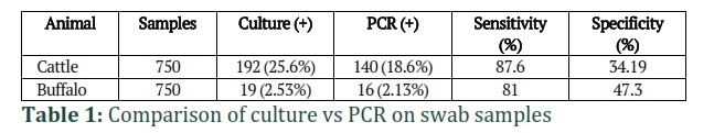
Methods: Animals were randomly monitored, and selected in each union council regardless of age, sex and species, but lameness and hyperthermia were the selection parameters. A proforma was used to record the experimental readings of each animal. A total of 1500 swab samples from bovines (cattle and buffaloes) were processed through conventional PCR and culture sensitivity tests to examine the comparative diagnostic efficacy and suitability of the test.
Results: Sensitivity and specificity of culture tests were 87.6% and 34.19% respectively, whereas PCR demonstrated 81% and 47.3% results for the said aspects, respectively. Alpha toxin gene (Ccta) was amplified at 52.2oC yielding an amplicon size of 1400 base pairs. Area wise and season wise prevalence of each animal was also determined. The prevalence of BQ in cattle was significantly higher (P<0.05) in Muzaffargarh (25.6%), Layyah (24.8%), Rahim Yar Khan (23.2%), and Bhakkar (29.6%) that belonged to the arid and dessert zones of Punjab-Pakistan whereas, it was non-significant (P > 0.05) in Lahore (0%) and Nankana Sahib (8%) which belonged to the canal irrigated zones.
Conclusion: There was higher prevalence of BQ in different areas of Punjab, Pakistan that show the higher risk of disease transmission. A systematic surveillance system is essential to regularly monitor the disease incidence and prevalence in these areas of Pakistan.
Keywords: Black Quarter; Prevalence; Pathological; Ruminants; Zones
Introduction![]()
Large ruminants confront various managemental and disease related problems. Clostridia species normally inhabit the intestinal microflora of various animals and humans, while many of these species are also pathogenic that cause pseudo-membranous colitis, food-borne illnesses, gas gangrene, and botulism [1]. Black Quarter (BQ) or Blackleg is a lethal disease of ruminants caused by an anaerobic, gram positive and spore forming bacteria, Clostridium chauvoei [2]. The disease is severe in young ones of ruminants which are well-fed and possess good health [2]. Myocarditis, rhabdomylitis, toxemia, per-acute, and acute mortality in ruminants are the characteristic features of this disease [2]. In the region of Indo-Pak and other geographical regions, BQ is highly prevalent by maintaining its endemic level with the 2nd most reported bacterial disease in animals [2]. Cytotoxin A (CctA), neuraminidase, hyaluronidase (γ), DNase (β), hemolysin (α) and oxygen labile hemolysin (δ) are the soluble antigens of Clostridium chauvoei strains. These are involved in pathogenesis by stabling oxygen level in host (animal) [3,4]. Molecular epidemiology of BQ has been performed on the basis of virulence and pathogenic factors [5]. The whole bacterial next generation sequencing (NGS) application has been given substantial importance to find molecular epidemiology of C. chauvoei [6]. The present study has been aimed to find prevalence of BQ in different ecological ones of Punjab, and to know efficacy of disease detection methods through comparison of histopathology, culture, and molecular methods.
Methods![]()
For this study, only 6 districts of Punjab were selected that comprise of 3 diverse zones between June 2018 to June 2019 using random selection method. The criteria of keeping sex, species, and age were not kept as standard method. Each district was further sub divided into union councils and animals were randomly spread. The criterion for selection of animals was based on signs of lameness and hyperthermia. All the clinical parameters were recorded from each animal selected from the study population and written in a proforma. Confirmation was made on the basis of PCR amplification of Clostridium chauvoei from swab samples (250 samples from each zone) taken from clinically suspected animals, while prevalence on the basis of seasons and areas were also calculated.
Statistical Analysis
One way ANOVA was used as standard method to analyze the available data from the research study. All those variables that responded as significant (P<0.05) through statistical methods were additionally assessed through Duncan multiple range test (DMRT) using SAS (SAS Int. Cary, North Carolina, V 9.1).
Results![]()
Culture and PCR identification
Each swab sample was processed to culture and PCR test to identify percentage of positive results. A comparison of isolation methods and biochemical identification through PCR was performed to know the efficacy of each test (Table 1).
Prevalence of BQ in different areas of Punjab
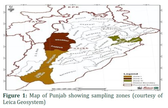
Zone-I comprised of district Muzaffargarh and Rahim Yar Khan (Plate 4.10). Muzaffargarh lies between 30°12′0″ North and 71°25′0″ East on map with an average rainfall of 21 mm. Rahim Yar khan lies between 28.42° North and 70.30° East. The average rain fall is about 100 mm.
Zone-II comprised of district Layyah and Bhakkar (Plate 4.10). Layyah is situated between 30.58° North and 70.56° East with average rain fall of 18.7 mm/annum. District Bhakkar lies between 31°38′North and 71°04′ East with an average rain fall of 19 mm/annum.
Zone-III comprised of Lahore and Nankana Sahib (Plate 4.10). Lahore is situated 31°32′59″ North and 74°20′37″ East with rain fall ranging 12-609 mm/annum. Nankana Sahib is situated between 31°27′ North and 73°42′ East with rain fall ranging 12-609 mm/annum (Geological Survey of Pakistan).
Zone-wise thorough prevalence:
The tables shows that the highest values were existed in II zone and then subsequently in I and III zone (Table 2), while non-significant difference was observed for the case of prevalence in buffaloes (P >0.05).
District-wise annual prevalence
A significant higher value (P<0.05) of BQ prevalence in cattle was observed in Bhakkar, Rahim Yar Khan, Muzaffar Garh, and Layyah. In a similar fashion, a bit similar trend of prevalence was observed in these districts (Table 3).
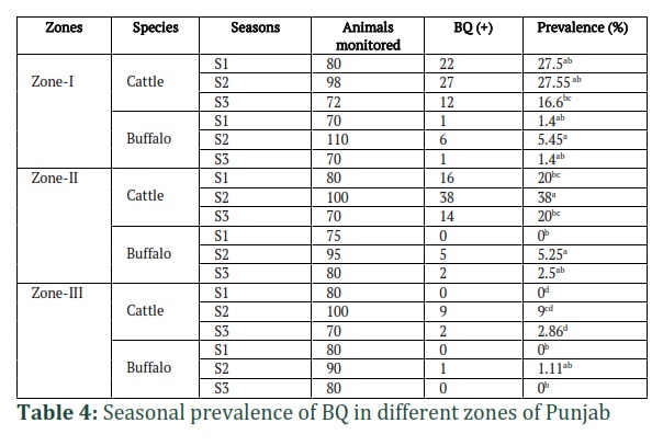
Zone-wise seasonal prevalence:
A significant variation (P < 0.05) was observed amongst all the zones regarding prevalence of BQ in cattle (Table 4.6). A significantly higher prevalence (P<0.05) was noted in Zone-I and II during January-April (S1), but non-significant (P>0.05) in Zone III. Similarly, there was no significant difference (P>0.05) noted in prevalence during May-August (S2) in Zone-I and II, while it was meaningfully lower (P <0.05) in III-Zone. The I and II zones were not found significantly different (P>0.05) during September-December (S3), while in Zone-III it was significantly lower (P<0.05) (Table 4). Prevalence in column having different superscripts indicates significant difference among seasons in buffaloes. A significantly higher prevalence (P<0.05) of BQ was observed during S2 in Zone-I and II. There was no significant difference (P>0.05) among S2 and S3 in Zone- I and Zone- III (Table 4).
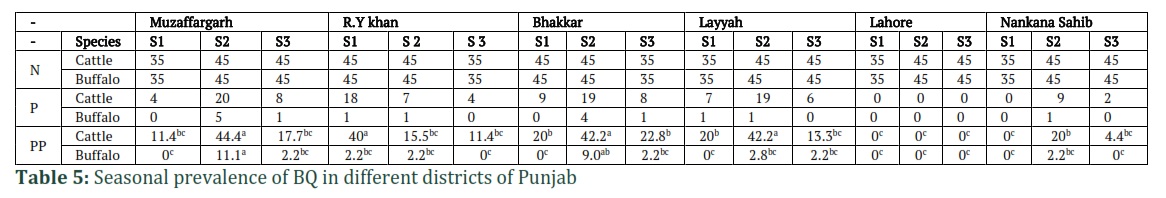
District-wise seasonal prevalence:
Seasonal prevalence of BQ was calculated in cattle among different districts and a significant difference (P < 0.05) was also noticed between the seasons and districts. A meaningfully higher prevalence (P < 0.05) was observed in district Muzaffargarh and Layyah belonging to different zones during S2. Prevalence having same superscripts is not significantly different (P > 0.05) from each other (Table 5). A considerable variation was recorded among various districts in different seasons. Meaningly higher (P < 0.05) was found during S2 in district Muzaffargarh followed by S3 and S1, respectively. There was no significant difference in prevalence (P > 0.05) during S1 and S2 in district Rahim Yar Khan, S3 in Layyah and Bhakkar. A noteworthy higher prevalence was observed in Layyah during S2 followed by S3 and S1. There was no case of BQ observed in district Lahore during the study period (Table 5).
Polymerase Chain Reaction (PCR)
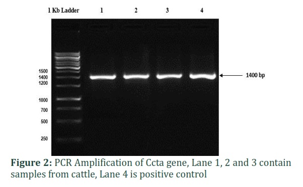
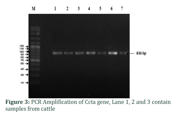
Variable results were obtained from PCR reaction. After the extraction of DNA, the product was mixed with the master mix and run-on agarose gel under optimum conditions of voltage. The Ccta was successfully amplified at 52.2oC which was an annealing temperature as shown in the figure (Fig. 2). The universal primers were corresponded to 16SrRNA region which were amplified at temperature of 50.2oC (Fig. 3).
Histopathology
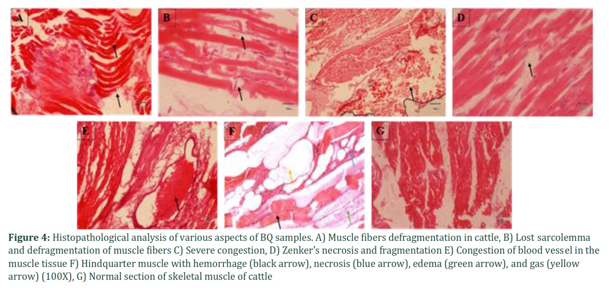
Necrosis and defragmentation of the muscle tissue were the prominent findings along with edema and hemorrhage under histology, while there was also infiltration of mononuclear cells. More sections showed localized congestion and emphysema (Fig. 4A, 4B & 4C). Gas bubbles and exudate were also observed that segregated the fiber bundles as well as individual muscle fibers. Granular and fatty degeneration towards the periphery of the sections were also evident (Fig. 4B, 4C & 4F). The diffusing toxins degenerated the leukocytes which were apparently absent in the sections, and only dispersed remains were seen at the peripheral areas of the lesion.
Lesion Scoring
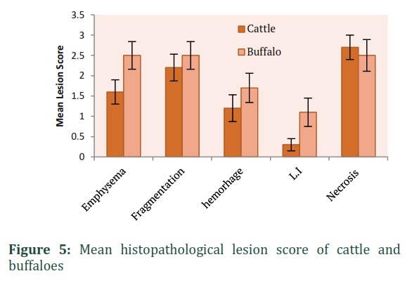
Histopathological examination was categorized by assigning them proper score. Means with same superscripts in the table were not significantly different (P>0.05) and vice versa (Table 6). Comparative trend of each lesion both in cattle and buffalo suffering from BQ was constructed (Fig. 5). Necrosis came out to the significant findings both in buffaloes and cattle followed by fragmentation, emphysema, hemorrhage, and leukocyte infiltration in cattle. In buffaloes, necrosis was the most significant (P<0.05) parameter followed by emphysema, fragmentation, hemorrhage and leukocytic infiltration.
Figures & Tables
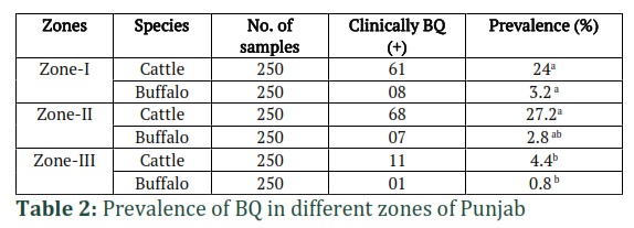


A total of 3 regional zones comprising of 2 districts of the Punjab indicate prevalence of BQ among large ruminants through cross sectional epidemiology. The order of prevalence was found to be 27.2%>24%>4.4% in zone II, I, and III respectively. However, buffaloes showed highest prevalence in zone I, II and III with values of 3.2%, 2.8%, 0.004% respectively.
The highest (27.5%) prevalence of BQ was observed in the initial months of the year among zone-I under study which then dropped down to zone number II, and III respectively. Nevertheless, the 38% prevalence of the 2nd quarter of the year was found to be highest that belonged to season 2. A comparable study was conducted by [7] who observed the highest prevalence of BQ (33.3%) in Cholistan, a desert area of Punjab province. However, this study was confined to one district only. Moreover, this study was based on scanning-based surveillance using participatory appraisal. Haqq et al. [8] and Ghafar et al. [9] also assessed financial crisis in the following year due to BQ in Dera Ismail Khan, Pakistan. The higher risk of BQ in zone-II could be due to the fact that the pathogen could remain more resilient to environmental effects in the said regions [10].
The present study endorses the fact that the association of BQ is more linked to the weather patterns or fluctuations in the seasons as the highest value of prevalence was observed in season 2 among all the zones. Most important months during the risk periods of a single year may range from May, June, July, and August. According to the best of author’s knowledge, such sorts of studies had not been conducted previously to conduct the prevalence of BQ which confers with the previous study [11] in which the authors observed prevalence in Chittagong of Bangladesh using 8 union counsels and reported 25 cases of BQ in their area under investigation. In the present research, the significant achievements included isolation of Clostridium chauvoei using two mediums i.e. blood and RCM. Later, biochemical techniques were used for further identification of this organism. Moreover, PCR successful amplified the DNA of the Clostridium chauvoei using different primers. The amplification of Ccta gene was amplified at1400 bp product [3,4]. Using these diversified techniques, the Clostridium chauvoei can be identified. The identification of local strain would be useful in studying the prevalence and development of vaccine of BQ disease. It is a matter of great concern that there are several vaccination strategies that are in pipeline in Pakistan, but still the pathogenesis of BQ disease can't be eradicated. For this purpose, the present research provided useful insights through biochemical tests, PCR to identify the Clostridium chauvoei strains and those can be used in further strategies to develop specialized vaccines to combat with this fatal disease.
The molecular test was also used as a confirmatory tool in the present study to further identify and confirm diagnosis of the Clostridium chauvoei at molecular level. The amplification of 836 bp was obtained, and 16SrDNA sequence revealed that the product was Clostridium chauvoei. A similar study was conducted preserved using formalin [12]. Similarly, culture of the earlier in which different primers were used to detect Clostridium chauvoei in samples that were earlier BQ pathogen also exhibited Clostridium chauvoei in gel documentation system in which primers successfully amplified a 509 bp product [13]. The 16S UNI-R and 16S UNI-L primers did not yield positive results despite the reaction conditions for PCR were kept similar [14]. It could be due to different serovars, or the PCR reaction conditions or the master mi concentrations. The findings of PCR are in accordance with the previous study [15], but the said study was focused only on tissues samples of the muscle fibers, biogas, and manure samples. Alpha toxin is one of the primary important toxins produced by C. chauvoei and is responsible for the virulence of this pathogenic bacterium in different ways. During this phase of experiment, partial amplification of Ccta gene was obtained by yielding 1400bp in previous study [3,4].
Histopathology of the organs exhibiting descriptive lesions has been previously applied to a number of contagious diseases including BQ. Nature and extent of hemorrhages, necrosis and degenerative lesions can be elaborated in the muscle tissues. The lesions were ordered in the following manner; necrosis, emphysema, and then the fragmentation of the sections. These
changes were also supported by earlier evidence [16] in which the authors observed similar gross and microscopic pathology in BQ infected cows. The infiltration of the leukocyte was observed in case of cattle only as supported by earlier credible published studies [10,17,18], while defragmentation and emphysematous changes were not significantly evident (P>0.05) in buffaloes. A more intercellular spaces among muscle fibers, and diffused hemorrhages were found in previous study [19] in BQ infected elephants. This may arise due to different environmental impact and subsequent degenerative changes in different species of the animals. It is important to mention that the accumulation of neutrophils around the necrotic fibers were also observed in the presence study which had already been endorsed earlier [17]. All of this evidence-based studies suggest supporting evidence that BQ cause a reasonable number of salient histopathological changes in animals.
In present study, the cross-sectional epidemiological tool of disease measurement, laboratory culture sensitivity test, and the molecular diagnosis were performed in parallel ways to validate different results for the confirmation of BQ disease in animals. The study period was selected between 2018 and 2019, and the project yielded significant findings for the molecular diagnosis as compared to the conventional culture sensitivity tests. The prevalence is also higher which indicates that the Alpha toxin gene or Ccta has been circulating among small and large ruminants in the said areas. The disease causes economic and production losses in developing state like Pakistan, therefore, strict sentinel or sero-surveillance is necessary to monitor, prevent and control the concurrent infections among animals.
Author Contributions
AI and ZIC conceived and designed this study; AI, ZIC and AA performed the experiments; MY, MAN, MR and WA analyze the data/results; MAN, WA and AK wrote the manuscript. All authors read and approved the final manuscript.
![]()
The authors declare that there is no conflict of interest.![]()
References
- Hatheway CL. Toxigenic clostridia. Clinical Microbiology Reviews, (1990); 3(1): 66-98.
- Prajapati A, Yogisharadhya R, Mohanty NN, Mendem,SK, Nizamuddin A, Chanda MM, Shivachandra SB. Whole-genome sequence analysis of Clostridium chauvoei isolated from clinical case of black quarter (BQ) from India. Archives of Microbiology, (2022); 204(6): 1-14.
- Frey J, Johansson A, Bürki S, Vilei EM, Redhead K. Cytotoxin CctA, a major virulence factor of Clostridium chauvoei conferring protective immunity against myonecrosis. Vaccine, (2012); 30(37): 5500-5.
- Frey J, Falquet L. Patho-genetics of Clostridium chauvoei. Research in Microbiology, (2015); 166(4): 384-92.
- Ziech RE, Gressler LT, Frey J, Vargas AC. Blackleg in cattle: current understanding and future research needs. Ciência Rural, (2018); 48.
- Rychener L, In-Albon S, Djordjevic SP, Chowdhury PR, Nicholson P, Ziech RE, de Vargas AC, Frey J, Falquet L. Clostridium chauvoei, an evolutionary dead-end pathogen. Frontiers in Microbiology, (2017); 8:1054.
- Khan FM. Participatory appraisal and scanning surveillance based contagious diseases risk profile of district Rahim Yar Khan (Pakistan). Pakistan Veterinary Journal, (2010); 30(4):198-202.
- Niamatullah M. Economic losses due to high incidence of black quarter disease in cattle and buffaloes and its treatment in district dera ismail khan. Pakistan Journal of Science, (2011); 63(2).
- Ghafar A, McGill D, Stevenson MA, Badar M, Kumbher A, Warriach HM, Gasser RB, Jabbar A. A participatory investigation of bovine health and production issues in Pakistan. Frontiers in Veterinary Science, (2020); 7: 248.
- Radostitis OM, Gay CC, Hinchcliff KW, Constable PD. Diseases associated with Clostridium species. Veterinary Medicine, (2006); 10: 841-3.
- Sultana M, Ahad A, Biswas PK, Rahman MA, Barua H. Black quarter (BQ) disease in cattle and diagnosis of BQ septicaemia based on gross lesions and microscopic examination. Bangladesh Journal of Microbiology, (2008); 25(1):13-6.
- Uzal FA, Hugenholtz P, Blackall LL, Petray S, Moss S, Assis RA, Miyakawa MF, Carloni G. PCR detection of Clostridium chauvoei in pure cultures and in formalin-fixed, paraffin-embedded tissues. Veterinary Microbiology, (2003); 2;91(2-3): 239-48.
- Sasaki Y, Yamamoto K, Kojima A, Tetsuka Y, Norimatsu M, Tamura Y. Rapid and direct detection of Clostridium chauvoei by PCR of the 16S-23S rDNA spacer region and partial 23S rDNA sequences. Journal of Veterinary Medical Science, (2000); 62(12):1275-81.
- Kuhnert P, Selja E, Capaul, Jacques Nicolet, Jochim Frey. Phylogenetic positions of Clostridium chauvoei and Clostridium septicurn based on 16s rRNA gene sequences. International Journal of Systematic Bacteriology, (1996); 46(4): 1174-1176.
- Bagge E, Lewerin SS, Johansson KE. Detection and identification by PCR of Clostridium chauvoei in clinical isolates, bovine faeces and substrates from biogas plant. Acta Veterinaria Scandinavica, (2009); 51(1): 1-9.
- Abreu CC, Edwards EE, Edwards JF, Gibbons PM, Leal de Araújo J, Rech RR, Uzal FA. Blackleg in cattle: a case report of fetal infection and a literature review. Journal of Veterinary Diagnostic Investigation, (2017); 29(5): 612-21.
- Sojka JE, Bowersock TL, Parker JE, Blevins WG, Irigoyen L. Clostridium chauvoei myositis infection in a neonatal calf. Journal of Veterinary Diagnostic Investigation, (1992); 4(2):201-3.
- Ambhore SR, Khan MA, Chavhan SG, Bhikane AU, Gaikwad NZ, Bhonsle AV. Clinico-pathological studies on black quarter in cattle. Indian Journal of Veterinary Pathology, (2018); 42(3): 155-159.
- Rahman H, Chakraborty A, Rahman T, Sharma R, Shome BR, Shakuntala I. Clostridial myonecrosis clinically resembling black quarter in an Indian elephant (Elephas maximus). Revue Scientifique et Technique, (2009); 28(3):1069.
This work is licensed under a Creative Commons Attribution-Non Commercial 4.0 International License. To read the copy of this license please visit: https://creativecommons.org/licenses/by-nc/4.0