

Full Length Research Article
Mixed Bacteriological Isolation Percentages from the Uterus of Cows Slaughtered due to Infertility
Ali Risvanli1,4, Hakan Kalender2, Tarik Safak3, Burak Fatih Yuksel4*, Burcu Karagulle2, Oznur Yilmaz5, Mehmet Akif Kilinc6
Adv. life sci., vol. 9, no. 4, pp. 521-525, December 2022
*- Corresponding Author: Burak Fatih Yuksel (Email: bfyuksel@firat.edu.tr)
Authors' Affiliations
2. Department of Microbiology, Faculty of Veterinary Medicine, University of Firat, Elazig- Turkey
3 Department of Gynecology and Obstetrics, Faculty of Veterinary Medicine, University of Kastamonu, Kastamonu- Turkey
4. Department of Obstetrics and Gynecology, Faculty of Veterinary Medicine, University of Firat, Elazig- Turkey
5. Department of Obstetrics and Gynecology, Faculty of Veterinary Medicine, University of Siirt, Siirt- Turkey.
6. Department of Obstetrics and Gynecology, Faculty of Veterinary Medicine, University of Bingol, Bingol- Turkey
[Date Received: 21/07/2022; Date Revised: 10/11/2022; Date Published: 31/12/2022]
Abstract![]()
Introduction
Methods
Results
Discussion
References
Abstract
Background: Intrauterine mixed infections are an important problem in cattle breeding. In this study, we aimed to determine the mixed bacteriological isolation rates from the uterus of cows with clinical metritis and to reveal the relationships between the bacteria that isolated together.
Methods: For this purpose, sterile swabs were taken from 490 uterus obtained from cattle slaughtered due to infertility in three slaughterhouses and used them to perform microbiological tests. After evaluating the data, it was determined that the bacterial isolation rate from uterus with clinical metritis was 76.14% (n = 268).
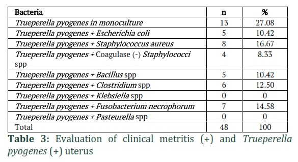
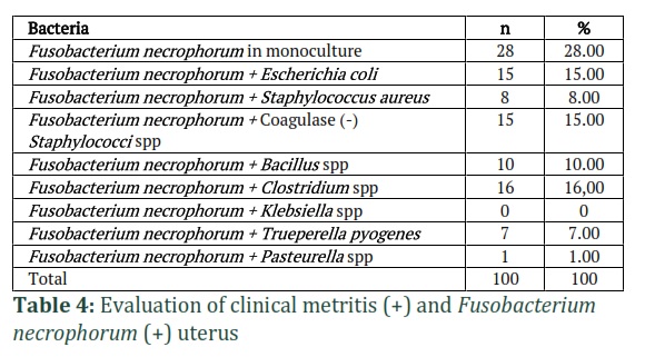
Results: The rates for 1, 2, 3, 4, and 5 bacteria isolated from the uterus with clinical metritis were 48.13%, 33.96%, 15.30%, 2.24%, and 0.37%, respectively. The isolation rates for Trueperella pyogenes, Fusobacterium necrophorum, and Escherichia coli alone from the uterus with clinical metritis were 27.08%, 28.00%, and 16.51%, respectively. In uterus with clinical metritis, Staphylococcus aureus (16.67%) was the most commonly isolated bacteria with T. pyogenes, Clostridium spp. (16%) co-isolated with F. necrophorum, and coagulase-negative staphylococci (16.51%) and Bacillus spp. (16.51%) were detected with E. coli.
Conclusion: As a result, it was concluded that when evaluating the microbiological results for cows with clinical metritis, mixed infections should be taken into more consideration, and the characteristics of the bacteria that isolate together should be considered during the treatment of mixed infections.
Keywords: Cattle; Uterine; Isolation; infertility
Introduction![]()
It has been reported that infectious agents are detected in the uterus of 93% of cows at 15 days postpartum, 50% at 30–45 days postpartum, and 9% at 45–55 days postpartum [1-3]. The most commonly detected microorganisms in the uterus after delivery are Trueperella pyogenes, Coliform spp., Pseudomonas aeruginosa, α-haemolytic streptococci, and Gram positive (+) and negative (-) anaerobic bacteria. Most cows eliminate intrauterine bacterial contamination in the first two weeks postpartum [4,5]. Failure to eliminate bacterial contaminations leads to puerperal metritis or clinical endometritis [6]. The most commonly isolated bacteria in animals with endometritis are Escherichia coli, T. pyogenes, Prevotella melaninogenica, and Fusobacterium necrophorum [6,7]. Adiguzel et al. [8] isolated E. coli (21.8%), T. pyogenes (44.9%), F. necrophorum (7.4%), and P. melaninogenica (6.9%) in their study on cows in the first 10 days postpartum. Pascottini et al. [9] have suggested that the uterine microbiota of cows with subclinical endometritis is similar to that of healthy cows, but that the microbiome in cows with clinical endometritis differs.
The bacteria that generally cause puerperal metritis are E. coli, P. melaninogenica, and F. necrophorum [7]. Among them, E. coli plays the most important role, as its presence increases the risk of subsequent infection of the uterus by other pathogens. The most important virulence factor related to E. coli causing metritis is seen as FimH, an adhesive protein that allows bacteria to adhere and colonize epithelial surfaces. It has been suggested that cows with E. coli expressing FimH in the uterus during the first two days postpartum are more likely to be infected with F. necrophorum 8–10 days after birth [10].
Trueperella pyogenes is often isolated from uterus with puerperal metritis or clinical endometritis. It has been reported that T. pyogenes has synergistic effects with bacteria such as F. necrophorum and P. melaninogenica [3,6,11]. Coagulase-negative staphylococci and α-haemolytic streptococci are also frequently isolated from cow uterus after calving. However, there is no information about whether they have the potential to predispose to infections with pathogenic bacteria in the postpartum period [11,12].
In this study, we aimed to reveal the mixed bacteriological isolation rates from the uterus of cows and the relationships of these isolated bacteria with each other.
Methods![]()
In this study, 490 uteruses were obtained from cattle slaughtered due to infertility in three separate slaughterhouses in eastern Turkey. The cattle whose uterus were removed were of different ages, breeds, and lactation periods. The study was performed between September 1st and October 30th of 2021. Sterile swabs were used to obtain samples from the uterus for microbiological tests following the standard rules of asepsis and antisepsis. The swabs were sent to the microbiology laboratory in a transport medium via cold chain technology.
Bacterial Isolation and Identification
Bacterial isolation and identification were performed in the Microbiology Laboratory of Fırat University, Faculty of Veterinary Medicine. Uterine swab samples placed into transport medium tubes (Cary Blair Medium, LAB505, Lab M) under sterile conditions were brought to the microbiology laboratory in the cold chain. The samples were inoculated on 5% blood agar (Oxoid) and MacConkey agar (Oxoid) and then incubated for 48 hours at 37 °C under aerobic conditions and 5% CO2. For the isolation of anaerobic bacteria, the swab samples were inoculated into Cooked Meat Medium (Oxoid) and incubated for 72 hours at 37 °C. An anaerobic environment was provided by using an anaerobic jar (Merck 1.16387.0001 CE) together with anaerobic gas kits (Anaerocult_A® Merck 1.13829 CE). To confirm the anaerobic environment, Anaerocult test sticks (Merck 1.15112.001 CE) with one blue-coloured end were placed in the jars. The colony morphology, Gram stain appearance, and oxidase, catalase, haemolysis, and other biochemical properties of the isolated microorganisms were examined, with the results used for identification. In addition, PCR testing was used to confirm the identification of F. necrophorum [12,13].
Determination of Clinical Metritis
The presence of clinical metritis in the uterus was determined as described by Şenünver and Nak [14]. Accordingly, uterus with symptoms such as cervical discharge, asymmetric uterine horns, thick walls, oedema, discoloration, increased tone, and a volume increase were defined as clinical metritis. These results were compared with those of the bacteriological tests. If the animals had other infertility and sterility problems, they were not included in the study. Animals that were clinically healthy and free of other infectious diseases were included in the study. Examination was carried out as soon as the animals were slaughtered.
Results![]()
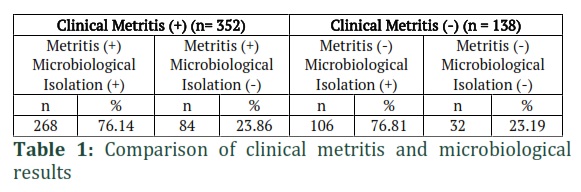
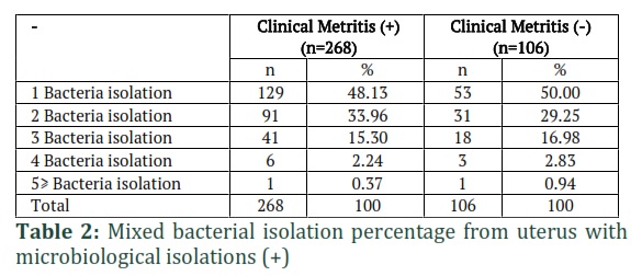
After evaluating the data, it was determined that the bacterial isolation rate from animals with clinical metritis was 76.14% (n = 268), while that from animals without clinical metritis was 76.81% (n = 106; Table 1). The proportions of uterus with 1, 2, 3, 4, and 5 bacterial isolates were 48.13%, 33.96%, 15.30%, 2.24%, and 0.37%, respectively (Table 2). The isolation rate for T. pyogenes alone from uterus with clinical metritis was 27.08% (n = 13). Staphylococcus aureus (n = 8, 16.67%) was found to be the bacteria with which T. pyogenes isolated most frequently with for uterus with clinical metritis, while Klebsiella spp. and Pasteurella spp. could not be detected (Table 3).The isolation rate for F. necrophorum alone from uterus with clinical metritis was 28.00% (n = 28). In these uteruses, Clostridium spp. (n = 16, 16%) were the bacteria with which F. necrophorum co-isolated the most, while Klebsiella spp. could not be detected (Table 4).
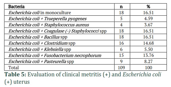
The isolation rate for E. coli alone from uterus with clinical metritis was 16.51% (n = 18). In these uteri, the bacteria that E. coli most co-isolated with were coagulase-negative staphylococci (n = 18, 16.51%) and Bacillus spp. (n = 18, 16.51%). Staphylococcus aureus (n = 4, 3.67%) was the bacteria with which it co-isolated the least (Table 5).
Figures & Tables
Although culture-based studies have revealed basic knowledge on the uterine microbiota of cows, the use of molecular techniques to characterize the uterine microbiota of cows with metritis and endometritis has led to a better understanding of the subject in recent years [15]. Results from culture-dependent studies indicate that T. pyogenes is a critical pathogen involved in the development of clinical endometritis, and Gram-negative anaerobes such as F. necrophorum, Porphyromonas levii, and P. melaninogenica may act synergistically with it. Culture-dependent studies also indicate that E. coli acts either as the main pathogen or as a precursor pathogen that sets the stage for T. pyogenes and Gram-negative anaerobes [16]. Wang et al. [17] stated that Fusobacterium, which causes clinical endometritis in cows, has positive correlations with other pathogens such as Porphyromonas, Parvimonas, Bacteroides, and Helcococcus, and therefore, that Fusobacterium acts synergistically with Trueperella, Porphyromonas, Parvimonas, and other bacteria to cause clinical endometritis suggests cows play a role in the microbiological status of uterus. It has been suggested that Trueperella and Gram-negative anaerobes (Fusobacterium, Bacteroides, and Porphyromonas) cause metritis and endometritis synergistically [10,18,19]. It is also widely believed that T. pyogenes promotes the isolation and colonization of F. necrophorum by producing an unknown growth factor [20]. It is thought that uterine pathogens interact to help each other avoid uterine defence mechanisms and facilitate colonization in the endometrium [10,15]. Bacteria such as Trueperella spp. and Fusobacterium spp. have also been detected in the uterus of heifers and even pregnant cows. [21,22]. Collectively, the co-occurrence of uterine pathogens can be considered to be of great importance in the development of uterine infections. However, the mechanisms behind the synergisms are not fully clarified. Ordell et al. [23] reported that bacteria were isolated from 76 of 79 samples taken from cows with acute puerperal metritis. In the same study, 68% of the samples had mixed isolations, 90% of which included Gram-negative bacteria; 45% had at least one Gram-positive bacterial species, and in 4 samples, more than four bacterial species were isolated. In a study by Werner et al. [12], after examining the uterus of 72 cows at 24 ± 1 days postpartum, the number of animals for which T. pyogenes cultured alone was 13, while 3 had T. pyogenes + E. coli, 1 had T. pyogenes + coagulase-negative staphylococci or α-haemolytic streptococci, and 10 had T. pyogenes + other bacteria. In the same study, the number of animals for which E. coli cultured alone was 1, while 2 had E. coli + coagulase-negative staphylococci or α-haemolytic streptococci, and 15 had E. coli + other bacteria. In our study presented here, S. aureus (16.67%) was the bacteria with which T. pyogenes co-isolated most often in uterus with clinical metritis, while Clostridium spp. (16%) co-isolated with F. necrophorum, and coagulase-negative staphylococci (16.51%) and Bacillus spp. (16.51%) co-isolated with E. coli. Our results agreed with some previous studies, but there were some differences, possibly due to the influence of different conditions (e.g. contamination) on the microbiological test results.
Moges et al. [24] found that the most commonly isolated bacteria from cows with clinical endometritis was T. pyogenes (25%), followed by Streptococcus spp. (20.8%), E. coli (20.8%), S. aureus (12.5%), Klebsiella spp. (8.3%), and Campylobacter fetus (4.2%). Another study by Takamtha et al. [25] reported the bacterial species isolated from cows with clinical endometritis were E. coli (24%), Corynebacterium spp. (18%), T. pyogenes (14%), Staphylococcus spp. (11%), and Streptococcus spp. (9%). In the present study, the rates for T. pyogenes, F. necrophorum, and E. coli isolated alone from uterus with clinical metritis were 27.08%, 28.00%, and 16.51%, respectively. As can be seen, our data agree with those from previous studies.
Appiah et al. [26] state that the microbiological status of the genital tract should be monitored to better understand uterine infections and thus provide more appropriate treatments.
We conclude that it would be beneficial to consider mixed infections apart from the bacteria that isolate alone during the evaluation of clinical metritis in cows.
![]()
Author Contributions
Ali Risvanli: conceptualization, project administration, investigation, resources, data curation, formal analysis, writing – original draft, review & editing. Hakan Kalender: Investigation, resources, writing-review & editing. Tarik Safak: Investigation & methodology. Burak Fatih Yuksel: Investigation & editing. Burcu Karagulle: Investigation. Mehmet Akif Kilinc: Investigation. Oznur Yilmaz: Investigation.
![]()
The authors declare that there is no conflict of interest.
Acknowledgment
This study was supported by the Scientific and Technological Research Council of Turkey (TUBITAK-TEYDEB No: 7200977).
References
- Sheldon IM, Dobson H. Postpartum uterine health in cattle. Animal reproduction science, (2004); 82295-306.
- Risco CA, Youngquist RS, Shore MD. Postpartum uterine infections. Current therapy in large animal theriogenology: Elsevier. (2007); pp. 339-344.
- Azawi O. Postpartum uterine infection in cattle. Animal reproduction science, (2008); 105(3-4): 187-208.
- Bondurant R. Inflammation in the bovine female reproductive tract. Journal of animal science, (1999); 77(suppl_2): 101-110.
- LeBlanc S, Duffield T, Leslie K, Bateman K, Keefe GP, et al. Defining and diagnosing postpartum clinical endometritis and its impact on reproductive performance in dairy cows. Journal of dairy science, (2002); 85(9): 2223-2236.
- Földi J, Kulcsar M, Pecsi A, Huyghe B, De Sa C, et al. Bacterial complications of postpartum uterine involution in cattle. Animal reproduction science, (2006); 96(3-4): 265-281.
- Sheldon IM, Cronin J, Goetze L, Donofrio G, Schuberth H-J. Defining postpartum uterine disease and the mechanisms of infection and immunity in the female reproductive tract in cattle. Biology of reproduction, (2009); 81(6): 1025-1032.
- Adıgüzel MC, Cengiz S, Cengiz M, Hayırlı A. Pathogenic Bacteria Present in the Lochia First 10− Day Postpartum Prolongs Days Open in Dairy Cows. Atatürk Üniversitesi Veteriner Bilimleri Dergisi, (2021); 16(1): 32-40.
- Pascottini OB, Van Schyndel S, Spricigo J, Rousseau J, Weese J, et al. Dynamics of uterine microbiota in postpartum dairy cows with clinical or subclinical endometritis. Scientific reports, (2020); 10(1): 1-11.
- Bicalho M, Machado V, Oikonomou G, Gilbert R, Bicalho R. Association between virulence factors of Escherichia coli, Fusobacterium necrophorum, and Arcanobacterium pyogenes and uterine diseases of dairy cows. Veterinary microbiology, (2012); 157(1-2): 125-131.
- Williams EJ, Fischer DP, Pfeiffer DU, England GC, Noakes DE, et al. Clinical evaluation of postpartum vaginal mucus reflects uterine bacterial infection and the immune response in cattle. Theriogenology, (2005); 63(1): 102-117.
- Werner A, Suthar V, Plöntzke J, Heuwieser W. Relationship between bacteriological findings in the second and fourth weeks postpartum and uterine infection in dairy cows considering bacteriological results. Journal of Dairy Science, (2012); 95(12): 7105-7114.
- Ozgen EK, Cengiz S, Ulucan M, Okumus Z, Kortel A, et al. Isolation and identification of Dichelobacter nodosus and Fusobacterium necrophorum using the polymerase chain reaction method in sheep with footrot. Acta Veterinaria Brno, (2015); 84(2): 97-104.
- Senunver A, Nak Y (2012) Infertility. In: Semacan A, Kaymaz M, Fındık M, Rişvanlı A, Köker A, editors. Obstetrics and Gynecology in Farm Animals. Malatya: Medipres. pp. 409-467.
- Jeon SJ, Cunha F, Vieira-Neto A, Bicalho RC, Lima S, et al. Blood as a route of transmission of uterine pathogens from the gut to the uterus in cows. Microbiome, (2017); 5(1): 1-13.
- Galvão KN, Bicalho RC, Jeon SJ. Symposium review: The uterine microbiome associated with the development of uterine disease in dairy cows. Journal of dairy science, (2019); 102(12): 11786-11797.
- Wang M-L, Liu M-C, Xu J, An L-G, Wang J-F, et al. Uterine microbiota of dairy cows with clinical and subclinical endometritis. Frontiers in microbiology, (2018); 92691.
- Bonnett BN, Martin SW, Gannon V, Miller RB, Etherington WG. Endometrial biopsy in Holstein-Friesian dairy cows. III. Bacteriological analysis and correlations with histological findings. Canadian Journal of Veterinary Research, (1991); 55(2): 168.
- Prunner I, Pothmann H, Wagener K, Giuliodori M, Huber J, et al. Dynamics of bacteriologic and cytologic changes in the uterus of postpartum dairy cows. Theriogenology, (2014); 82(9): 1316-1322.
- Dadarwal D, Palmer C, Griebel P. Mucosal immunity of the postpartum bovine genital tract. Theriogenology, (2017); 10462-71.
- Karstrup CC, Klitgaard K, Jensen TK, Agerholm JS, Pedersen HG. Presence of bacteria in the endometrium and placentomes of pregnant cows. Theriogenology, (2017); 9941-47.
- Moore S, Ericsson A, Poock S, Melendez P, Lucy M. Hot topic: 16S rRNA gene sequencing reveals the microbiome of the virgin and pregnant bovine uterus. Journal of dairy science, (2017); 100(6): 4953-4960.
- Ordell A, Unnerstad HE, Nyman A, Gustafsson H, Båge R. A longitudinal cohort study of acute puerperal metritis cases in Swedish dairy cows. Acta veterinaria Scandinavica, (2016); 58(1): 1-8.
- Moges N, Regassa F, Yilma T, Unakal CG. Isolation and antimicrobial susceptibility of bacteria from dairy cows with clinical endometritis. Journal of Reproduction and Infertility, (2013); 4(1): 04-08.
- Takamtha A, Phanaratkitti V, Adirekkiet O, Panyapornwitaya V, Boonyayatra S, et al. Prevalence of isolated bacteria from clinical endometritis uterine and antimicrobial susceptibility in postpartum dairy cows. Chiang Mai Vet J, (2013); 11(3): 237-245.
- Appiah, MO, Wang J, Lu W. Microflora in the reproductive tract of cattle: a review. Agriculture, (2020); 10(6): 232.
This work is licensed under a Creative Commons Attribution-Non Commercial 4.0 International License. To read the copy of this license please visit: https://creativecommons.org/licenses/by-nc/4.0

