

Full Length Research Article
Molecular Identification of Cystic Echinococcosis in Mosul, Iraq
Sinan K. Abood1, Najah S. N. AL-Omar2*
Adv. life sci., vol. 12, no. 3, pp. 460-465, August 2025
*- Corresponding Author: Najah S. N. AL-Omar (Email: najahsbio26@uomosul.edu.iq)
Authors' Affiliations
2. Department of Biology, College of Science, University of Mosul – Iraq
[Date Received: 28/09/2024; Date Revised: 18/02/2025; Available Online: 31/10/2025]
Abstract![]()
Introduction
Methods
Results
Discussion
References
Abstract
Background: Cystic echinococcosis (CE) is a significant parasitic disease with substantial global economic and public-health impacts. It is caused by the larval form of Echinococcus granulosus.
Methods: The aim of this study was to identify the Echinococcus species in humans and livestock within Mosul, Iraq. This research work comprised fifty-five samples of viable Echinococcus cysts, including thirty from humans and twenty-five sheep. Human specimens were acquired from the main hospital in Mosul. Animal specimens were collected in the Al-Saadoon abattoir from June 2022 to March 2023.
Results: DNA samples were extracted from the protoscolex and blastoderm of each cyst sample. The mitochondrial 12S rRNA and cox1 genes were amplified by polymerase chain reaction. Gene sequencing and phylogenetic analyses were performed on 10 PCR-positive samples (5 from humans and 5 from sheep) selected by cyst size for laboratory feasibility.
Conclusion: The findings revealed that genotypes G1 and G3 were present in human echinococcosis cysts in Mosul while G1 genotypes were found in sheep. These findings highlight the need for integrated public-health and veterinary control measures.
Keywords: Echinococcus granulosus, Cystic echinococcosis, sensu stricto, Nineveh, Iraq
Introduction![]()
Hydatid disease is caused by the larval (metacestode) stage of the tapeworm Echinococcus granulosus, a parasite belonging to the family Taeniidae [1]. This adult tapeworm is an intestinal parasite found solely in dogs and various wild canids. Humans and ungulates act as intermediate hosts, becoming infected through the ingestion of infectious eggs, harboring asexual larvae (metacestodes) [2]. Human cystic echinococcosis is caused accidentally by ingesting the eggs of Echinococcus granulosus. Once matured, the larvae typically invade the liver in over 65% of cases, while they may also affect the brain, kidneys, lungs, and spleen to a lesser extent [3]. To accurately diagnose the condition, it is essential to genetically identify the species and genotype responsible for CE in individuals, as well as to understand the vectors used for the transmission of the parasite and implement targeted measures of control. The causative agent of CE is a cryptic species complex known as Echinococcus granulosus. At present, G1, G3, G4, G5, G6/7, G8, and G10 genotypes that belong to Echinococcus granulosus have been identified [4-6].
Based on these genotypes, four unique species have been identified: E. granulosus sensu stricto (s.s.) (G1 and G3), previously known as the “sheep strain” and “buffalo strain,” respectively; E. equinus (G4), which was formerly called the “horse strain”; E. ortleppi (G5), which is called “cattle strain”; E. canadensis G6/7 clusters, which were identified “camel and pig strain”; E. canadensis G8 and G10, recognized as “cervid strain”; and E. felis, as well as referred to “lion strain” [7]. Among the E. sensu lato (s.l.) species complex, E. granulosus (s.s.) (G1 and G3) stands out as the species posing the greatest public health risk, accounting for the vast majority of human cases confirmed at the molecular level (88.5%) [8]. The most prevalent species within the E. granulosus sensu lato (s.l.) complex are E. ortleppi (G5) and E. canadensis (G8) [8-10]. Additionally, E. canadensis (G6/7) has been found to be more common in humans than previously thought, representing approximately 11% of all documented cases with genetic confirmation [8].
Cystic echinococcosis poses a significant issue globally, including in Iraq, where published epidemiological data is limited. Previous research [11-14] indicates that E. granulosus is prevalent in the cities of Erbil, Duhok, Baghdad, and Mosul.
Molecular testing in Iraq has only been conducted on E. canadensis (G6) and E. granulosus (s.s.) (genotypes G1 and G3). In the Duhok governorate, individuals have been found to carry both G1 and G3 [15]. Erbil has revealed the presence of G1 in sheep and cattle, along with some of its microvariants [16,17]. In the Kirkuk Governorate, G1 was identified in humans, goats, sheep, and cattle [18]. G1 and G3 were found in both humans and animals within the Sulaymaniyah Governorate [19]. The Missan Governorate has shown the presence of G1 and G3 in humans, as well as in cattle, buffaloes, goats, camels, and sheep [20]. In Qadisiya, Najaf, and Diwaniya governorates, G1, G3, and G6 were identified in humans and sheep, as well as in cattle and camels [21]. In this study, the objective was to molecularly identify the strains of Echinococcus granulosus present in the Ninewa Governorate to inform public-health control and prevention strategies.
Methods![]()
Sample Collection
The current study involved 55 samples, consisting of 30 human Echinococcus cysts and 25 from animals (sheep). Human samples were sourced from the main hospital in Mosul, while animal samples were obtained from the Al-Saadoon slaughterhouse between June 2022 and March 2023. For sequencing, five human and five sheep isolates were selected based on cyst size, as larger cysts were expected to yield more viable protoscolices and higher-quality DNA for sequencing under consistent extraction conditions. Echinococcus eggs were gathered, and the eggshells were disinfected using 70% alcohol. A sterile medical syringe with a capacity of 50 mL was utilized to extract the cystic fluid, and the protoscolices were collected in aseptic conditions.
Protoscolices and cystic fluid that were recovered were placed into a 250 mL beaker. The germ layer was extracted and transferred to a sterile plate containing 0.9% NaCl, where it was rinsed periodically with PBS. To assess viability, the fluid from each Echinococcus cyst underwent centrifugation at 3000 × g for 5 minutes. Subsequently, 0.1% Eosin Y solution was employed [22]. The resulting protoscolex and blastoderm were preserved in 70% ethanol, and the majority of the DNA was then utilized for identifying the primary parasite species through PCR and gene sequencing.
DNA extraction
DNA extraction was performed using 25 mg of protoscolices, along with an additional 10 mg of germ layers, employing a genomic DNA extraction kit from Geneaid. To eliminate the alcohol, both the germ layers and protoscolices were rinsed three times in PBS (pH 7.2). The concentration and quality of the extracted DNA were assessed using a Nanodrop spectrophotometer (THERMO, USA).
Preparation of Agarose Gel Electrophoresis
To prepare a 2% agarose gel, start by combining 2 g of agarose in the beaker with 100 mL of buffer (1x TBE). The mixture was microwaved for 2 minutes until dissolved and allowed to cool to 50 °C. Incorporate 5 µL of RedSafe into the melted gel to stain the DNA, stirring gently. Ensure that the gel tray is thoroughly cleaned and dried, then attach a comb to one side of the tray and secure it using tape. Carefully pour the melted agarose into the dish and let it set at room temperature for 20 minutes.
The tape and comb were carefully removed to prevent damage to the gel. Position the gel tray in the electrophoresis tank so that the gel wells are oriented towards the cathode, and then add TBE 1x buffer to the tank until it reaches a depth of approximately 1 cm, ensuring it fully covers the gel.
PCR Product Detection
Gradually add 5 µL of the PCR product to each well. Add 5 µL of the DNA ladder (Bio-labs) (100-1000 bp) to the first well; this will serve as a molecular-weight size marker for estimating the size of the PCR products. Cover the device and activate the power at 80 V for 50 minutes. After the designated time had elapsed, the gel was carefully removed and photographed using a digital camera.
The molecular identification of E. granulosus is based on a portion of the mitochondrial 12S rRNA gene
A 490 bp portion of the mitochondrial 12S rRNA gene was amplified using primers described by Rostami et al. [23]. 12SR (F) the forward primer (5′-AGGGGATAGGAC ACAGTGCCAGC-3′), was used, while 12SR (R) (5′-CGGTGTACATGAGCTAAAC-3′) was the reverse primer [23].
Primers were utilized to amplify a segment of the 12S rRNA gene, measuring around 490 bp in length. The PCR products were loaded onto a 2% agarose gel prepared with 5 µL of RedSafe per 100 mL.
cox1 gene (partial Mitochondrial Cytochrome Oxidase) detection in Echinococcus granulosus parasite based on the PCR technique
A segment of the cox1 gene from the mitochondria, encompassing the entire length of the gene along with a species-specific portion, was amplified utilizing primers that were previously established (Spotin et al., [24]). JB3 (F): (5′-TTTTTTGGGCATCCTGAGGTTTAT-3′), the forward primer was used, while JB4.5 (R): (5′-AAAGAAAGAACATAATGAAAATG-3′) was the reverse primer [24].
DNA Sequencing analysis
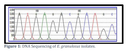
DNA sequencing was used to identify E. granulosus genotypes from positive samples (Figure 1) [25].
Sequencing of the PCR amplicons to determine their nucleotide composition
The nucleotide sequence of the PCR products was determined to identify the Echinococcus granulosus genotype. At the SomaGene Center in the U.S., genetic tree analysis was conducted to identify the strains, utilizing known Echinococcus granulosus strains from the National Center for Biotechnology Information (NCBI) as a reference. Phylogenetic tree analysis, which involved DNA sequence analysis, was performed through molecular genetic analysis using MEGAX software, along with a sequence alignment analysis based on convergence analysis. The captured sequences underwent manual curation, and the resulting sequences were compared to reference sequences in GenBank using the BLAST method (https://blast.ncbi.nlm.nih.gov).
Results![]()
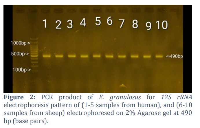
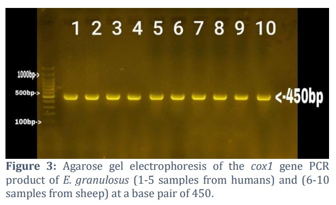
All isolates were identified as Echinococcus granulosus through analysis of partial gene sequences of 12S rRNA (490 bp) and cox1 (450 bp). From patients admitted to the 9th ward of the General Hospital in Mosul, five human isolates were selected based on cyst size for laboratory feasibility, as larger cysts typically provide sufficient DNA for sequencing, while five animal isolates were obtained from sheep slaughtered at the AL-Saadoon slaughterhouse in Mosul. Agarose gel electrophoresis confirmed the presence of the target gene for the mitochondrial 12S rRNA of Echinococcus granulosus, which produced a 490 bp band, as illustrated in Figure 2. Furthermore, PCR results confirmed the existence of a 450 bp fragment corresponding to the mitochondrial cytochrome c oxidase subunit 1 gene (cox1) (Figure 3). PCR successfully amplified both 12S rRNA and cox1 mitochondrial fragments from protoscolices and blastoderm.
The ten selected PCR products were dispatched to the SomaGene Center in the U.S. for sequencing of 12S rRNA and a partial cox1 gene, aimed at identifying the parasite of E. granulosus. All the obtained sequences were aligned and compared utilizing the BLAST tool from GenBank. While the products of (PCR) underwent sequencing, two samples have been submitted to (GenBank): one from sheep (accession no. LC757382), and one from human (accession no. LC757383).
MEGAX software has been employed to study the phylogenetic relationships. The isolates from human and sheep were compared with other published NCBI-BLAST isolates.
These 5 human isolates were contrasted with some other isolates published in the NCBI-BLAST. The human isolates demonstrated the greatest similarity to the Iranian and Turkish isolates (Table 1).
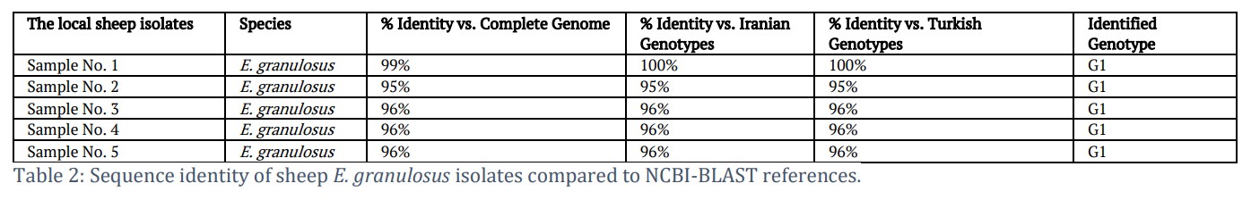
Consequently, all five sheep isolates were identified as micro variants of the G1 common sheep strain (see Table 2).
Figures & Tables
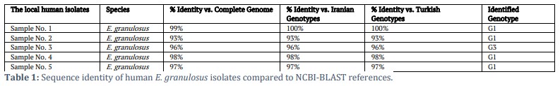
The human infection with echinococcosis in Mosul was likely caused by the strains G1 and G3, a conclusion supported by most of the samples, which ranged from 93% to 99%. Notably, strain (G1) was found to be predominant among these samples. Additionally, four of the five human samples exhibited strains with sequence identities between 96% and 99%.
The examination of five samples from sheep infected with echinococcosis cysts revealed that the strain identified as G1 was consistent across all samples when compared to other strains in the gene bank. According to NCBI-BLAST, all isolates were closely related to those from Iran and Turkey, and they all fell under the same strain (G1).
TThis study confirms the presence of the common sheep strain (G1) and introduce a novel approach to classify Echinococcus granulosus. In Mosul, E. granulosus (s.s.) was identified, aligning with recent research indicating that this species constitutes the majority (88.5%) of all species that are characterized molecularly as E. granulosus (s.l.) species worldwide [8]. A specific group of cysts was identified as having either the G1 or G3 genotype. The analysis revealed that both genotypes were widespread, with G1 being the most prevalent; however, there was no significant correlation found with sex, age, or occupation.
The findings were consistent with similar research conducted in various regions of Iraq, as the provinces predominantly harbor individuals with the G1 and G3 genotypes, particularly from Al-Najaf and Al-Diwaniyah [21], Dohuk [15], Sulaymania [19], Kirkuk [18], and Misan [20]. Identical genotypes have also been found in neighboring nations, including Turkey [26,27], Iran [28], Jordan [29], and Saudi Arabia [30,31]. Furthermore, this study’s results align with those of a prior comprehensive investigation that identified G1 as the most prevalent genotype circulating in Europe [32].
In Nineveh Province, our research revealed the existence of E. granulosus cluster genotypes G1 and G3. To determine the variations in pathogenicity and susceptibility of the G1 and G3 genotypes towards humans and various animals, thorough epidemiological investigations are essential. Given that a significant number of animals act as intermediate hosts for these genotypes, it is important to integrate epidemiological strategies with these findings. A limitation of this study is the non-random selection of sequenced isolates based on cyst size. This criterion may have introduced selection bias if cyst growth characteristics correlate with genotype. Consequently, the results should be interpreted cautiously, and future studies should employ random or stratified sampling to ensure representative genotypic analysis.
Acknowledgement
The authors appreciate the assistance of the Department of Biology, the Faculty of Science, and the University of Mosul.
Author Contributions
Sinan K. Abood conceived and designed the study, collected and analyzed the data, and drafted the manuscript. Najah S.N. AL-Omar supervised the research, contributed to data interpretation, and critically reviewed and edited the manuscript.
The authors declare that there is no conflict of interest in the publication of this article.![]()
References
- Gessese AT. Review on epidemiology and public health significance of hydatidosis. Veterinary medicine international, (2020); 2020(1): 8859116.
- Thompson R. The taxonomy, phylogeny and transmission of Echinococcus. Experimental parasitology, (2008); 119(4): 439-446.
- Botezatu C, Mastalier B, Patrascu T. Hepatic hydatid cyst–diagnosis and treatment algorithm. Journal of medicine and life, (2018); 11(3): 203.
- Nakao M, Lavikainen A, Yanagida T, Ito A. Phylogenetic systematics of the genus Echinococcus (Cestoda: Taeniidae). International journal for parasitology, (2013); 43(12-13): 1017-1029.
- Romig T, Ebi D, Wassermann M. Taxonomy and molecular epidemiology of Echinococcus granulosus sensu lato. Veterinary parasitology, (2015); 213(3-4): 76-84.
- Vuitton DA, McManus DP, Rogan MT, Romig T, Gottstein B, et al. International consensus on terminology to be used in the field of echinococcoses. Parasite, (2020); 27(2020): 1-41.
- Nakao M, Yanagida T, Okamoto M, Knapp J, Nkouawa A, et al. State-of-the-art Echinococcus and Taenia: phylogenetic taxonomy of human-pathogenic tapeworms and its application to molecular diagnosis. Infection, Genetics and Evolution, (2010); 10(4): 444-452.
- Rojas CAA, Romig T, Lightowlers MW. Echinococcus granulosus sensu lato genotypes infecting humans–review of current knowledge. International Journal for Parasitology, (2014); 44(1): 9-18.
- Kim HJ, Yong TS, Shin MH, Lee KJ, Park GM, et al. Phylogenetic characteristics of Echinococcus granulosus sensu lato in Uzbekistan. The Korean Journal of Parasitology, (2020); 58(2): 205.
- Macin S, Orsten S, Samadzade R, Colak B, Cebeci H, et al. Human and animal cystic echinococcosis in Konya, Turkey: molecular identification and the first report of E. equinus from human host in Turkey. Parasitology research, (2021); 120(2021): 563-568.
- Al-Mukhtar A, K Qasim I. Serological survey of hydatid disease in asymptomatic peoples in Mosul City, Iraq. Rafidain Journal of Science, (2017); 26(1): 1-8.
- Al-Marsomy WA. Epidemiology of Hydatid Disease in Iraq: A Study of Hydatidosis Patients in Baghdad Province. Indian Journal of Forensic Medicine & Toxicology, (2021); 15(2): 3525-3530.
- Al-Saeed A, Al-Mufty KSA. Human Hydatidosis in Duhok-Kurdistan Region–North of Iraq. Medical Journal of Babylon, (2016); 13: 125-133.
- Saida LA, Nouraddin AS. Epidemiological study of cystic echinococcosis in Man and slaughtered Animals in Erbil province, Kurdistan Regional-Iraq. Tikrit Journal of Pure Science, (2011); 16(4): 45-50.
- AHMED BD, MERO WM, Salih A, NING X, CASULLI A, et al. Molecular Characterization of Echinococcus Granulosus Isolated from Human Hydatid Cyst Using Mitochondrial cox1 Gene Sequencing in Dohuk Province-Kurdistan Region, Iraq. Science Journal of University of Zakho, (2013); 1(1): 72-80.
- Hassan ZI, Meerkhan AA, Boufana B, Hama AA, Ahmed BD, et al. Two haplotype clusters of Echinococcus granulosus sensu stricto in northern Iraq (Kurdistan region) support the hypothesis of a parasite cradle in the Middle East. Acta Tropica, (2017); 172(2017): 201-207.
- Abdulla RG, Mageed SN, Obed CE, Jumaa JA. Molecular characterization of fertile hydatid cysts from the liver of the sheep and cows and associated environmental influence factors. Iraqi Journal of Veterinary Sciences, (2020); 34(2): 321-327.
- Hasan HF, Fadhil MH, Fadhil ZH. Molecular characterization of Echinococcus granulosus isolated from human and domestic animals in Kirkuk, Iraq. Animal Research International, (2016); 13(3): 2544-2547.
- Hama A, Hassan Z, Mero WS, Interisano M, Boufana B, et al. A morphologically unusual Echinococcus granulosus (G1 genotype) cyst in a cow from Kurdistan-Iraq. Epidemiology (sunnyvale), (2015); 5(2015): 1-3.
- Mhouse Alsaady HA, Naeem Al-Quzweeni HA. Molecular Study of Echinococcus Granulosus in Misan Province, South of Iraq. Indian Journal of Public Health Research & Development, (2019); 10(9): 1062.
- Mahdi ZMS, Al-Hamairy AK, Al-Rubaiey HM. Genotyping of Echinococcusgranulosus Isolates from Human, Sheep and Cattles Hydatid Cysts in Some Central Euphrates Provinces, Iraq. Medico-legal Update, (2020); 20(2): 570-575.
- Esfandiari B, Youssefi M. Comparison of eosin and trypan blue staining in viability of hydatid cyst protoscoleces. Global Veterinaria, (2010); 4(5): 456-458.
- Rostami S, Salavati R, Beech RN, Sharbatkhori M, Babaei Z, et al. Cytochrome c oxidase subunit 1 and 12S ribosomal RNA characterization of Coenurus cerebralis from sheep in Iran. Veterinary Parasitology, (2013); 197(1-2): 141-151.
- Spotin A, Mahami-Oskouei M, Harandi MF, Baratchian M, Bordbar A, et al. Genetic variability of Echinococcus granulosus complex in various geographical populations of Iran inferred by mitochondrial DNA sequences. Acta tropica, (2017); 165(2017): 10-16.
- Ansorge WJ. Next-generation DNA sequencing techniques. New biotechnology, (2009); 25(4): 195-203.
- Vural G, Baca AU, Gauci CG, Bagci O, Gicik Y, et al. Variability in the Echinococcus granulosus cytochrome C oxidase 1 mitochondrial gene sequence from livestock in Turkey and a re-appraisal of the G1–3 genotype cluster. Veterinary parasitology, (2008); 154(3-4): 347-350.
- Kurt A, Avcioglu H, Guven E, Balkaya I, Oral A, et al. Molecular characterization of Echinococcus multilocularis and Echinococcus granulosus from cysts and formalin-fixed paraffin-embedded tissue samples of human isolates in northeastern Turkey. Vector-Borne and Zoonotic Diseases, (2020); 20(8): 593-602.
- Nikmanesh B, Mirhendi H, Ghalavand Z, Alebouyeh M, Sharbatkhori M, et al. Genotyping of Echinococcus granulosus isolates from human clinical samples based on sequencing of mitochondrial genes in Iran, Tehran. Iranian journal of parasitology, (2014); 9(1): 20-27.
- Issa HS, Abdel-Hafez SK, Hijjawi NS, Al-Qaoud KM. Molecular Characterization of Echinococcus granulosus sensu stricto Cysts of Domestic Ruminants in Jordan. Jordan Journal of Biological Sciences, (2018); 11(3): 301-306.
- Metwally DM, Qassim LE, Al-Turaiki IM, Almeer RS, El-Khadragy MF. Gene-based molecular analysis of cox1 in Echinococcus granulosus cysts isolated from naturally infected livestock in Riyadh, Saudi Arabia. PLoS One, (2018); 13(4): e0195016.
- Al-Mutairi N, Taha H, Nigm A. Molecular characterization of Echinococcus granulosus in livestock of Al-Madinah (Saudi Arabia). Journal of Helminthology, (2020); 94(2020): e157.
- Casulli A, Massolo A, Saarma U, Umhang G, Santolamazza F, et al. Species and genotypes belonging to Echinococcus granulosus sensu lato complex causing human cystic echinococcosis in Europe (2000–2021): a systematic review. Parasites & Vectors, (2022); 15(1): 109.
This work is licensed under a Creative Commons Attribution-Non Commercial 4.0 International License. To read the copy of this license please visit: https://creativecommons.org/licenses/by-nc/4.0

