

Full Length Research Article
Molecular report of resistant isolates of Theileria annulata by targeting the cytochrome b gene in cattle and ticks in Babylon, Al-Qasim city of Iraq
Ahmed Hassan Salah1, Muthanna Hadi Hussain2*
Adv. life sci., vol. 12, no. 3, pp. 528-533, August 2025
*- Corresponding Author: Muthanna Hadi Hussain (Email: muthanna.hussain@qu.edu.iq)
Authors' Affiliations
2. Department of Internal and Preventive Medicine, College of Veterinary Medicine, Al-Qadisiyah University, Al-Qadisiyah – Iraq
[Date Received: 23/09/2024; Date Revised: 18/02/2025; Available Online: 31/10/2025]
Abstract![]()
Introduction
Methods
Results
Discussion
References
Abstract
Background: Ticks are the carriers of the deadly animal illness theileriosis. Theileria annulata in cattle is an obligatory intracellular protozoan parasite carried by ticks (Hyalomma spp.) that infects hosts with mild, severe, and fatal illnesses. Theileria annulata, also known as tropical theileriosis, is a lymphoproliferative illness that causes severe mortality and morbidity in calves. Currently available antiparasitic medications work well in animals, but owing to developing resistance, animals may die or continue to be carriers. Treatment works best while the disease is still in its early stages.
Methods: In the current study, 2.5 mL of blood was drawn from the jugular vein and placed in an EDTA anticoagulant tube. Tick samples from each animal suspected of having Theileria annulata infection were placed in plastic tubes and sent straight to the laboratory for microscopic diagnosis using a blood smear and, later, PCR testing, which yielded a positive result.
Results: PCR targeting the 18S rRNA gene showed amplification in all tested blood samples (100%), confirming infection with T. annulata. In ticks, 81.25% (13/16) was positive for T. annulata. Furthermore, the cytochrome b gene (resistant gene) was detected in 100% of the calves’ samples and 81.25% of the tick samples tested using PCR.
Conclusion: The cytochrome b gene was detected in all (100%) calf samples and 81.25% of tick samples, which may be a contributing factor to mortality in infected calves. Sequencing analysis is a necessary method for the identification of mutations in the ticks and calves' blood samples.
Keywords: Theileria annulata, Cytochrome b gene, Tick, Buparvaquone, Sequencing
Introduction![]()
Theileria annulata is widely dispersed in the Middle East, Russia, China, and Africa [1]. Various Theileria species from the phylum Apicomplexa produce the major tick-borne disease known as bovine theileriosis, which affects domesticated cattle in tropical and subtropical regions [2]. Certain species’ effects on cattle mortality and morbidity cause major economic losses in several states of the United States [3].
The infected animals had pyrexia (40.5 – 41.5 °C), nasal and ocular discharges, swelling of the superficial lymph nodes, salivation, anemia, respiratory distress, and eye lesions [4]. Animals that recover from acute illness typically have, in order to maintain the parasite population, Theileria piroplasms that act as reservoirs [5].
The development of the superficial lymph nodes was linked to these symptoms. Icterus, anemia, and sporadic hemoglobinuria are symptoms of theileriosis because the parasites are killing the erythrocytes [6]. Theileria annulata is thought to be the most virulent Theileria species, with a global distribution, and is the cause of tropical bovine theileriosis [7]. Anorexia, a lingering fever, enlarged lymph nodes, diarrhea, and anemia are the signs of clinical tropical theileriosis [8].
Thin blood smears stained with Giemsa and lymph node biopsy smears are mostly analyzed microscopically to determine the diagnosis of acute theileriosis. Although microscopy is the 'gold standard' for the diagnosis of acute infections, it is not sensitive enough to demonstrate the low parasitemia that is usually observed in carrier animals [9]. However, the technique is only reliable for acute case diagnosis. Furthermore, due to a low parasitemia and the tiny number of piroplasms that may be detected in the peripheral blood in preclinical instances, it might be difficult to discriminate between various pathogenic and non-pathogenic piroplasms. Therefore, substantial expertise is required [10]. PCR assays for the diagnosis of T. annulata are more precise and dependable than other conventional procedures, and because of this, it has become the method of choice for diagnosing bovine and ovine theileriosis in epidemiological investigations [11-12]. RFLP (Restriction Fragment Length Polymorphism) assay was employed as a diagnostic tool to enable direct, concurrent, highly specific, and sensitive identification of Theileria spp. [13].
Buparvaquone, an antitheilerial medication, has been used successfully in the field to treat tropical theileriasis [14] . Currently, the main medication used to treat tropical theileriosis is buparvaquone. The hydroxy naphthoquinone compounds parvaquone and buparvaquone, which operate primarily on cytochrome b (Cyto b) of Theileria mitochondria, were first presented as active medications in the 1970s. Since that time, buparvaquone has been the theileriosis therapy of choice on a global scale [15].
Phylogenetic models could suggest that novel resistance mutations can start from a single source, become immobile through collection, and then spread through parasites. This situation would probably result from the transfer of hosts that had medication-resistant alleles in their genes. Contrarily, as a result of host migration, resistance mutations may continually develop from many sources, become fixed by selection, and transfer amongst parasites [16].
Methods![]()
Clinical examination
Most of the information was taken from the owner before the samples were collected from the suspected infected calves. The information, including the breed of calves, is recorded; the calves are selected from two to ten months of age, breed, and sex, and the signs, like the swollen lymph nodes, anemia, state of mucous membranes, and the presence of ticks on the body, are considered. Research was conducted between April and August 2022, when tick activity was at its peak, with all samples selected from Babylon, Al-Qasim city.
Blood and tick sample collection
The samples were collected between April and August 2022, when tick activity was at its peak. Sixteen blood samples from calves and sixteen tick samples were randomly selected from various pastoral areas in Babylon, Al-Qasim city. The sample size was limited due to restricted outbreak cases during the study period. Approximately 2.5 ml of blood was drawn from the jugular vein into an EDTA anticoagulant tube from each animal suspected of Theileria annulata infection and immediately transported to the laboratory. The sample size was limited due to restricted outbreak cases during the study period. Blood samples were kept at -20 °C until they were used in this study. Later, ticks were also collected from the same infected calves in a beaker with the help of tongs through the direct use of ethanol on the ticks and were transported to the lab. To make DNA extraction for PCR, DNA was stored at -20 °C until it was used in the study.
Microscopic detection of Theileria annulata
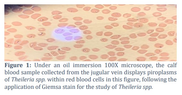
Peripheral blood smears were prepared on glass slides, which were then dried and fixed with methanol for roughly five minutes, then stained with 10% Giemsa stain for about 30 minutes, and studied under 100x oil immersion magnification [17].
DNA extraction and PCR conditions
Using a DNA isolation kit, total DNA was isolated from tick samples and EDTA blood (Genomic DNA mini kit, blood/cultured, Korea) to prepare the sample for PCR using two primers, 18srRNA for the detection of T. annulata and the cytochrome b gene for the detection of gene resistance. Amplification of the partial sequence of the cytochrome b gene of Theileria annulata was performed using two primers (Tcytb F, R). Thiel-cyto F (5' CAGGGCTTTAACCTACAAATTAAC3') and Thiel-cytoR (5' CCCTCCACTAAGCGTCTTTCGACAC3') produce 1092 bp according to Mhadhbi et al., [18].
Molecular detection
The cytochrome b gene for the detection of resistant genes in both Theileria annulata and tick samples was used for the detection of the parasite's resistance to antiparasitic drugs (buparvaquone). The total volume of reaction for the PCR was 25 µL and is composed of a DNA template of 2 µL, 1 µL of each primer, 12.5 µL of master mix, and 8.5 µL of distilled water.
Results![]()
Examination of Blood Films by Microscopy
The microscopic examination was used to determine whether piroplasms were present. All sixteen blood samples taken from the calves gave a positive result due to the presence of piroplasms in the RBCs. Figure 1 shows the T. annulata under 100X oil immersion.
PCR test result
Detection of the cytochrome b gene in T. annulata blood sample
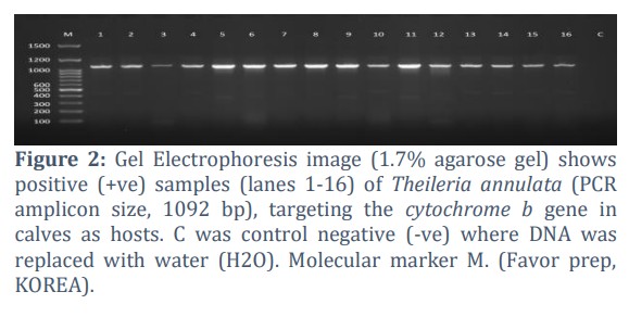
A 1092 bp fragment of the cytochrome b gene was used to detect the resistant gene of Theileria annulata in blood samples of calves, using the PCR test; C was the control (-ve), in which water (H2O) was added instead of DNA, as shown in Figure 2.
Detection of T. annulata by Cytochrome b in tick samples
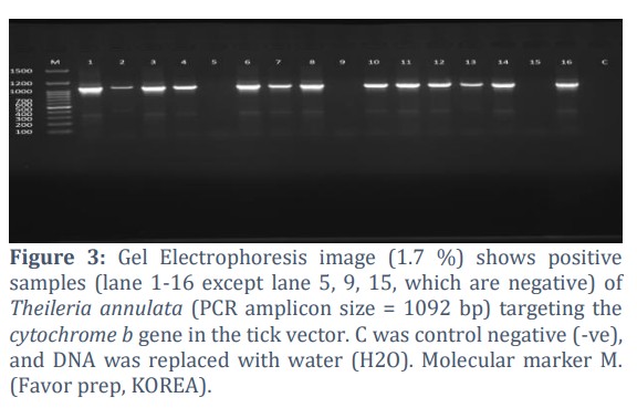
The cytochrome b gene was used on tick samples, a 1092 bp fragment, to detect the resistant gene of Theileria annulata in tick samples using PCR tests. C was control negative (-ve), in which water (H2O) was added instead of DNA. Figure 3 shows the positive result (1-16), except (5, 9, 15), which give a negative (-ve) result.
Cytochrome b Gene Amplification, Analysis, and Sequencing
A subset of 20 positive PCR products was selected for DNA sequencing and molecular evolutionary analysis. The screened sequences were cleaned of background noise and submitted to the GenBank database. Based on acquired accession numbers, a comparative investigation was further conducted with other global strains: DNA sequencing analysis, e.g., building of a phylogenetic tree, was carried out with multiple alignment of sequences using Clustal W as well as Molecular Evolutionary Genetics Analysis version 11 (MEGA X). Sequences thus obtained were submitted to alignment with susceptible as well as resistant global strains to identify similarities as well as discrepancies. In building a phylogenetic tree, reference sequences thus obtained using NCBI-BLAST were utilized. Analysis was carried out using the software quoted accordingly [19].
Amino acid sequence analysis
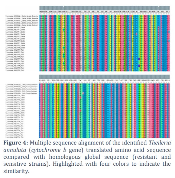
Most isolates in Figure 4 showed T mutations that evolved into A in sequences with accession numbers (OQ627715, OQ627723) in cattle and (OQ627726, OQ627727, OQ627730) in ticks. As shown in Figure 4, the substitutions observed in our dataset occur at codons 175, 177 (F→C in tick isolates), 200 (V→M in cattle), and 224 (W→G in cattle); no change is evident at codon 119 in the alignment. “A” was present in every sequence; however, the accession number (OQ627717) in the cattle sequences at position 175 was different. A mutation in the accession number (OQ627728, OQ627731) at position 177 caused the amino acid (C) to emerge in the tick isolate rather than (F). Cattle with the accession numbers OQ627716 and OQ627720 had a mutation where the amino acid M was observed rather than V. Cattle with accession numbers OQ627715 and OQ627721 have a mutation where G was observed rather than W.
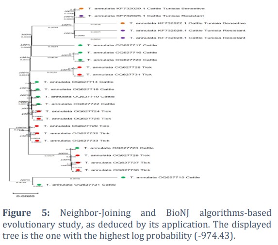
Phylogenetic analysis
Tree(s) for heuristic search were generated automatically by execution of the Neighbor-Joining and BioNJ algorithms on a matrix of pairwise distances calculated using the JTT model and shown in Figure 5. As a tree topology suitable for further investigation, a tree topology with the maximum value of log likelihood was selected. Below each branch, branch lengths have units of substitutions per site, and the tree is displayed at scale. There were 25 amino acid sequences in this investigation. The final dataset contained 302 locations in total. MEGA11 software was used to perform evolutionary analyses [19].
Figures & Tables
The most hazardous species for ruminants of all sizes is Theileria annulata. Theileria parasites can be identified by microscopic analysis of thin blood smears, lesions, clinical signs, and case history, but a similarity in the clinical signs was observed. The physical resemblance of piroplasms, false-negative diagnoses caused by the parasite's rarity (particularly in carrier animals), and difficulties recognizing mixed infections are some of the problems associated with microscopic detection [20]. Ticks present the main factor of transmitting disease, and they seem endemic in Iraq, with widespread distribution due to the suitable climatic circumstances during nine months of the year, because of the appropriate temperature and moisture [21].
Therefore, precise and sensitive diagnostic tests, such as PCR, could be used to diagnose and differentiate between different types of piroplasms [13]. The stained blood smears contained intraerythrocytic stages of Theileria annulata. Theileria parasite infection can be diagnosed through microscopic examination of thin blood smears, as shown in Figure 1. Several molecular markers, including the cytochrome b genes, have been employed in recent years to ascertain the phylogenetic relationships among the piroplasm population [22]. For assessing evolutionary relationships between species at the levels of families and genera, the mitochondrial cytochrome b gene is frequently used, as reported by Castresana et al., [23].
In location 192, a mutation was noted in accession number (OQ627714, OQ627715, OQ627716, OQ627717, OQ627718, OQ627719, OQ627720, OQ627721, OQ627722, OQ627723) in calves and (OQ627723, OQ627724) in the tick V amino acid, while the sensitive strain showed the similar mutation in the accession number (KF732022.1 and KF732029.1) and resistant strain (KF732025.1, KF732026.1, KF732028.1) in cattle in Tunisia. In position 200, there is a mutation in the accession number (OQ627716, OQ627720) in cattle, where V mutated into M, and all the samples of V amino acid appeared. There is a mutation at position 224 in accession numbers (OQ627715, OQ627721) in which a G amino acid was observed in cattle in the position of W, and all the samples have a G amino acid. There is a mutation in accession number KF732022.1 in a resistant strain of cattle in Tunisia that mutated into S, while other samples take the leucine (L) amino acid.
These results are consistent with findings made by Mhadhbi et al., [24] and Chatanga et al., [25]. They discovered an identical mutation at codon 143. They also discovered that fifty isolates had mutations at codons 129 (18/50) and 227 (3/50), but some positions in all examined sequences had no alterations. The resistant isolate (JQ308839) had the proline (P) 253 and serine (S) mutation, but the sensitive isolate (JQ308837) had no mutations. All discovered mutations suggest a more error-prone nature of this gene, according to Goodman et al., [26], who underlined that the cytochrome b gene is encoded by many copies on the mitochondrial genome, and the mitochondrial polymerase enzyme has low fidelity.
These results support that the cytochrome b gene can be utilized as a genetic marker to differentiate resistant T. annulata isolates. Small sample sizes were one of our study's limitations due to a shortage of clinical cases that may be used as examples and a lack of finances. Therefore, we suggest that future studies consider using a larger sample size and an open time to involve all seasons. Additionally, we recommend that field researchers check for buparvaquone resistance in various cattle breeds. The phylogenetic tree between various species of Theileria parasites using the cytochrome b gene sequence was very useful. It should be noted that a bootstrap value is used to determine how reliable a phylogenetic tree is. Values between 70% and 89% are considered to be quite significant, and those above 90% are considered extremely significant. Furthermore, numbers that are less than 70% are thought to be less significant and consistent [27].
Acknowledgement
This research is supported by Professor Dr. Amjed Alsultany of the University of AL-Qadisiyah for molecular work on blood and tick samples.
Author Contributions
All authors developed the concept, designed the methodology, collected, analyzed the data, and prepared the original draft of the manuscript.
As stated by the authors, they have no conflicting interests.![]()
References
- Uilenberg G, Perie NM, Lawrence JA, De Vos AJ, Paling RW, et al. Causal agents of bovine theileriosis in southern Africa. Tropical animal health and production, (1982); 14(3): 127-140.
- Ota N, Mizuno D, Kuboki N, Igarashi I, Nakamura Y, et al. Epidemiological survey of Theileria orientalis infection in grazing cattle in the eastern part of Hokkaido, Japan. Journal of Veterinary Medical Science, (2009); 71(7): 937-944.
- Mohamed SB, Alagib A, AbdElkareim TB, Hassan MM, Johnson WC, et al. Molecular detection and characterization of Theileria spp. infecting cattle in Sennar State, Sudan. Parasitology Research, (2018); 117(4): 1271-1276.
- Osman SA, Al-Gaabary MH. Clinical, haematological and therapeutic studies on tropical theileriosis in water buffaloes (Bubalus bubalis) in Egypt. Veterinary parasitology, (2007); 146(3-4): 337-340.
- Abid K, Bukhari S, Asif M, Sattar A, Arshad M, et al. Molecular detection and prevalence of Theileria ovis and Anaplasma marginale in sheep blood samples collected from Layyah district in Punjab, Pakistan. Tropical Animal Health and Production, (2021); 53(4): 439.
- Maxie G. Jubb, Kennedy & Palmer's pathology of domestic animals. 2015; 2. Elsevier health sciences
- Anupama R, Srinivasan SR, Parthiban M. Molecular studies on theileriosis and identification of Theileria orientalis in India using PCR. Indian Veterinary Association, (2015); 92(2): 9-11.
- Radostits OM, Gay C, Hinchcliff KW, Constable PD. Veterinary Medicine: A textbook of the diseases of cattle, horses, sheep, pigs and goats. 2007; 10: 2045-2050. SAUNDERS Elsevier
- Böse R, Jorgensen WK, Dalgliesh RJ, Friedhoff KT, De Vos AJ. Current state and future trends in the diagnosis of babesiosis. Veterinary parasitology, (1995); 57(1-3): 61-74.
- Alsaad KM, Suleiman EG, Al-Obaidi QT. Theileriosis in newborn calves in Mosul, Iraq. Basrah Journal of Veterinary Research, (2013); 12(1): 265-274.
- d'Oliveira C, Van Der Weide M, Habela MA, Jacquiet P, Jongejan F. Detection of Theileria annulata in blood samples of carrier cattle by PCR. Journal of Clinical Microbiology, (1995); 33(10): 2665-2669.
- Aktas M, Altay K, Dumanli N. A molecular survey of bovine Theileria parasites among apparently healthy cattle and with a note on the distribution of ticks in eastern Turkey. Veterinary Parasitology, (2006); 138(3-4): 179-185.
- Jalali SM, Khaki Z, Kazemi B, Rahbari S, Shayan P, et al. Molecular detection and identification of Theileria species by PCR-RFLP method in sheep from Ahvaz, Southern Iran. Iranian Journal of Parasitology, (2014); 9(1): 99-106.
- Chaudhry U, Redman EM, Raman M, Gilleard JS. Genetic evidence for the spread of a benzimidazole resistance mutation across southern India from a single origin in the parasitic nematode Haemonchus contortus. International Journal for Parasitology, (2015); 45(11): 721-728.
- Neelam N, Rakha NK, Ricky Jhambh RJ, Meenakshi Virmani MV, Parveen Goel PG, et al. Investigations into the haemato-biochemical profile and oxidative stress indices in cattle naturally infected with bovine tropical theileriosis. Haryana Veterinarian, (2017); 56(2): 129-134.
- Chaudhry U, Redman EM, Ashraf K, Shabbir MZ, Rashid MI, et al. Microsatellite marker analysis of Haemonchus contortus populations from Pakistan suggests that frequent benzimidazole drug treatment does not result in a reduction of overall genetic diversity. Parasites & vectors, (2016); 9(1): 1-11.
- Barcia JJ. The Giemsa stain: its history and applications. International journal of surgical pathology, (2007); 15(3): 292-296.
- Mhadhbi M, Naouach A, Boumiza A, Chaabani MF, BenAbderazzak S, et al. In vivo evidence for the resistance of Theileria annulata to buparvaquone. Veterinary Parasitology, (2010); 169(3-4): 241-247.
- Stecher G, Tamura K, Kumar S. Molecular evolutionary genetics analysis (MEGA) for macOS. Molecular biology and evolution, (2020); 37(4): 1237-1239.
- Shayan P, Rahbari S. Simultaneous differentiation between Theileria spp. and Babesia spp. on stained blood smear using PCR. Parasitology Research, (2005); 97(4): 281-286.
- Sajid MS, Iqbal Z, Khan MN, Muhammad G. In vitro and in vivo efficacies of ivermectin and cypermethrin against the cattle tick Hyalomma anatolicum anatolicum (Acari: Ixodidae). Parasitology research, (2009); 105(4): 1133-1138.
- Afshari A, Tavassoli M, Esmaeilnejad B, Esmaeilnia K. Molecular characterization and phylogenetic analysis of pathogenic Theileria spp. isolated from cattle and sheep based on cytochrome b gene in Iran. Archives of Razi Institute, (2021); 76(2): 243-252.
- Castresana J. Cytochrome b phylogeny and the taxonomy of great apes and mammals. Molecular Biology and Evolution, (2001); 18(4): 465-471.
- Mhadhbi M, Chaouch M, Ajroud K, Darghouth MA, BenAbderrazak S. Sequence polymorphism of cytochrome b gene in Theileria annulata Tunisian isolates and its association with buparvaquone treatment failure. PloS one, (2015); 10(6): 1-11.
- Chatanga E, Mosssad E, Abubaker HA, Alnour SA, Katakura K, et al. Evidence of multiple point mutations in Theileria annulata cytochrome b gene incriminated in buparvaquone treatment failure. Acta tropica, (2019); 191(2019): 128-132.
- Goodman CD, Buchanan HD, McFadden GI. Is the mitochondrion a good malaria drug target?. Trends in parasitology, (2017); 33(3): 185-193.
- Shaffer HB, Meylan P, McKnight ML. Tests of turtle phylogeny: molecular, morphological, and paleontological approaches. Systematic Biology, (1997); 46(2): 235-268.
This work is licensed under a Creative Commons Attribution-Non Commercial 4.0 International License. To read the copy of this license please visit: https://creativecommons.org/licenses/by-nc/4.

