

Full Length Research Article
The therapeutic benefit of epicatechin in induced cytokine storm in mice
Zeena Tariq1, Ahmed Abu Raghif2*, Ahmed Abed Mansoor3
Adv. life sci., vol. 12, no. 3, pp. 589-593, August 2025
*- Corresponding Author: Ahmed R. Abu-Raghif (Email: ar_armat1967@nahrainuniv.edu.iq)
Authors' Affiliations
2. Department of Pharmacology, College of Medicine, Al-Nahrain University, Baghdad, Iraq
3. College of Pharmacy, National University of Science and Technology, Dhi Qar, Iraq
[Date Received: 04/08/2024; Date Revised: 16/01/2025; Available Online: 31/10/2025]
Abstract![]()
Introduction
Methods
Results
Discussion
References
Abstract
Background: Cytokine storm is an unbalanced systemic inflammatory response defined by the release of massive quantities of proinflammatory cytokines and chemokines that damage tissue and lead to multiple organ failure. Several compounds are being investigated as potential modulators of excessive cytokine release that leads to systemic malfunction. This study aims to assess the therapeutic efficacy of Epicatechin in Swiss albino male mice suffering from cytokine storm induced by lipopolysaccharides (LPS).
Methods: Fifty healthy male Swiss albino mice were used for the animal experiment. The mice were separated into 5 groups, each group containing 10 mice. The study involved various groups, including control (AH), lipopolysaccharides (LPS), dimethyl sulfoxide (DMSO), methylprednisolone (MT) and epicatechin (ET), which received a single injection of 5 mg/kg lipopolysaccharide solution, except the AH group. The groups were injected with various treatments. First, a 1% dimethyl sulfoxide (DMSO) solution was injected into the DMSO group. 50 mg/kg methylprednisolone solution was injected into the MT group. 25 mg/kg epicatechin solution was injected into the ET group. This experiment was conducted till day 8; on the 8th day, certain cytokine storm-related parameters were studied, which included the serum IL-6, TNF-α and IL-1β levels, along with lung tissue pathological changes.
Results: The treated groups showed a significant decrease in all measured serum cytokine levels, along with improved lung histopathological changes compared to non-treated groups.
Conclusion: Epicatechin effectively reduces all studied serum cytokine levels in mice, thereby mitigating lipopolysaccharide (LPS)-induced cytokine storm and lung injury.
Keywords: 1% DMSO, Lipopolysaccharides, Methylprednisolone, Epicatechin, Cytokines, Cytokine Storm
Introduction![]()
Small molecules called cytokines are secreted by cells for intercellular communication and signalling. By binding to their respective receptors, cytokines activate endocrine, autocrine and paracrine processes that, depending on the cytokine and the target cell, trigger various responses, including angiogenesis, inflammation and cell proliferation and differentiation. Interleukins are a large group of proteins that modulate cells' growth, differentiation and activation during inflammation and immune response by binding to receptors with high affinity [1]. Macrophages, T-lymphocytes and natural killer cells are the fundamental creators of TNF-α, which is primarily absorbed by TNFR1 and TNFR2 at the cell membrane and cytoplasm, respectively. TNF-α, binding to receptors, activates NF-κB and MAPK pathways, promoting homeostatic bioactivities like tissue regeneration, cell proliferation, survival, host defence and inflammation [2]. IL-6 is a pleiotropic cytokine that has roles in hematopoiesis, bone metabolism, embryonic development, immunological responses and other essential processes. The COVID-19 cytokine storm, linked to chronic inflammation, autoimmune disorders, and cancer, also includes IL-6 [3]. The release of the proinflammatory cytokine IL-1β, activated by caspase-1 and NLRP3 inflammasome, is a key factor in promoting inflammation [4]. B cells and macrophages are the primary producers of IL-1β, which can trigger large inflammatory events, causing acute-phase response, fever, and low blood pressure [5].
Cytokine storm is a kind of inflammation in which the production of inflammatory cytokines is rapidly and enormously produced in a dysregulated way; it is characterised by systemic inflammation, multiorgan dysfunction and symptoms that, if left untreated, can progress to multiorgan failure. The cytokine storm's onset and duration change depending on the etiological agent and the timing of the treatment. The immune system should be able to identify pathogens, react appropriately to the pathogen burden and then restore homeostasis. To effectively combat the pathogen, this immune response must be in a balance, avoiding a hyperinflammatory reaction, in which an excess of cytokines results in clinically significant collateral damage. The balance can be achieved if the immune system produces just enough cytokines to deal with an infection and not more than that threshold [6].
In this case, the ability of epicatechin to modulate the resulting hyperinflammatory cytokine storm was investigated. Epicatechin is a flavan-3-ol compound belonging to the flavonoid class of plant metabolites [7]. The extract of epicatechin from Acacia catechu is also present in food and beverages like apples, plums, cocoa and tea. The antioxidant, anti-inflammatory and anti-apoptotic effects of Epicatechin are extremely diverse [8-10].
Methods![]()
Animals
The study was approved by the College of Medicine, Al-Nahrain University’s institutional review board on 3rd August 2022, as per Document IRB/170 and approval number UNCOMIRB202206161, following a review of the latest instalment, topic information and research plan.
A week of habituation was required before work started. Fifty healthy Swiss male albino mice, raised under sterile conditions at the age of 7-8 weeks and weighing 25-30 g, were positioned in plastic enclosures in the Al-Nahrain University animal room. The cages were lined with wood chips and maintained under a 12-hour light–dark cycle in a well-ventilated, pathogen-free environment at 21–25 °C, with ad libitum access to food and water.
Chemicals and drugs
Epicatechin and methylprednisolone were acquired in powdered form from Haibo Chemicals, Inc. (China). The powdered lyophilised LPS was purchased from Sigma Aldrich Chemical Co., Ltd. (USA). DMSO was provided by Chem-Lab nv in Belgium. Loba Chemie Pvt. India was the manufacturer of chloroform (99%). ELISA kits for mice were acquired from Xinlong Technology Co., Ltd. in China.
Lipopolysaccharide solution preparation
10 mg of freeze-dried lipopolysaccharide powder was dissolved in 10 mL of normal saline for 15 minutes to create a lipopolysaccharide solution.
Drugs preparation
Epicatechin and methylprednisolone were dissolved in 1% DMSO to prepare the epicatechin and methylprednisolone solution. Then, to achieve the needed volume, they were diluted with distilled water.
Experimental design
An animal experiment was conducted with fifty healthy male Swiss Albino mice with cytokine storm. They were put into five groups of ten mice (n=10). The Control (AH) group was of healthy mice without any intervention. A single injection was given to the LPS induction group, having a dose of 5 mg/kg without any therapy. After that, the DMSO group was given a similar dose of LPS injection and after 1 hour, this group was given 1% DMSO injection daily for 7 successive days. The methylprednisolone (MT) group was given a single LPS injection of 5 mg/kg dose and after 1 hour, a 50 mg/kg/day dose of methylprednisolone was injected in the MT group mice for 7 successive days. The epicatechin (ET) group was again given a single LPS injection at a 5 mg/kg dose and, after 1 hour, received a 25 mg/kg/day dose of epicatechin injection for 7 successive days. All injections were given through the intraperitoneal route.
Assessment of Histopathological Score
The experiment was terminated on day 8. Blood was collected by jugular venipuncture under mild chloroform anaesthesia and centrifuged for 20 minutes at 3000 rpm. Due to the instructions by the manufacturer, ELISA assays were used to analyse the levels of proinflammatory cytokines (IL-1β, IL-6 and TNF-α). Then, the serum was stored at -20 °C. Lastly, the reader was calibrated at 450 nm for 5 minutes.
Scissors and forceps were employed to remove the mice’s lungs after their sacrifice and necropsy. Next, they were placed in a 10% formalin solution. Before staining with lung hematoxylin and eosin (H&E) for analysis, they were dehydrated, embedded in paraffin and deparaffinized. The slides were analysed and assessed to identify histological alterations under a bright-field light microscope. The tissue samples were evaluated blindly by a histopathologist, with findings recorded on a scale of 0 to 3, with 0 being normal, 1 being mild, 2 being moderate and 3 being severe.
Statistical analysis
Statistical analysis was performed using the Statistical Package for the Social Sciences (SPSS) software. Data are presented as mean ± standard deviation (SD). One-way analysis of variance (ANOVA) was employed to compare differences between groups. Statistical significance was set at p < 0.05, and values of p < 0.001 were considered highly significant.
Results![]()
Influence on proinflammatory cytokines
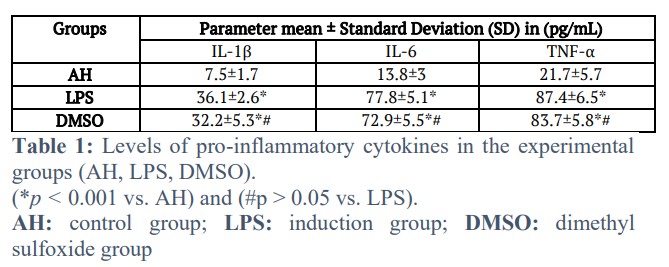
The proinflammatory cytokine levels in the blood serum of the control group (AH), lipopolysaccharide group (LPS) and DMSO group are shown in Table 1. The table shows a highly significant increase in the LPS and DMSO group values compared with those of the AH group (p < 0.001). The DMSO group showed no considerable difference from the LPS group.
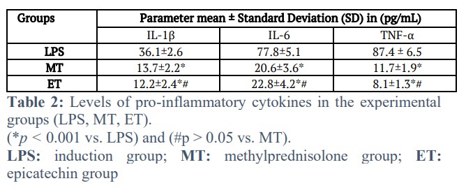
When comparing pro-inflammatory cytokine levels in the serum of the MT and ET groups with LPS group, as shown in Table 2, a highly significant reduction is recorded (P < 0.001). The results of the ET group were comparable to the MT group, and no noteworthy variance is revealed between them.
Influence on lung histopathological changes
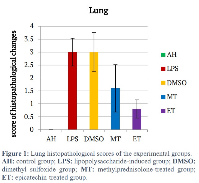
The lung histopathological scores of all experimental groups — control (AH), lipopolysaccharide (LPS), DMSO, methylprednisolone (MT), and epicatechin (ET) are presented in Figure 1. The LPS and DMSO groups exhibit significantly higher scores of lung tissue damage compared with the AH group (p < 0.001). In contrast, both epicatechin and methylprednisolone treatments markedly improved the histopathological scores relative to the LPS model group (p < 0.001).
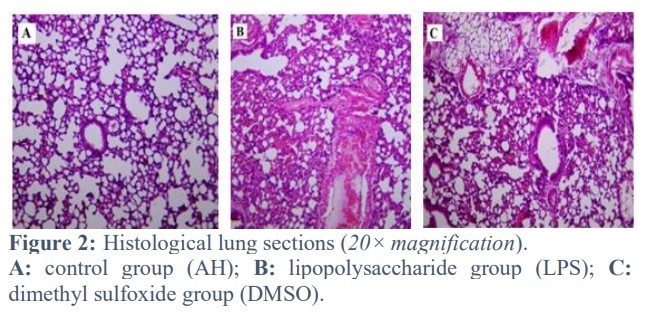
The lung sections in the histological images of the lipopolysaccharide (LPS) and dimethyl sulfoxide (DMSO) groups revealed clear alterations and damage, including alveolar congestion and bleeding, protein compounds, extreme infiltration of inflammatory cells and disruption of alveolar septa. A diffuse alveolar injury with emphysema alterations is observed in comparison to the control group in Figure 2 (AH) (P < 0.001).
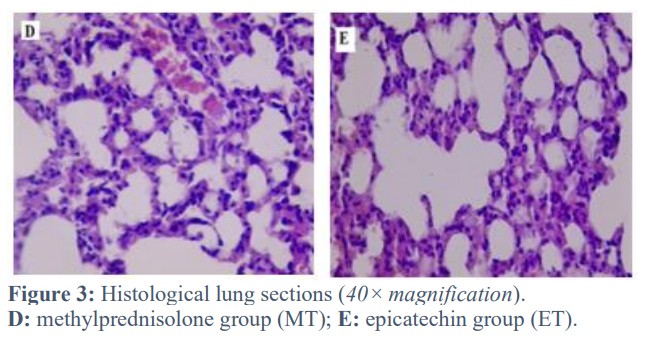
Compared with the lipopolysaccharide (LPS) group, the methylprednisolone (MT) and epicatechin (ET) groups showed markedly reduced histopathological damage, with milder alveolar capillary obstruction and less interstitial inflammatory cell infiltration, as shown in Figure 3.
Figures & Tables
The outer membrane of Gram-negative bacteria contains lipopolysaccharides, which strongly stimulate the mammalian immune system to produce inflammation. Toll-like receptors (TLRs) activate the innate immune system, with the LPS lipid A moiety generally influencing immunological activation and response [12]. The study found that lipopolysaccharides stimulate an immune response with increased cytokine levels as compared with the AH group. The results of the current study correspond with other studies that mentioned similar effects of LPS [13,14]. The immune response is induced by LPS through its interaction with the membrane receptor TLR4. Accordingly, the proinflammatory cytokines are released, and the NF-κB and MAPK pathways are activated through cellular mechanisms [15].
In contrast to the LPS group, epicatechin demonstrated a considerable decrease in serum levels of IL-1β, IL-6 and TNF-α in the current study. Under LPS circumstances, Xing et al. (2019) also found a similar decrease in inflammatory cytokines [9]. By preventing LPS-induced TLR4 overexpression and/or by promoting TLR4 degradation, epicatechin suppressed TLR4 expression and had an anti-inflammatory impact. Additionally, epicatechin controls LPS-mediated TLR4 activation by preventing TLR4 access, preventing lipid rafts from being sealed off and preventing TLR4 dimerisation [16]. Additionally, by inhibiting the phosphorylation of ERK, p38, JNK, p65, IκB, and expression in the MAPK and NF-κB signaling pathways, epicatechin efficiently lowers the production of inflammatory mediators [17,18]. The discovery that NLRP3 inflammasome activation and the emergence of inflammatory disorders of the airways are related has been unique in recent years [19]. Epicatechin has strong antioxidant capabilities since it may deactivate the cell’s ROS. Tian et al. (2021) found that epicatechin inhibits oxidative stress, preventing the maturation and secretion of IL-1β and IL-18 through caspase-1 action [20]. Previous studies show that PGE2 can induce IL-6, IL-1β and TNF-α in various cell types through EP2 and EP4 receptors and their cAMP-dependent pathway [21-23]. Epicatechin inhibited the activity of COX-2 and iNOS enzymes in LPS-stimulated macrophages, suppressing the release of NO and PGE2 [24].
Cytokine storms lead to significant histopathological damage, especially in the lungs, following LPS interaction with TLRs by releasing many proinflammatory cytokines, which stimulates neutrophil sequestration and activation that further releases many ROS, protease, leukotrienes and other inflammatory mediators such as platelet-activating factors to recruit other inflammatory cells which in turn release proinflammatory cytokine that lead to cytokine storm and parenchymal tissue damage such as infiltration of monocyte cell, damaged or decreased alveolar septa that may lead to the destruction of the alveolar compartment and decrease surfactant synthesis [25,26]. Epicatechin inhibits the TLR4 response and inflammatory cytokine production, reducing tissue damage caused by LPS and thus reducing inflammation [27]. Dickerhof et al. (2014) demonstrated that epicatechin significantly inhibits macrophage migration inhibitory factor (MIF), a crucial part of the inflammation response [28]. Similarly, its direct binding to the active site of p-p38 in the MAPK pathway is essential for the regulation of neutrophil chemotaxis [9]. Epicatechin lowers oxidative stress by limiting elevated ROS and NOX-4 levels [29].
Mice's cytokine storm induced by lipopolysaccharides can be effectively influenced by epicatechin since it can decrease the IL-1β, IL-6 and TNF-α levels in serum and ameliorate the damage to the mice’s lungs. Future studies should explore the molecular mechanisms of epicatechin’s interaction with TLR4 and NLRP3 pathways using gene-expression and protein-profiling approaches
Acknowledgement
The authors are thankful to Professor Dr. Salim Rasheed Al-Obaidy for his assistance with the histopathological aspects of the experiment.
Author Contributions
Conceptualisation, data collection and manuscript preparation: Zeena T. Tariq
Supervision: Ahmed R. Abu-Raghif
Support and assistance with experimental procedures and documentation: Ahmed F. Abed Mansoor
No conflicts of interest are disclosed by the authors.![]()
References
- Brocker C, Thompson D, Matsumoto A, Nebert DW, Vasiliou V. Evolutionary divergence and functions of the human interleukin (IL) gene family. Human genomics, (2010); 5(2010): 1-26.
- Jang DI, Lee AH, Shin HY, Song HR, Park JH, et al. The role of tumor necrosis factor-alpha (TNF-α) in autoimmune disease and current TNF-α inhibitors in therapeutics. International journal of molecular sciences, (2021); 22(5): 2719.
- Hirano T. IL-6 in inflammation, autoimmunity, and cancer. International immunology, (2021); 33(3): 127-148.
- Zalinger ZB, Elliott R, Weiss SR. Role of the inflammasome-related cytokines Il-1 and Il-18 during infection with murine coronavirus. Journal of Neurovirology, (2017); 23(2017): 845-854.
- Wang Y, Che M, Xin J, Zheng Z, Li J, et al. The role of IL-1β and TNF-α in intervertebral disc degeneration. Biomedicine & Pharmacotherapy, (2020); 131(2020): 110660.
- Mahmudpour M, Roozbeh J, Keshavarz M, Farrokhi S, Nabipour I. COVID-19 cytokine storm: The anger of inflammation. Cytokine, (2020); 133(2020): 155151.
- Wan J, Zhang L, Ruan Z. Dietary Supplementation with Epicatechin Improves Intestinal Barrier Integrity in Mice. Foods, (2022); 11(20): 3301.
- Shariati S, Kalantar H, Pashmforoosh M, Mansouri E, Khodayar MJ. Epicatechin protective effects on bleomycin-induced pulmonary oxidative stress and fibrosis in mice. Biomedicine & Pharmacotherapy, (2019); 114(2019): 108776.
- Xing J, Yu Z, Zhang X, Li W, Gao D, et al. Epicatechin alleviates inflammation in lipopolysaccharide-induced acute lung injury in mice by inhibiting the p38 MAPK signaling pathway. International Immunopharmacology, (2019); 66(2019): 146-153.
- Wu H, Xie Y, Xu Y, Hu Z, Wan X, et al. Protective effect of Epicatechin on APAP-induced acute liver injury of mice through anti-inflammation and apoptosis inhibition. Natural product research, (2020); 34(6): 855-858.
- Daniel WW. Biostatistics: a foundation for analysis in the health sciences. 1978; 129: 1-504. Wiley
- Brodzikowska A, Ciechanowska M, Kopka M, Stachura A, Włodarski PK. Role of Lipopolysaccharide, Derived from Various Bacterial Species, in Pulpitis—A Systematic Review. Biomolecules, (2022); 12(1): 138.
- Abed Mansoor AF, Abu Raghif AR. Attenuated effects of rivastigmine in induced cytokine storm in mice. Journal of Emergency Medicine, Trauma & Acute Care, (2022); 2022(3): 12.
- Sahib HB, Kathum OA, Alanee RS, Jawad RA, Al-Shammari AM. The Anti‐Cytokine Storm Activity of Quercetin Zinc and Vitamin C Complex. Advances in Virology, (2022); 2022(1): 1575605.
- Skrzypczak-Wiercioch A, Sałat K. Lipopolysaccharide-induced model of neuroinflammation: Mechanisms of action, research application and future directions for its use. Molecules, (2022); 27(17): 5481.
- Prince PD, Fischerman L, Toblli JE, Fraga CG, Galleano M. LPS-induced renal inflammation is prevented by (−)‐Epicatechin in rats. Redox biology, (2017); 11(2017): 342-349.
- Li Z, Fu X, Fan Y, Zhao C, Wang Q, et al. Effect of Epicatechin on inflammatory cytokines and MAPK/NF-κB signaling pathway in lipopolysaccharide-induced acute lung injury of BALB/c mice. General Physiology and Biophysics, (2022); 41(4): 299-308.
- Wu C, Li F, Zhang X, Xu W, Wang Y, et al. (−)-Epicatechin Ameliorates Monosodium Urate-Induced Acute Gouty Arthritis Through Inhibiting NLRP3 Inflammasome and the NF-κB Signaling Pathway. Frontiers in pharmacology, (2022); 13(2022): 799552.
- Leszczyńska K, Jakubczyk D, Górska S. The NLRP3 inflammasome as a new target in respiratory disorders treatment. Frontiers in Immunology, (2022); 13(2022): 1006654.
- Tian X, Xue Y, Xie G, Zhou Y, Xiao H, et al. (−)-Epicatechin ameliorates cigarette smoke-induced lung inflammation via inhibiting ROS/NLRP3 inflammasome pathway in rats with COPD. Toxicology and applied pharmacology, (2021); 429(2021): 115674.
- Cho JS, Han IH, Lee HR, Lee HM. Prostaglandin E2 induces IL-6 and IL-8 production by the EP receptors/Akt/NF-κB pathways in nasal polyp-derived fibroblasts. Allergy, Asthma & Immunology Research, (2014); 6(5): 449-457.
- Zasłona Z, Pålsson-McDermott EM, Menon D, Haneklaus M, Flis E, et al. The induction of pro–IL-1β by lipopolysaccharide requires endogenous prostaglandin E2 production. The Journal of Immunology, (2017); 198(9): 3558-3564.
- Lewis A, Elks PM. Hypoxia induces macrophage tnfa expression via cyclooxygenase and prostaglandin E2 in vivo. Frontiers in immunology, (2019); 10(2019): 2321.
- Wang H, Cao Z. Anti-inflammatory effects of (-)-Epicatechin in lipopolysaccharide-stimulated raw 264.7 macrophages. Tropical Journal of Pharmaceutical Research, (2014); 13(9): 1415-1419.
- Wang H, Ma S. The cytokine storm and factors determining the sequence and severity of organ dysfunction in multiple organ dysfunction syndrome. The American journal of emergency medicine, (2008); 26(6): 711-715.
- Ye R, Liu Z. ACE2 exhibits protective effects against LPS-induced acute lung injury in mice by inhibiting the LPS-TLR4 pathway. Experimental and molecular pathology, (2020); 113(2020): 104350.
- Xing J, Yu Z, Zhang X, Li W, Gao D, et al. Epicatechin alleviates inflammation in lipopolysaccharide-induced acute lung injury in mice by inhibiting the p38 MAPK signaling pathway. International Immunopharmacology, (2019); 66(2019): 146-153.
- Dickerhof N, Magon NJ, Tyndall JD, Kettle AJ, Hampton MB. Potent inhibition of macrophage migration inhibitory factor (MIF) by myeloperoxidase-dependent oxidation of epicatechins. Biochemical Journal, (2014); 462(2): 303-314.
- Cilleros DÁ, López-Oliva ME, Martín MÁ, Ramos S. (−)-Epicatechin and the colonic metabolite 2, 3-dihydroxybenzoic acid protect against high glucose and lipopolysaccharide-induced inflammation in renal proximal tubular cells through NOX-4/p38 signalling. Food & function, (2020); 11(10): 8811-8824.
This work is licensed under a Creative Commons Attribution-Non Commercial 4.0 International License. To read the copy of this license please visit: https://creativecommons.org/licenses/by-nc/4.0

