

Full Length Research Article
Molecular Detection of Toxoplasma gondii In Local and Imported Red Meat and Meat Products in Al-Diwaniyah City Markets, Iraq
Yuser J. Mahmood1, Hadi M. Almayali2*
Adv. life sci., vol. 12, no. 3, pp. 490-497, August 2025
*- Corresponding Author: Hadi M. Al-Mayali (Email: hadi.hamza@qu.edu.iq)
Authors' Affiliations
[Date Received: 28/09/2024; Date Revised: 12/01/2025; Available Online: 31/10/2025]
Abstract![]()
Introduction
Methods
Results
Discussion
References
Abstract
Background: Toxoplasmosis is a prevalent parasitic disease that affects both humans and many domestic animals and causes abortion and death of fetuses and neonates. It results from infection with Toxoplasma gondii.
Methods: During this study, 300 samples (150 local and 150 imported meat and product samples like steak, minced meat, sausages, burgers, and livers) were examined by microscopy on digested tissue juice prepared from the peptic digestion technique and molecularly by polymerase chain reaction (PCR). The objective was to detect Toxoplasma gondii infection through the B1 gene, which is of amplicon length 469 bp.
Results: The results of local meat examined through microscope and PCR technique showed that samples taken from steak meat gave the highest positive infection (22%), Minced meat (20%), burgers (12%), and liver 8%, while the lowest percentage of positive infection was in sausages; it was only one positive sample (4%). The total infection rate was 14.66% and the results did not show any significant differences in infection at infection probability (P > 0.05). Imported meat showed only 7 positive samples (4.66%).
Conclusion: The higher prevalence of Toxoplasma gondii in local fresh meat and meat products makes them more harmful to human health than imported meat, whereas freezing, salting, and other processes make imported meat products less contaminated than fresh meat. Based on genetic tree analysis and genotyping, the Indian strain of the parasite registered in the NCBI gene bank under the accession number DQ872518.1 is prevalent in both imported and local meat in Iraq, with a molecular similarity ranging with local strains from 99.60% to 99.96%.
Keywords: Toxoplasmosis, Meat Products, B1 gene
Introduction![]()
Toxoplasmosis, a common parasitic disease in humans as well as domesticated animals, can cause fetal abortion as well as death among newborns, brought about by the Eimeriidae family protozoan Toxoplasma gondii [1]. Notably, the causative agent is the only species belonging to its own genus in the Apicomplexa class that infects nearly one-third of the world’s humans [2]. Although felids, such as cats, are definitive hosts for Toxoplasma gondii, humans as well as other animals serve as intermediate hosts [1]. Human infections mostly result from the consumption of raw or undercooked meat containing tissue cysts, contaminated food, as well as water with oocysts, and mother-to-baby transmission [3,4]. Extremely widespread in humans as well as animals globally, Toxoplasma gondii tends to confer asymptomatic latent infections, yet it is responsible for substantial morbidity as well as mortality in humans and animals [5,6]. Toxoplasma gondii has a broad spectrum of hosts [7]. Toxoplasmosis is the third leading cause of foodborne illness-related hospitalizations worldwide. Analysis of determinants of risk has shown that 30-63% of human infections are caused by eating undercooked meat, with meat type as well as eating behavior playing a central role [8]. Pregnant women, as well as anemic individuals, tend to be particularly interested in eating meat as well as organ meats such as liver on the pretext of their therapeutic value. Sheep as well as goat meat, commonly used, are estimated to be infected by 24.5-33% by toxoplasmosis [9,10]. Determinants of transmission of toxoplasmosis involve eating behavior, as well as contact with cats, area, and age. Confirmatory evidence from serological experiments has shown that animals infected by Toxoplasma gondii, as well as slaughtered for consumption, can transmit the infection to humans by means of their meat, making raw meat a means of disease transmission [11]. This foodborne parasite causes infection in humans by ingestion of contaminated food as well as water containing mature oocysts, as well as eating raw or undercooked meat or meat products containing tissue cysts and infected pets [6]. The detection of Toxoplasma gondii is done by means of microscopic biopsy, serological tests, and molecular methods. Among these, molecular methods are being reported as the most sensitive and specific [6], thus proving effective as well as appropriate for detecting this disease on account of their own advantageous nature.
The bioassay method, employing laboratory cats or mice, is considered a gold standard for the detection of the parasite in meat samples. It is a labor-intensive and ethically demanding method, especially in the analysis of multiple samples and animals. Due to these challenges, the Polymerase Chain Reaction (PCR) method was created as a highly sensitive method for the detection of Toxoplasma gondii DNA in meat samples with a maximum requirement of 50 mg of sample for DNA extraction, as compared to the requirement of 500 gram or more of meat to be fed to a single cat or 50-100 grams for mouse injection following sample digestion [12-14].
Since little has been studied regarding the prevalence of Toxoplasma gondii in meat and meat products in Iraq, particularly in Diwaniyah city, the purpose of this study is to examine the occurrence of the parasite in different types of local meat as well as imported meat, including their products that are sold to humans in local markets, butcher shops, and food stores. The aim is to assess the level of contamination of fresh as well as frozen meat with Toxoplasma gondii.
Methods![]()
Samples collection
This study was carried out in the city of Al-Diwaniyah, Iraq, from June to December 2022. The study sought to determine the infection rate of Toxoplasma gondii in local and imported red meat and meat products. A total of 300 samples were obtained (150 local red meat products and 150 imported). The products covered a variety ranging from steaks, minced meat, sausages, burgers, and livers of slaughtered animals.
Isolation of T. gondii bradyzoite
The peptic digestion method (Al-Salhy 2013) was used to lyse the tissue and release the bradyzoites of Toxoplasma gondii from tissue, as well as to destroy tissue cysts for genomic DNA extraction. The digestive solution contains 2.6 g pepsin, 5 g NaCl, 7 mL HCl, and 485 mL distilled water. Using scissors, each sample was cut into pieces and pooled with approximately 50 grams of beef from each specimen before being inserted into a sterile test tube. Thereafter, the tube was combined with 10 mL of digesting solution and incubated at 40 °C for 30 minutes.
They were passed through two layers of gauze into a flask, the filtrate into a test tube, and then spun in a centrifuge at 5000 rpm for 10 minutes. The precipitate was removed, and 5 mL of sodium bicarbonate solution was added to it, which was mixed until dissolved and centrifuged again for 10 minutes at 5000 rpm. After the supernatant was discarded again, the precipitate was resuspended in 600 μL of normal saline and well mixed. After the removal of normal saline, DNA from these samples was collected [15].
Genomic DNA Extraction
Each meat and meat product sample was extracted with the gSYNC DNA extraction Kit (Geneaid, USA) in accordance with the manufacturer’s instructions, and genomic DNA was extracted from each sample. As detailed below:
Up to 100 mg of meat was weighed and transferred into a sterile 1.5 mL microcentrifuge tube. 200 µL of meat slurry was prepared, then 20 µL of Proteinase K was added, mixed by pipetting, and incubated at 60°C for 5 minutes. After that, 200 µL of GSB buffer was added and the sample was vortexed, followed by incubation at 60°C for 30 minutes. Finally, the sample was incubated at 60°C for 5 minutes, inverting the tube every 2 minutes.
After that, the samples of these meat products were incubated, and then a 2-minute centrifugation was conducted at 14,000 rpm. A fresh tube containing 200 μL of GSB buffer was added to the lysate and mixed, followed by the addition of the supernatant to a 1.5 mL Eppendorf tube by vortexing. During the binding/lysis step, the meat and meat product samples were incubated at 70 °C for 15 minutes and inverted 4 times over 3 minutes. Then, 100% ethanol was added to the meat product lysate mixture, and the solution was vigorously agitated by vortexing for 10 seconds. All the mixture was transferred into the GS column situated in a 2 mL collecting tube, which was placed within the GS columns (DNA filter columns). After 2 mL collection tubes had collected a flow-through, the latter was removed from the tubes, and the GS columns were put into new 2 mL collection tubes. After which, 400 μL of W1 Buffer was loaded onto the GS columns and spun for 30 seconds at 14,000 rpm. After discharging the flow-through, the 2 mL collection tubes and the columns were reinserted into the instrument, and the flow-through was discarded. This was followed by 600 µL of Wash Buffer that was added to the column, which was centrifuged for 30 seconds at 14,000 rpm, and the flow-through was discarded. After filtration, the GS columns were transferred without using any additives to 2 mL collection tubes and centrifuged for an additional 3 minutes at 14,000 rpm. The desiccated GS column was placed in a sterile 1.5 mL Eppendorf tube, and 80 μL preheated elution buffer was added in the middle of the column matrix. After that, the elution was allowed to stand for 3 minutes to complete the absorption of the elution. Pure DNA was obtained by conducting a final centrifugation at 14,000 rpm for 30 seconds.
Estimation of Genomic DNA
The extracted genomic DNA was measured for concentration (ng/μL) and purity using the NanoDrop spectrophotometer (THERMO, USA). DNA purity was determined with relation to protein contamination and overall DNA quality by measuring the absorbance ratio of 260 nm/280 nm.
Polymerase Chain Reaction (PCR)
Conventional PCR was conducted to identify T. gondii by amplifying the B1 gene, using the methodology outlined by [16] in the subsequent steps:
Preparation of PCR Reaction
The whole reaction was developed in the Maxime PCR PreMix kit, using the recipe of the manufacturer for master mix preparation:
DNA template was added at 5 μL, forward primer (10 pmol) and reverse primer (10 pmol) were added to the total of 1.5 μL each, and nuclease-free water was added to the total of 12 μL to bring the final volume to 20 μL. The PreMix Kit is a regular PCR formulation or mixture of Taq DNA polymerase, dNTPs, TrisHCl (pH 9.0), KCl, MgCl2 stabilizer, and other chemicals combined. All tubes were prepared, put into an Exispin vortex centrifuge, centrifuged at 3000 rpm for 3 minutes, transferred to the PCR thermocycler, and amplified.
Primers
In previous research by Ortega-Pacheco et al. (2013), a PCR primer was established to identify the B1 gene of T. gondii [17]. The Scientific Researcher Company, Iraq, provided the primer at an amplicon of 469 bp and has the sequence: F = AAAAATGTGGGAATGAAAGAG and R = ACGAATCAACGGAACTGTAAT.
Detection of T. gondii Strains
To determine the genetic sequence of the isolated T. gondii strains, PCR products from fourteen positive samples were subjected to DNA sequencing using the AB DNA sequencing system and submitted to Bioneer Company in Korea.
Nucleotide Sequences of Local Isolates of Toxoplasma gondii Compared with Strains Registered in the International Gene Bank Using the B1 Gene
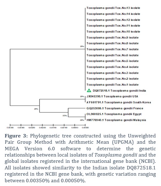
Nucleotide sequencing was carried out to investigate the presence of new genotypes of T. gondii genes. This was achieved through phylogenetic tree analysis of the local isolates from this study and their comparison with globally registered genotypes available in the international gene bank (NCBI-GenBank). The phylogenetic tree data were analyzed using MEGA version 6.0 software, applying the Unweighted Pair Group Method with Arithmetic Mean (UPGMA).
The analysis identified the presence of substitution mutations between the genes of the local isolates and the corresponding genotypes of globally registered isolates. The sequences were analyzed and compared with entries in the International GenBank (NCBI), showing high similarity to strain DQ872518.1.
Statistical Analysis
All data from this study were calculated and statistically tested using the SPSS software, applying the Chi-square (X²) distribution. The probability value was determined as follows: P ≤ 0.05 was considered statistically significant.
Results![]()
Microscopic examination
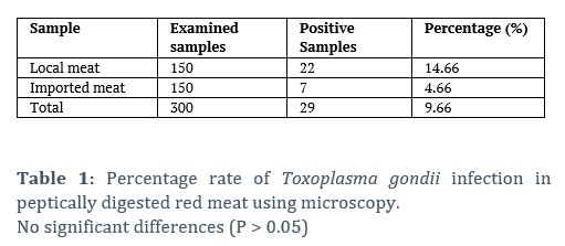
150 different samples of local meat and 150 different samples of imported meat were examined microscopically during the study; 22 samples (14.66%) and 7 samples (4.66%), respectively, were positive by microscopic examination using the peptic method (Table 1).
Local Meat
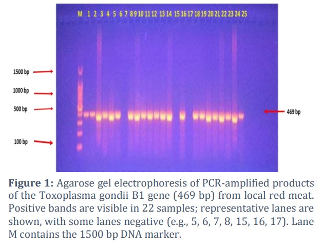
All 300 samples were examined by polymerase chain reaction. The results of PCR were consistent with those of microscopic examination; 22 samples (14.66%) were positive for Toxoplasma gondii by using the B1 gene at a molecular size of 469 bp (Figure 1).
Imported meat
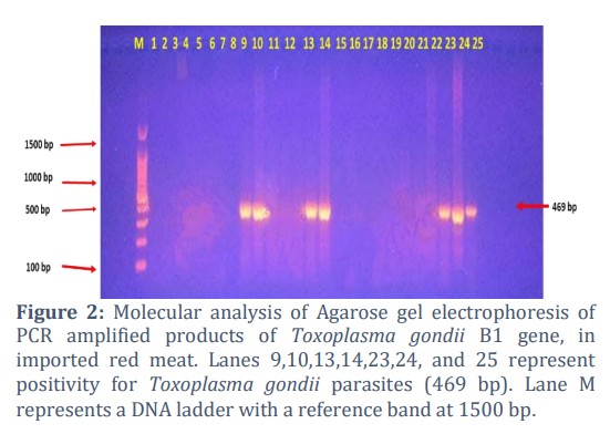
A molecular study of 150 samples of imported meat showed that there were only 7 positive samples for infection with Toxoplasma gondii, with a total infection rate of 4.66%, as shown in Figure 2.
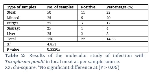
The results of the molecular study of local meat examined according to the source of the sample to detect infection with the Toxoplasma gondii showed that samples taken from steaks gave the highest positive result (11 positive samples) with a percentage of 22%, while samples taken from minced local meat recorded the appearance of 5 positive samples and a percentage of 20%, and the samples taken from burgers (3 positive samples, 12%), and the samples taken from the liver (2 positive samples, 8%), while the lowest percentage of positive infection was sausages, which is only one positive sample. With a percentage of 4%, the results did not show any significant differences in infection at the probability level (P > 0.05), while the total infection rate was 14.66%, as shown in Table 2.
Imported meat
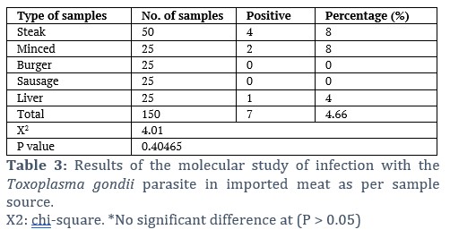
The results of the molecular study to detect infection with Toxoplasma gondii according to the source of the sample showed that samples taken from steaks gave the highest positive result (4/50 positive samples) with a percentage of 8%, while samples taken from minced meat recorded (2/25 positive samples) with a percentage of 8%, and the samples taken from the liver recorded one sample out of a total of 25 with a rate of 4%, while the samples of sausages and burgers did not show any positive result, and the statistical analysis did not show any significant differences at the level of probability (P > 0.05) as shown in Table 3.
Genetic sequencing
The results of the multiple sequence alignment (MSA) analysis of the nucleotide sequences obtained from the polymerase chain reaction (PCR) products of the Toxoplasma gondii gene revealed that all the studied samples were consistent with the global isolates of the species, previously identified in India, the United States, Korea, France, Egypt, and Malaysia, and registered in the National Center for Biotechnology Information (NCBI). The genetic variations detected ranged between 0.00350%–0.00050%, as illustrated in Figure 3.
Phylogenetic Tree Analysis of Toxoplasma gondii Isolated from Local and Imported Meats
The phylogenetic tree was constructed using the Unweighted Pair Group Method with Arithmetic Mean (UPGMA) and the MEGA 6.0 software to illustrate the phylogenetic relationships of Toxoplasma gondii. The analysis was based on the B1 gene in order to determine the molecular sequence of nitrogenous bases and to identify the genetic relationship between local isolates of the parasite obtained from local and imported meats and their products in Al-Diwaniyah, and the global isolates of the parasite registered in the international gene bank (NCBI) using the same gene, as shown in Figure 3.
The phylogenetic data analysis of the molecular sequences of the genes from the local isolates obtained from meats and their products revealed that all locally identified T. gondii isolates were identical to the Indian strain of the parasite registered in the NCBI gene bank under the accession number DQ872518.1, with a molecular similarity ranging between 99.60% and 99.96%, as demonstrated in Figure 3.
Figures & Tables
Due to its significance to the human diet, especially beef, meat plays an important part in our diet [18]. As with other animals, cattle can be infected with Toxoplasma gondii and develop significant health complications, especially abortions in both cattle and people [19]. T. gondii infection in cattle in Iraq has been the subject of little research, with only two studies in Mosul and Al-Diwaniyah [16,20]. In Mosul and Al-Diwaniyah, one study used the Latex Agglutination Test (LAT) for the identification of T. gondii in meat juices, whereas the Al-Diwaniyah study used Polymerase Chain Reaction (PCR) to improve the sensitivity of detection. On the other hand, numerous studies have been carried out in Iraq about the frequency of T. gondii in many of the hosts, such as sheep [21], poultry [22], stray cats [22], and women who had abortions [21], which indicates the importance of this parasite in diverse hosts, notably, humans. In this study, 300 meat samples, including locally produced and imported minced meat, steaks, sausages, burgers, and liver, were evaluated for T. gondii infection. Digested tissue samples were investigated microscopically for the pepsin digestion technique, and an overall infection rate of 9.66% was determined. The prevalence of local and imported meat was quite dissimilar, with local meat having 14.66% infection and imported meat only 4.66%. Together, the findings are consistent with the published result from the study of Zakaria et al., [20] using the LAT technique and Asgari et al., [16], including the analysis of 100 digested beef juice specimens analyzed by PCR, with T. gondii positive rates of 17% and 22%, respectively. In Tunisia, the same results were obtained in the molecular tests as T. gondii DNA was found in 19.3% (29/150) of cow meat samples. The findings of the present investigation are in accord with previous publications that reported the public health risks of eating beef infected with T. gondii [23]. PCR identification of the B1 gene in the current study showed lower infection rates than northern Portugal (50%) and Colombia (38 out of 180 samples were identified as infected with T. gondii in chicken, pig, and cattle using the same genetic target) [24,25]. In contrast, the rate found in this investigation exceeded those previously reported in Brazil and Switzerland, which were 2 and 3.8% respectively [26]. Additionally, following research of Anvari et al., [27] pointed out that imported meat had a considerably higher infection rate (26 percent) than locally produced meat (six percent), differing from the results obtained in the current research, where locally produced meat possessed a higher frequency of T. gondii. However, our findings support the claims made by Asgari et al. [16] that imported meat has been subjected to processing techniques (salting, freezing, hot air drying), combined with preservatives likely to diminish T. gondii infection. Meat products show that hot smoking and grilling meat reduce the risk of transmitting parasites in processed meat products [28]. Factors such as climatic conditions, sample size, livestock management practices, hygiene standards at the slaughterhouse, processing method, environmental cat density, human dietary habits, and geographical factors like altitude and the climate condition of the region could be responsible for the variation in T. gondii prevalence among different studies [29]. It turns out that 15 percent of samples of local and international meat products were found to be infected, for steak it was 15 percent and for minced beef it was 14 percent, for liver 6 percent, for burgers 6 percent, and for sausages 2 percent. Overall, the infection rate in all local and imported samples was 9.66 percent. The findings are in agreement with those from Fallah et al., [30] in Iran, in which the rate of T. gondii infection in burger samples was 15%. Burger, sausages, and kebabs gave infection rates of 16.66%, 19.1%, and 56.6%, respectively. Consistent with Aspinall et al., [31] from the United Kingdom, 71 samples of pork, beef, and lamb were surveyed by PCR of samples and found that overall 19% of the samples were infected with T. gondii. On the other hand, previous research by Rahdar et al., [32] studying 90 different samples of burgers and sausages through PCR found no T. gondii infections, whereas this survey revealed T. gondii infections in the samples studied. These variances suggest that a few of the inconsistencies are due to geographical variation, differences in food processing methods, or differences in sample selection criteria. Out of a total of 150 samples of local and imported meat products, the results showed that the highest infection rate was in steak meat, with a rate of 15%, minced meat (14%), burgers (6%), liver (6%), and sausages 2%. As for the total infection rate with Toxoplasma gondii, it was 9.66% in both local and imported types. These results were in agreement with the results recorded by Fallah et al., [30] in Iran, when it was found that the infection in burgers was also 15%, in addition to other products in his study, such as burgers, sausages, and kebabs. The infection rates were 16.66%, 19.1% and 56.6%, respectively. It also agrees with the study of Aspinall et al., [31] in the United Kingdom, where they recorded infection with Toxoplasma gondii in 71 samples of pork, lamb, and beef using the polymerase chain reaction technique and showed that the infection rate was 19%. Study by Rahdar et al., [32] differs from the current study, as they recorded no infection in 90 samples of burgers and sausages that were examined by polymerase chain reaction (PCR). The results of the current study showed that steak (15 percent) and minced beef (14 percent) gave the highest infection rates, followed by liver (6 percent), burgers (6 percent), and sausages (2 percent). This suggests that sheep meat and liver are consumed more often in the research region, as no variation was found in infection rates for other kinds. The finding of T. gondii inside cow veal indicated no preferred localization within the tissue of the calf studied, and thus, this is consistent with the study of Burrells et al., [33]. Many studies have shown the prevalence of Toxoplasma gondii infection in different types of meat [34]. The application of serum methylation and PCR methods was used to investigate the incidence of T. gondii in persons who consume sheep meat in Tunisia, as illustrated by Boughattas et al., [35]. Of the lambs under six months, forty-eight percent were seropositive; seventy-three point six percent of older lambs; forty-one point two percent of sheep tissue samples also tested positive in PCR analysis. Also, a case of T. gondii prevalence done in Ethiopia using the Direct Agglutination Test (DAT) noted a prevalence of 17.68 percent (111 out of 628) in sheep and goats, noting a higher rate of infestation among the female adult sheep and goats in the rainy season [36].
The Toxoplasma infection in locally provided fresh meat is higher than that of internationally supplied fresh meat and consequently presents a higher health risk to humans. However, beef products imported are not as infected because of preservation procedures such as freezing and salting, which diminish the parasite's lifetime. Analysis using a genetic tree and genotyping of T. gondii from domestically supplied and imported meat from Iraq revealed that the Indian strain was the main type of T. gondii strain in Iraq. This similarity or genetic closeness to strains from other countries, such as India, Korea, Egypt, and France, may be attributed to the global distribution of these strains, which are also present in neighboring countries or those used for comparison, as well as to the origins of the imported meats into Iraq.
The strong similarity with the Indian strain, particularly in imported meats and their products, may be because the majority of imported meats are of Indian origin. As for the local meats, the resemblance may be explained by the predominance of the Indian strain in Iraq, in addition to the issue of commercial fraud in processed meat products, where Indian meat is commonly used, especially in the manufacturing of meat products.
Acknowledgement
We thank the College of Education at Qadisiyah University for providing support to researchers.
Author Contributions
Yuser J. Mahmood: Methodology and writing.
Hadi M. Al-Mayali: reviewing and editing.
There is no conflict of interest regarding this manuscript.![]()
References
- Webster JP. Dubey, J.P. Toxoplasmosis of Animals and Humans. Parasites & Vectors, (2010); 3(2010): 1-2.
- Shapiro K, Bahia-Oliveira L, Dixon B, Dumètre A, de Wit LA, et al. Environmental transmission of Toxoplasma gondii: Oocysts in water, soil and food. Food and waterborne parasitology, (2019); 15(2019): e00049.
- Dubey JP. History of the discovery of the life cycle of Toxoplasma gondii. International journal for parasitology, (2009); 39(8): 877-882.
- Innes EA, Bartley PM, Maley S, Katzer F, Buxton D. Veterinary vaccines against Toxoplasma gondii. Memórias do instituto oswaldo cruz, (2009); 104(2009): 246-251.
- Sroka J, Bilska-Zając E, Wójcik-Fatla A, Zając V, Dutkiewicz J, et al. Detection and molecular characteristics of Toxoplasma gondii DNA in retail raw meat products in Poland. Foodborne pathogens and disease, (2019); 16(3): 195-204.
- Robert-Gangneux F, Dardé ML. Epidemiology of and diagnostic strategies for toxoplasmosis. Clinical microbiology reviews, (2012); 25(2): 264-296.
- Dubey JP, Jones JL. Toxoplasma gondii infection in humans and animals in the United States. International journal for parasitology, (2008); 38(11): 1257-1278.
- AJC C. Sources of toxoplasma infection in pregnant women: European multicentre case-control study. BMJ, (2000); 321(2000): 142-147.
- Hashemi-Fesharki R. Seroprevalence of Toxoplasma gondii in cattle, sheep and goats in Iran. Veterinary parasitology, (1996); 61(1-2): 1-3.
- Khamesipour F, Doosti A, Mobarakeh HI, Komba EV. Toxoplasma gondii in cattle, camels and sheep in Isfahan and Chaharmahal va Bakhtiary Provinces, Iran. Jundishapur Journal of Microbiology, (2014); 7(6): e17460.
- Dong H, Su R, Lu Y, Wang M, Liu J, et al. Prevalence, risk factors, and genotypes of Toxoplasma gondii in food animals and humans (2000–2017) from China. Frontiers in microbiology, (2018); 9(2018): 2108.
- Alkhaled MJ, Yakoob AY, Al-Hamadani AH. An investigation of toxoplasmosis in free range chickens, industrial chickens and duck in mid Euphrates area of Iraq. Al-Qadisiya Journal of Veterinary Medicine Sciences, (2012); 11(2012): 17-24.
- Santos SL, de Souza Costa K, Gondim LQ, da Silva MS, Uzêda RS, et al. Investigation of Neospora caninum, Hammondia sp., and Toxoplasma gondii in tissues from slaughtered beef cattle in Bahia, Brazil. Parasitology Research, (2010); 106(2010): 457-461.
- Mohammed AA, Abdullah SH. Diagnostic study of toxoplasmosis in domestic chickens in Sulaimani Province. AL-Qadisiyah Journal of Veterinary Medicine Sciences, (2013); 12(2): 63-69.
- Al-Salhy FMS (2013) Molecular Diagnosis of Toxoplasma gondii in Local And Imported Meat And Meat Products Of Cattle In AL- Diwaniyah Province. University of AL-Qadisiyah.
- Asgari Q, Sarnevesht J, Kalantari M, Sadat SJ, Motazedian MH, et al. Molecular survey of Toxoplasma infection in sheep and goat from Fars province, Southern Iran. Tropical Animal Health and Production, (2011); 43(2011): 389-392.
- Ortega-Pacheco A, Acosta Viana KY, Guzmán-Marín E, Segura-Correa JC, Alvarez-Fleites M, et al. Prevalence and risk factors of Toxoplasma gondii in fattening pigs farm from Yucatan, Mexico. BioMed research international, (2013); 2013(1): 231497.
- Dubey JP. A review of toxoplasmosis in cattle. Veterinary parasitology, (1986); 22(3-4): 177-202.
- Saadatnia G, Golkar M. A review on human toxoplasmosis. Scandinavian journal of infectious diseases, (2012); 44(11): 805-814.
- Zakaria EG. Detection of Toxoplasma gondii antibodies in different meat juices. Rafidain journal of science, (2011); 22(2011): 17-25.
- Mohammed NS, Al-A'ssie AH, Al-saqur IM. Genotyping of Toxoplasma gondii isolated from aborted Iraqi women. Diyala Journal of Medicine, (2015); 9(1): 44-52.
- A'aiz NN. Determination of Toxoplasma gondii lineages of sheep in Wasit, Iraq. Iraqi Journal of Veterinary Sciences, (2016); 30(2): 23-26.
- Amdouni Y, Rjeibi MR, Rouatbi M, Amairia S, Awadi S, et al. Molecular detection of Toxoplasma gondii infection in slaughtered ruminants (sheep, goats and cattle) in Northwest Tunisia. Meat science, (2017); 133(2017): 180-184.
- Lopes AP, Vilares A, RODRIGUES A, MARTINS T, FERREIRA I, et al. Genotyping characterization of Toxoplasma gondii in cattle, sheep, goats and swine from the North of Portugal. Iranian Journal of Parasitology, (2015); 10(3): 465.
- Franco-Hernandez EN, Acosta A, Cortés-Vecino J, Gómez-Marín JE. Survey for Toxoplasma gondii by PCR detection in meat for human consumption in Colombia. Parasitology Research, (2016); 115(2016): 691-695.
- Berger-Schoch AE, Herrmann DC, Schares G, Müller N, Bernet D, et al. Prevalence and genotypes of Toxoplasma gondii in feline faeces (oocysts) and meat from sheep, cattle and pigs in Switzerland. Veterinary parasitology, (2011); 177(3-4): 290-297.
- Anvari D, Saadati D, Nabavi R, Eskandani MA. Epidemiology and molecular prevalence of toxoplasma gondii in cattle slaughtered in Zahedan and Zabol Districts, South East of Iran. Iranian journal of parasitology, (2018); 13(1): 114.
- Guo M, Buchanan RL, Dubey JP, Hill DE, Lambertini E, et al. Qualitative assessment for Toxoplasma gondii exposure risk associated with meat products in the United States. Journal of food protection, (2015); 78(12): 2207-2219.
- DHM J, TG W. Toxoplasmosis: a comprehensive clinical guide. Hong Kong Medical Journal, (2003); 9(3): 230.
- Fallah E, Hajizadeh M, Farajnia S, Khanmahammadi M. SAG2 locus genotyping of Toxoplasma gondii in meat products of East Azerbaijan Province, North West of Iran During 2010-2011. African Journal of Biotechnology, (2011); 10(62): 13631-13635.
- Aspinall TV, Marlee D, Hyde JE, Sims PF. Prevalence of Toxoplasma gondii in commercial meat products as monitored by polymerase chain reaction–food for thought?. International journal for parasitology, (2002); 32(9): 1193-1199.
- Rahdar M, Samarbaf ZA, Arab L. Evaluating the prevalence of Toxoplasma gondii in meat and meat products in Ahvaz by PCR method. Jundishapur Journal of Microbiology, (2012); 5(4): 570-573.
- Burrells A, Taroda A, Opsteegh M, Schares G, Benavides J, et al. Detection and dissemination of Toxoplasma gondii in experimentally infected calves, a single test does not tell the whole story. Parasites & vectors, (2018); 11(2018): 1-8.
- Plaza J, Dámek F, Villena I, Innes EA, Katzer F, et al. Detection of Toxoplasma gondii in retail meat samples in Scotland. Food and waterborne parasitology, (2020); 20(2020): e00086.
- Boughattas S, Ayari K, Sa T, Aoun K, Bouratbine A. Survey of the parasite Toxoplasma gondii in human consumed ovine meat in Tunis City. PLoS One, (2014); 9(1): e85044.
- Gebremedhin EZ, Abdurahaman M, Hadush T, Tessema TS. Seroprevalence and risk factors of Toxoplasma gondii infection in sheep and goats slaughtered for human consumption in Central Ethiopia. BMC research notes, (2014); 7(2014): 1-6.
This work is licensed under a Creative Commons Attribution-Non Commercial 4.0 International License. To read the copy of this license please visit: https://creativecommons.org/licenses/by-nc/4.0

