

Full Length Research Article
Combination of zinc nanoparticles with chitosan scaffolds increased cytokine genes on wound healing of infected rats with methicillin-resistant Staphylococcus aureus (MRSA)
Mohammad Kazem Shahmoradi1, Mehrdad Amini Nogorani2, Fatemeh Mansouri3, Leila Zarei4*
Adv. life sci., vol. 10, no. 1, pp. 93-98, March 2023
*– Corresponding Author: Leila Zarei (Email: leilazarei652@yahoo.com)
Authors' Affiliations
2. Student Research Committee, Faculty of Medicine, Lorestan University of Medical Sciences, Khorramabad – Iran
3. Department of Genetics and Immunology, Faculty of Medicine, Urmia University of Medical Sciences, Urmia – Iran
4. Department of Anatomical Sciences, Faculty of Medicine, Lorestan University of Medical Sciences, Khorramabad – Iran
Abstract![]()
Introduction
Methods
Results
Discussion
References
Abstract
Background: The present study aims to determine the effect of zinc nanoparticles with chitosan in the expression of cytokine genes during wound healing of infected rats with methicillin-resistant Staphylococcus aureus (MRSA).
Methods: In this study, all male Wistar rats were divided into five groups. Group M1: 0.1 mL sterile saline 0.9% solution was added to the wounds with no infection. Group M2: the wounds were infected with methicillin-resistant Staphylococcus aureus (MRSA) and only treated with 0.1 mL the sterile saline (0.9%) solution. Group M3: Animals with infected wounds were treated with zinc nanoparticles. Group M4: animals with infected wounds were treated with chitosan. Group M5: animals with infected wounds were treated with 0.1 mL solution of zinc nanoparticles with chitosan. Skin biopsy samples were removed for the histological studies and quantitative assessment of IL-6, VEGF, TNF and TGF genes using real-time PCR in each group.
Results: Quantitative histological and neovascularization studies showed that there was significant difference between rats in group M5 compared to other groups. The infected group M5 exhibited a significant increase in the expression levels of VEGF: 8.02, TNF: 5.34, TGF: 7.98, and decrease of IL-6:-3.34 folds as compared to the other groups on the 21st day (P<0.05). Also, on the same day, the minimum wound surface area was observed in group M5. The surface area between each study group and other groups was statistically significant (P<0.05).
Conclusion: Our studies also show that the type of zinc nanoparticles with chitosan scaffolds has more effects than other types of compounds in wound healing.
Keywords: Nanozinc; chitosan; wound; meticillin-resistant infection; Inflammatory cytokine
Introduction![]()
Wound healing is an important public health challenges and numerous cellular responses, various growth factors and signaling pathways are the primary events in the etiology of wound healing. Delay during wound repair, antibiotic drug resistant and various infections have increased worldwide in the past few years [1]. Open wounds are susceptible to starting infection by bacteria and there are good entry sites for bacteria and facilitate systemic infections. Infectious wounds are slowly healing and lead to undesirable secretions and cause toxins from dead cells and new cell formation. As a result, we need to stimulate and restore the natural functions of the damaged skin and reduce irritation and pain of injury; and to activate tissue restoration processes [1]. Staphylococcus aureus is one of the most important hospital infections in many healthcare centers [2]. Methicillin-resistant staphylococcus aureus (MRSA) is the most widespread bacterial pathogen that leads to different infections of the skin and soft tissue. There are invasive severe infections such as pneumonia, endocarditis, bacteremia, and sepsis [3,4]. It has been suggested that staphylococcus aureus infection is resistant to drugs and leads to an increase in mortality and a rise in annual healthcare costs [5,6]. However, Methicillin was used as an important and widespread anti-bacterial drug in medicine [7].
Chitosan is a non-toxic biodegradable polymer prepared by alkaline de-acetylation of chitin. There is a wide range of chitosan in different proportions of de-acetylation [8]. Chitosan thin films have a wide application in medicine and biology. One of these applications is making wound dressings [9-11].
In recent years Some nanoparticles such as silver, gold, and copper as well as titanium and zinc oxide nanoparticles have been shown in treating skin injuries [12-15]. Nanoparticles including nanocapsules, polymersomes, and solid lipid nanoparticles are ideal strategies to enhance drug delivery and healing wounds. The functional effects of zinc oxide nanoparticles include increased re-epithelialization, antibacterial activity and fibroblast proliferation, and gene expression [16, 17]. It has been reported that a compound including chitosan and zinc oxide has been used successfully in making composite bonds for wound dressing and biofilms [16,18].
Scientists have revealed that various growth factors, such as insulin-like growth factor (IGF), vascular endothelial growth factor (VEGF), transforming growth factor (TGF), fibroblast growth factor (FGF), and keratinocyte growth factor (KGF) play a crucial role in the wound healing. Also, the role of macrophages, fibroblasts, mast cells are to clean the injury site through phagocytosis, repair and secretion of cytokines or growth factors [1, 2].
The main objective of this study was to investigate the expression pattern of inflammatory IL-6, VEGF, TNF and TGF genes on wound healing of infected rats to provide a standard therapeutic option during this study. Combination of zinc nanoparticles with chitosan scaffolds can act as a valuable option in the cell migration, stimulate the immune response in many microbial infections and an ideal dermal substitute for ulcers and wound regeneration. It acts as a valuable option to accelerate wound healing and effective antibiotics in future.
Methods![]()
Ethical Considerations
The research project was approved by the ethics committee of the Lorestan University of Medical Sciences, with IR.LUMS.REC.1401.067; registry grant number 267 for final thesis of residency students.
Animals
In this study, we used 60 male Wistar rats in the weight range of 180-200 g at Lorestan University of Medical Sciences, Khoramabad, Iran. These animals were held in standard cages under natural light and room temperature. Before starting studies, the animals have undergone 2 weeks of adaptation period to minimize the negative effects of new environmental stress. The method of making wound infectious, histopathology evaluation, and bacterial inoculation were performed according to a previous study by Dr Abbaszadeh et.al [14]. The tissue samples were removed on days 7, 14, and 21 after the surgery of wounds from all group animals.
After anesthesia and a small incision under sterile conditions, a sample of 7mm diameter was taken from all healed and intact tissues. The samples were immersed in buffered formalin 10% for preservation. After fixing and molding tissue in paraffin, sections of 5 µm thickness were prepared by microtome. The samples were stained through the hematoxylin-eosin method.
Measuring the wound surface area
To determine the role of zinc/chitosan in skin wound healing, we first assessed the difference in wound repair between control rats and infected rats with a zinc/chitosan bandage, using full-thickness excisional wounds introduced to the dorsal skin of each group. During the study, mortality was not observed in each animal group.
All wounds were photographed by a digital camera accompanied by a software ruler as a measuring scale. The wound surface area was measured using Adobe Acrobat 9 Pro Extended software (Adobe Systems Inc., San Jose, CA, USA). Day 0 was considered the baseline. This was repeated on days 7, 14, and 21.
The method for making the biodegradable layer of chitosan-nano-zinc
Chitosan-nano-zinc was prepared according to Lasshami et al [15]. Briefly, 10 mL aqueous polysaccharide (10 mL, 0.5% w/w) was mixed with 7.5mL acid ascorbic solution of 0.23 M and 5 mL acetic acid solution of 2.4 M. Then, 0.25 mL zinc solution of 0.51M was added to this prepared mixture. Changing the color of the reaction mixture from colorless to red is evidence of the formation of the chitosan-zinc nanoparticles. Finally, the solution was diluted with distilled water up to a final volume of 50mL. So, the concentration of chitosan-zinc nanoparticles was 200 mg/L. Then, 10 mL of prepared solution was dialyzed using permeable membranes with the permeability to molecules of 12 kDa molecular weight against 2 L of distilled water at room temperature for 2 hours [16].
Animal groups
All male rats were divided into five groups after the dorsal small incision. Group M1: 0.1 mL sterile saline 0.9% solution was added to the wounds with no infection. Group M2: the wounds were infected with methicillin-resistant Staphylococcus aureus (MRSA) and only treated with 0.1 mL sterile saline (0.9%) solution. Group M3: animals with infected wounds were treated with zinc nanoparticles Group M4. Animals with infected wounds were treated with chitosan Group M5: animals with infected wounds were treated with 0.1 mL solution of zinc nanoparticles with chitosan.
RNA extraction and quantitative real-time PCR (qRT-PCR) analysis of all samples
Cytokine mRNA expression level was assessed using qRT-PCR. Total RNA was extracted from skin samples using RNX-plus solution (Gene all, South Korea) according to the manufacturer’s instructions and then stored at −20°C. The quality and quantity of extracted RNAs were evaluated by the absorbance ratio at 280/260 nm using NanoDrop 2000 spectrophotometer (Thermo Fisher Scientific, USA). Complementary DNA (cDNA) was synthesized from 1μg RNA using First Strand cDNA Synthesis Kit (Takara, Japan). Quantitative PCR reaction was performed in triplicate on a real-time PCR detection system (ABI, USA) using SYBR Green Premix EX TaqTM II (Takara, Japan). The cycling conditions were: 10 seconds at 95 °C, then 40 cycles at 95°C for 10 seconds and 60 °C for 20 seconds. The melting conditions were 65-95 °C for 10 seconds. Specific sets of primers were designed. Besides, negative controls (nuclease-free water), beta-actin (as a reference gene) and melt curve analysis were employed in each run. Fold changes in gene expression were calculated by 2-ΔΔCT method.
Statistical Analysis
All histochemical and fold change expression data were completed by using Microsoft Excel 2013 software and SPSS (version 20.0; IBM Corp, USA). Differences among groups were evaluated by Kruskal–Wallis variance analysis. The statistical analyses are presented as the mean values ±standard deviations (SD) of three experiments. The variance in level of p < 0.05 was designated as significant. Comparison among days was assessed by Mann–Whitney U-test.
Results![]()
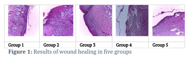
Surface area of wounds among all the study groups was statistically significant on days 7, 14 and 21 (p value < 0.001). The maximum area was observed in group 2 (open wound with methicillin-resistant infection) on day 14. The minimum surface area of wound in group 5 (infectious wound with chitosan-nano-zinc scaffold) was observed on day 14. The minimum surface area was seen in group 5 on day 21. The surface area between each study group and other groups was statistically significant (p value<0.001) (Figure 1).
Several gene expressions were assessed using the fold change values in the various groups, including TNF, TGF, VEGF, IL-6 and IL-1. TNF, TGF and VEGF genes exhibited undetectable levels in group M1 (normal healthy skin) and in the infected animal groups (M2-M5) on day 7, because samples were very small.
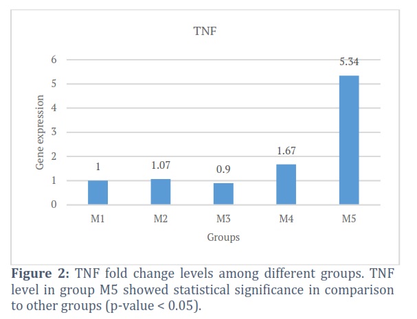
Grossly, TNF gene expression levels were different in all studied groups and controls on day 21. All fold changes showed statistical significance with p values < 0.05 of M1:1, M2:1.07, M3:0.09, M4:1.67 and M5:5.34. TGF gene expression levels were different in all studied groups and controls on day 21. All fold changes showed statistical significance with p values < 0.05 of M1:1, M2:1.18, M3:1.92, M4:2.67 and M5:7.98. VEGF gene expression levels were different in all groups studied and controls on day 21. All fold changes showed statistical significance with p values < 0.05 of M1:1, M2:1.08, M3:2.98, M4:4.07 and M5: 8.02. IL-6 gene expression levels were different in all studied groups and controls on day 21. All fold changes showed statistical significance with p values < 0.05 of M1:1, M2:1.07, M3:-0.96, M4:-1.92, and M5:-3.34 (Figure 2).
Figures & Tables
In recent years, due to their specific characteristics including low toxicity, bioavailability, penetration power, and wider distribution in various biological tissues compared to their macromolecular counterparts, metallic and metal oxide nanoparticles have attracted more attention [17].
Chitosan prevents dehydration and infection of wounds and enhances wound healing. Today, the use of nanoparticles prevents or reduces infections [18]. In this present study, we used chitosan as a vehicle for transferring zinc nanoparticles.
Bano et.al. performed chitosan and its derivatives in wound dressing in 2017 [19]. They claimed that chitosan is useful in treating injuries and burns in different forms. Due to its antimicrobial attributes, chitosan has been assessed in treating wounds. Histological studies suggested that chitosan has been effective in wound healing inducing structural renewal and epithelialization [19].
Also, we observed that the chitosan nano-zinc film group approached a more consistent formation of germinal tissue. Considering the importance of the existence of germinal tissue, we found that chitosan was a more efficient vehicle for zinc nanoparticles. And possibly, through epithelialization and formation of epithelial tissues, chitosan prevents microbial infections and can improve the wound healing process [20]. To determine the role of zinc/chitosan in skin wound healing, we first assessed the difference in wound repair between control rats and infected rats with a zinc/chitosan bandage, using full-thickness excisional wounds introduced to the dorsal skin of each group. The basic steps of skin repair include local cell accumulation, cytokine secretion, inflammatory or pro-inflammatory factors invasion [4]. As cytokines are candidate mediators of repair and are highly informative [5]. This study showed that wound healing was significantly different in treated animal groups and control groups. Molecular detection of cytokine may have potential value in detecting wound healing process [6]. In normal skin tissues, IL-6, VEGF, TNF and TGF are absent or expressed at very low levels [7]. The expression levels of IL-6 in the chitosan/zinc nanoparticle treated wound area decreased during the healing process compared to controls at day 21. Because inflammation decreases at day 21. Also, our findings indicate that VEGF, TNF and TGF mRNA levels are higher in experimental samples compared to controls at day 21. These genes play their roles in wound healing in different ways by affecting different cells in the wound region. Based on these results, various growth factors, such as vascular endothelial growth factor (VEGF), transforming growth factor (TGF), have increased in the wounds of experimental group M5. Fast wound healing process is observed in infected animals with zinc/chitosan scaffolds. This vascular proliferation was predominant in group M5 than other groups, and it was weak in group M1 at day 21. The high expression of cytokine leads to the accumulation of fibroblast in skin cells. VEGF is a marker for endothelial cells during the process of neovascularization and capillaries formation in the proliferative phase. In angiogenesis, VEGF expression at the site of injury accelerates the wound healing process. VEGF induces angiogenesis at the wound area. VEGF expression was increased 8.02-fold compared to the other groups. TNF levels are dramatically higher in group M5 than in other groups. The TGF expression level increases during the repair process. TGF is a very important cytokine in the wound healing process, which is produced by repair cells and enters the wound space. In fact, by increasing the expression of TNF and TGF gene levels (5.34 and 7.98 folds, respectively) compared to other groups, fibroblasts accumulate in wound area and produce and secrete extracellular matrix components. Increased expression of TNF gene causes differentiation of other cells into fibroblasts [8]. These findings are consistent with the results of Zhang et al. (2004) [9]. FGF, VEGF and TGF, as the potent angiogenic factors, are produced by mast cells (MCs)[10]. MCs have critical influence on neovascularization regulating homeostasis and wound-healing processes in the neoangiogenic region. It means that combination of zinc nanoparticles with chitosan scaffolds may induce an adaptive response by stimulating the cellular system. Medicinal plants are natural and useful medicinal substances [21, 22]. The use of nanoparticles as medicine in the treatment of diseases is recommended [23-28].
Thus, chitosan/zinc nanoparticles have been identified as an important player in wound physiological healing in methicillin-resistant Staphylococcus aureus-infected animal and may facilitate therapeutic strategies not only for animal but also for other chronic wounds in humans. Our studies also show that the type of zinc nanoparticles with chitosan scaffolds have more effects than other types of compounds in wound healing.
Author Contributions
F Mansouri: Conceptualization, Investigation, Writing – Review & Editing English, L Zarei: Methodology, Validation and Investigation.
All authors have no conflict of interests or personal relationships.
Acknowledgment
This study was financially supported by Lorestan University of Medical Sciences. The research project was approved by the ethics committee of the Lorestan University of Medical Sciences, IRAN with the number of IR.LUMS.REC.1401.067.
![]()
References
- Srinivas Reddy B, Kiran Kumar Reddy R, Naidu VG, Madhusudhana K, Agwane SB, Ramakrishna S, et al. Evaluation of antimicrobial, antioxidant and wound-healing potentials of Holoptelea integrifolia. J Ethnopharmacol, (2008);115(2):249-56.
- Khan HA, Ahmad A, Mehboob R. Nosocomial infections and their control strategies. Asian Pac J Trop Biomed, (2015);5(7):509-14.
- Calfee DP. Methicillin-resistant Staphylococcus aureus and vancomycin-resistant enterococci, and other Gram-positives in healthcare. Curr Opin Infect Dis( 2012);25(4):385-94.
- Gould IM, David MZ, Esposito S, Garau J, Lina G, Mazzei T, et al. New insights into meticillinresistant Staphylococcus aureus (MRSA) pathogenesis, treatment and resistance. Int J Antimicrob Agents,( 2012);39(2):96-104.
- 5 Klevens RM, Morrison MA, NadlJ, Petit S, Gershman K, Ray S, et al. Invasive methicillin-resistant Staphylococcus aureus infections in the United States. JAMA. (2007);298(15):1763-71
- Fischbach MA, Walsh CT. Antibiotics for emerging pathogens. Science. (2009);325(5944):1089-93.
- Braine T. Race against time to develop new antibiotics. Bull World Health Organ. (2011);89(2):88-9.
- Donohue, J.M., et al., Zinc Chloride Health Advisory. 1992, ENVIRONMENTAL PROTECTION AGENCY WASHINGTON DC OFFICE OF WATER.
- Roopan, S.M., et al., Biosynthetic trends and future aspects of bimetallic nanoparticles and its medicinal applications. Applied microbiology and biotechnology, (2014); 98(12): p. 5289-5300.
- Oyarzun-Ampuero F, Vidal A, Concha M, Morales J, Orellana S, Moreno-Villoslada I. Nanoparticles for the Treatment of Wounds. Curr Pharm Des, (2015);21(29): 4329-4341.
- Jayasuriya AC, Aryeai A, Jayatissa AH. ZnO nanoparticles induced effects on nanomechanical behavior and cell viability of chitosan films. Mater Sci Eng: C. (2013); 33(7): 3996-3688:
- 12.Saper RB, Rash R. Zinc: An essential micronutrient. Am Fam Physician, (2009); 79(9): 768-772.
- Kumar PT, Lakshmanan VK, Anilkumar TV, et al. Flexible and microporous chitosan hydrogel/nano-ZnO composite bandages for wound dressing: in vitro and in vivo evaluation. ACS Appl Mater Interfaces, (2012);4(5): 2618-2629
- Abbaszadeh, A., et al., Effects of Chitosan/Nano Selenium Biofilm on Infected Wound Healing in Rats; An Experimental Study. Bulletin of Emergency & Trauma, (2019); 7(3): p. 284
- Lakshmi SJ, RS RB, Sharanagouda H, Ramachandra CT, Nadagouda S, Nidoni U. Effect of biosynthesized zinc oxide nanoparticles coating on quality parameters of fig (Ficus carica L.) fruit. Journal of Pharmacognosy and Phytochemistry, (2018);7(3):10-4.
- Golbui Daghdari, S., et al., The effect of ZnO nanoparticles on bacterial load of experimental infectious wounds contaminated with Staphylococcus aureus in mice. Nanomedicine Journal, (2017); 4(4): p. 232-236.
- Jain, P.K., et al., Noble metals on the nanoscale: optical and photothermal properties and some applications in imaging, sensing, biology, and medicine. Accounts of chemical research, (2008); 41(12): 1578-1586
- Miller, C., et al., Gaseous nitric oxide bactericidal activity retained during intermittent high-dose short duration exposure. Nitric oxide, (2009); 20(1):16-23.
- Bano, I., et al., Chitosan: A potential biopolymer for wound management. International journal of biological macromolecules, (2017); 102: 380-383.
- Mirastschijski, U., et al., Zinc, copper, and selenium tissue levels and their relation to subcutaneous abscess, minor surgery, and wound healing in humans. Biological trace element research, (2013); 153(1): 76-83.
- Uroko R I, Aaron C F, Uche M E, Aguwamba C, Ogwo E U, Nweje-Anyalowu P C et al . Effect of Aju Mbaise on sperm morphology, semen quality, sex hormonal levels, gonadosomatic index and testicular histology of Avodart-induced rats. Plant Biotechnol Persa, (2022); 4 (2) :22-36
- AL- Ethawi MB, AL-Taae HH. First record at molecular level for Rhizoctonia solani causing Rot Root on Aleo vera plants in Iraq. Caspian Journal of Environmental Sciences, (2022); 1-11. doi: 10.22124/cjes.2022.5811
- Falih BT, Mohammed ST, Mohammed NJ. Effects of the silver nanoparticle synthesis from the leaves of the Capparis spinosa plant on the liver of mice infected with visceral leishmaniasis. Caspian Journal of Environmental Sciences, (2022); 20(4): 785-791.
- Bahmani M. A New Method for Promoting Biologic Synthesis and Reducing the Size of Titanium Dioxide Nanoparticles (Tio2 NPs) Synthesized by Origanum Vulgare. Plant Biotechnol Persa, (2019); 1 (1) :10-12
- Bozorgpanah Kharat Z, Mohammadi Galangash M, Ghavidast A, Shirzad-Siboni M. Removal of reactive black 5 dye from aqueous solutions by Fe3O4@SiO2-APTES nanoparticles. Caspian Journal of Environmental Sciences, (2018); 16(3): 287-301. doi: 10.22124/cjes.2018.3068
- Rahmati M, Shokri S, Ahmadi M, Ahmadi M, Marvi Moghadam N, seyfi S, et al . Comparison of Pesticide Effect of Copper Oxide Nanoparticles Synthesized by Green Chemistry and Plant Extracts on Anopheles Stephensi Mosquitoes. Plant Biotechnol Persa, (2022); 4 (1):89-96.
- Johari S, Sourinejad I, Asghari S, Bärsch N. Toxicity comparison of silver nanoparticles synthesized by physical and chemical methods to tadpole (Rana ridibunda). Caspian Journal of Environmental Sciences, (2015); 13(4): 383-390.
This work is licensed under a Creative Commons Attribution-Non Commercial 4.0 International License. To read the copy of this license please visit: https://creativecommons.org/licenses/by-nc/4.0