

Review Article
Negative staining: a forgotten technique in microbiology
Entesar Ahmed Alazazi1, Sarfraz Ahmed2, Mogana Das Murtey1*
Adv. life sci., vol. 10, no. 3, pp. 309-313, September 2023
*– Corresponding Author: Mogana Das Murtey (moganadasmurtey@usm.my)
Authors' Affiliations
2. Department of basic sciences, KBCMA College Of Veterinary and Animal Sciences, University of Veterinary and Animal Sciences, Lahore, Narowal – Pakistan
[Date Received: 08/11/2022; Date Revised: 10/06/2023; Date Published Online: 30/09/2023; Date Updated: 06/09/2025]
Abstract![]()
Introduction
Methods
Discussion
Conclusion
References
Abstract
Keywords: Negative staining; Electron microscopy; Microbiology; bacteria; Microscopy
Introduction![]()
The term “negative staining” first introduced by Brenner and Horne (1959) because this technique makes the cells appear lighter than the background. Since its introduction in the 1950s, the negative staining technique had been widely accepted and applied for the analysis of various substances; comprising microorganisms, viruses, macromolecules, and cell structures such as membranes and organelles. Primarily, the negative staining technique is used to examine the morphological characteristics of bacteria and viruses such as the shape and size, particularly in species that are difficult to stain, for example, Spirilla (bacteria).The technique is a simple and rapid approach that enables the characterization of small structural features of biological substances in a reproducible manner. Besides, it is also recognized as a suitable method for staining cells that are too delicate to undergo heat fixation. The application of this technique in microscopy allows researchers not only to determine the essential morphological characteristics of bacteria and viruses as described earlier but also to examine other details such as the presence and configuration of flagella (unipolar, bipolar, or peritrichous) or diagnosis of a virus in particular clinical situations as well as the analysis of virus entry and assembly [1]. Thus, this staining method is proven to be useful as it only requires minimal instrumentation but produces high-quality micrographs with remarkable contrast and resolution [2]. Although there are a few challenges that researchers can face during a negative staining procedure such as the presence of artifacts due to staining and improper dehydration [3,4].
Generally, the bacterial suspensions to be visualized are supported on a carbon film-coated grid. When the negative stain solution comprising of heavy metals is applied to the bacteria, the stain is not absorbed and repelled by the bacteria due to the surface is negatively charged. The deposited metal from the staining solution blocks off some of the electron beams, resulting in a darker area of observation around the bacteria. As a result of the exclusion of the stain by the bacterial cell, it would appear brighter due to the electron beam penetration through the cell[5].
For viruses, since they are generally too small for direct observation by light microscopy, transmission electron microscopy can be an obvious choice using fast and simple methodology such as negative staining. It gives relevant information on virus ultrastructure such as shape, surface structure, and features such as appendages [3]. Negative staining allows the observations of virus particulates in suspensions by preserving the structure of the virus particle[4].
Considering the benefits of this technique, this review demonstrates the values and uses of negative staining in investigating ultrastructural details of two species of bacteria, namely, Klebsiella pneumoniae and Staphylococcus aureus as well as bacteriophage (virus); Enterobacteria T6 phage.
Methods![]()
Literature search strategy and selection criteria
Short review of the literature synthesizing the findings of the literature retrieved from searches of computerized databases, hand searches and authoritative texts.
Sample Criteria
Inclusion criteria
Data sources: PubMed, Medline, Web of Science, Scopus and scientific
computerized databases.
Exclusion criteria
Data sources: Wikipedia or unknown sources.
The information will be searched using keywords negative staining, electron microscopy, microbiology, bacteria, microscopy. The article will be reviewed if it is related to the research title.
Research Tools
- Computerized databases
- Hand searches
- Authoritative texts
Method/Design
Negative staining for viruses and bacteria
This technique involves the use of a drop of the organism culture, which is examined by placing it onto a carbon film-coated, 400 mesh copper grid that is held with self-locking fine forceps. After allowing the particles or cells to attach to the film for between one to three minutes, the droplet is dried using filter paper. The grid is then left alone for another minute. Next, a drop of methylamine tungstate is dropped onto the surface of the grid. After one minute, the stain droplet is wicked to dry again with filter paper. Finally, the grid is placed in a filter paper-lined petri dish until it can be observed using TEM [6,7].
Negative staining for powdered (particulate) samples
The technique for powdered (particulate) samples involves preparing a particulate suspension using solvents like water, ethanol, acetone, or isopropyl alcohol. A drop of the prepared suspension is subsequently transferred onto a carbon film-coated, 400 mesh copper grid and allowed to be adsorbed for one to three minutes. Ideally, the drop should be pipetted out after the larger particles in the suspension have been sedimented to the bottom of the sample tube. The timing for adsorption to the film is dependent on the thickness of the suspension, thus it varies with specimens. After one minute, the stain droplet is wicked to dry again with filter paper. Subsequently, the grid is placed in a filter paper-lined petri dish until further electron microscopic examination [6,7].
Figures & Tables
Principle of Negative Staining in Microscopy
The negative staining technique involves the use of an acidic stain, for example, methylamine tungstate, uranyl acetate, phosphotungstic acid, or phosphomolybdic acid. Generally, an acidic stain readily donates a hydrogen ion (proton) resulting in the chromophore of the dye becoming negatively charged. As the cell surface of the majority of bacterial cells is negatively charged, therefore, it would repel and block the penetration of the negatively charged chromogen of the acidic stain. This results in the unstained bacterial cells being easily distinguished against the darker background.[6,7].
The practical application of this technique in microscopy has several benefits. Firstly, the stain helps to preserve the natural size and shape of the examined bacteria, as heat fixation is not required in this technique; resulting in minimal distortion effects caused by heat and chemicals. Secondly, it is particularly advantageous for the successful identification of bacteria that are difficult to stain, such as Spirilla. On the contrary, because heat fixation is not performed during the staining process, the organisms are still viable. Therefore, the laboratorian must practice caution and take necessary safety measures when handling the slides [6,7].
This technique involves the direct examination of the particulate or colloidal components using a specimen support grid specifically designed for Transmission Electron Microscope (TEM) after the cells have been embedded in an electron-dense ‘stain’. Besides, it relies on the production of an outline of the cell structure by the metallic stain, rather than reacting positively with the cell, thus, an overall detail on the size, shape, and flagella structure of the bacteria can be obtained. As the negative-staining electron microscopy technique is also quick and easier to conduct, the results of the analysis will be available within several minutes. Therefore, it is considered to be the most useful approach in electron microscopy technique, particularly, in terms of the number of samples that can be diagnosed. Further, in this technique, the particles of a suspension which are adsorbed onto the surface of the specimen support, are stabilized and contrasted with heavy metal stains, thus, enabling the visualization and categorization of the morphology of the cells at sub-nanometre size [6,7].
Additionally, due to the user-friendliness and relatively high throughput of this staining technique, it is routinely applied for quality assurance in microscopy techniques, for example, in examining virus cultures. This method allows the transfer of numerous virus particles into a suspension to preserve the morphological structure of the virus which is usually damaged by techniques such as freeze-thawing or by grinding in a potter. Moreover, the efficacy of sample preparation speed is also a vital criterion in diagnostic electron microscopy. Thus, it is inevitable to state that the combination of the negative-staining technique with electron microscopy examination serves as a first-line method in this field [8].
Concerning the microscopic techniques used, the ‘open view’ of electron microscopy offers first-hand information on all of the nanoparticles found in a sample. For example, virus particles are recognized based on various morphological characteristics, such as shape, size, surface structure, and other distinct features, for instance, the presence of appendages. Most often, the diagnosis from the identification process only leads to a systematic group of organisms rather than to a specific virus. However, because the structural characteristics of a virus are relatively constant during its development, identification and diagnosis are still made possible by this technique compared to other methods. This is possible even in the worst-case scenario when the nucleic acids have been significantly altered by mutation. Additionally, in other fields such as plant pathology and veterinary, the role of diagnostic electron microscopy is more crucial, as there is a lack of availability of other diagnostic tools [9].
In terms of its practical approach, negative staining uses a whole-mount preparation method which enables all the structural characteristics of the specimen of interest to be observed as a two-dimensional projection. Further, to examine the structure of bacteria in a more detailed and comprehensive manner, especially in the cellular context, ultrathin sections must be prepared. Most often, thicker samples are subjected to ultrathin sectioning, though the sample thickness is restricted to 200–300 nm, depending mainly on the density. With this, additional information can be obtained through a two-dimensional reconstruction of the negatively stained virus preparation. Currently, the advancement in microscopy techniques, such as the introduction of the immunolabeling technique allows researchers to combine structural details along with molecular information by incorporating immunolabeling with the negative staining technique. This immuno-negative staining method can contribute to a higher diagnostic specificity besides improving one’s understanding of the molecular topography of viruses [10-12].
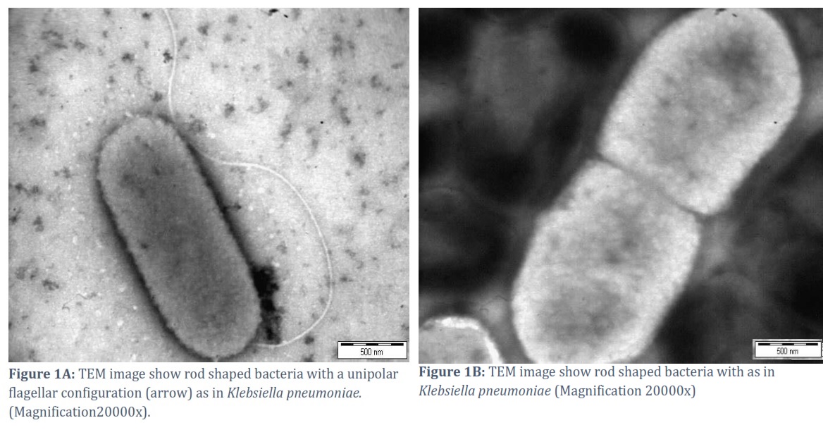
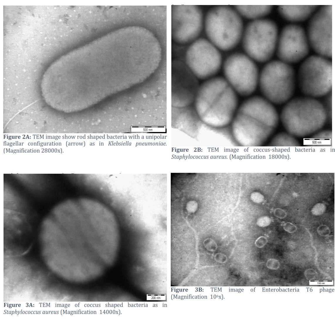
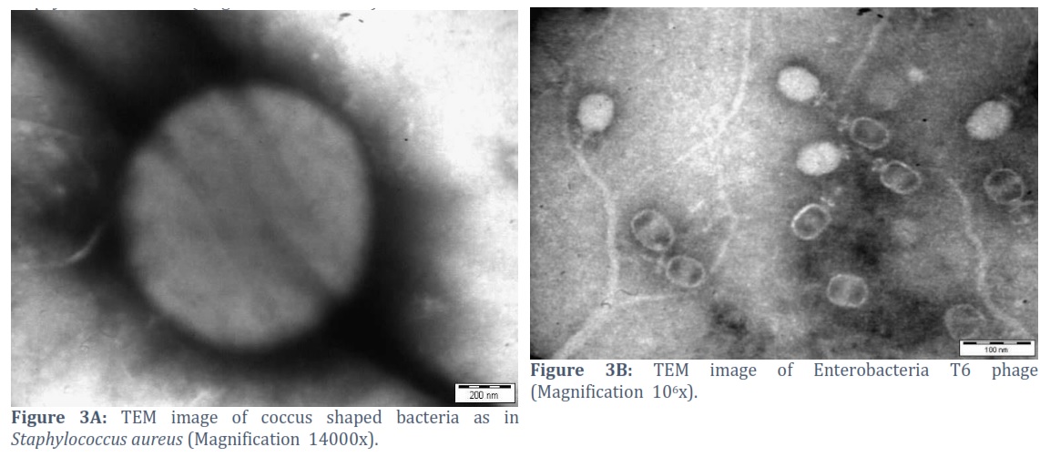
The images shown in Figures 1 to 3 are of Klebsiella pneumoniae and Staphylococcus aureus obtained from TEM examination, after being subjected to negative staining. The figures indicate great clarity and the contrast between the bacterial cells and the background is evident through this staining technique. The figures indicate great clarity and the contrast between the bacterial cells and the background is evident through this staining technique.
The negative staining technique offers several benefits, such as the utilization of a single stain and the elimination of heat fixation for the sample. By employing an acidic stain, negative staining takes advantage of the repulsion between the negative charges of the stain and the bacterial surface, preventing the dye from entering the cell. As a result, negative staining produces a distinct cell appearance against a dark backdrop, offering clear visualization [13]. As the negative staining technique is still valid and applicable to identify virus and bacteria, future studies should be undertaken to explore the use of this technique for various other microorganisms, which causes different types of diseases. Thus, it is believed that this paper will be of value to laboratorians and researchers, particularly, to provide an alternative method for the application ofthe negative staining technique using an electron microscope on various other bacteria and viruses of medical importance.
Conclusion
Negative staining is currently fast disappearing from mainstream microscopy techniques. Thus, it is important to highlight the advantages and applications of the negative staining technique among laboratorians, particularly in the current, fast-paced lab environment.
Author Contributions
MDM conceived the idea. SA designed the experiment. EAA performed the experiments. All authors contributed to the article and approved the submitted version.
![]()
The authors declare that there is no conflict of interest.
![]()
References
- Laue M.Electron microscopy of viruses. In Methods in cell biology, (2010); 96: pp. 1-20). Academic Press.
- Bremer A., Hiner M., and Aebi U. Negative staining In Cell Biology: A Laboratory Handbook (J. E. Celis, ed.), 1998; Vol. 3, pp. 277-284. Academic Press, San Diego.
- Harris R, Bhella D. and Adrian M. Recent developments in negative staining for transmission electron microscopy. Microscopy and Analysis, (2006); 113: 17.
- Cizmar P. and Yuana Y. Detection and characterization of extracellular vesicles by transmission and cryo-transmission electron microscopy. In Extracellular Vesicles,2017; pp. 221-232. Humana Press, New York, NY.
- Frank J. Three-Dimensional Electron Microscopy of Macromolecular Assemblies.1996. Academic Press, San Diego.
- Hayat MA. Principles and techniques of electron microscopy: biological applications, 2000; Cambridge University Press. Cambridge, United Kingdom.
- Dykstra MJ. Biological Electron Microscopy, Theory, Techniques and Troubleshooting. Plenum Press,1992;N.Y. pp. 218-222
- Ohi M, Li Y, Cheng Y. and Walz T.Negative staining and image classification—powerful tools in modern electron microscopy. Biological procedures online, (2004); 6(1): 23-34.
- Curry A, Appleton H. and Dowsett, B. Application of transmission electron microscopy to the clinical study of viral and bacterial infections: present and future, (2006); Micron, 37(2): 91-106.
- Mast J and Demeestere L. Electron tomography of negatively stained complex viruses: application in their diagnosis. Diagnostic Pathology, (2009); 4(1): 1-7.
- Deguchi N, Jorgensen PL, Maunsbach AB. Ultrastructure of the sodium pump: Comparison of thin sectioning, negative staining, and freeze-fracture of purified, membrane-bound (Na+,K+)-ATPase. J. Cell Biolology, (1977); 75: 619-634.
- Frank J and Radermacher M. Three-dimensional reconstruction of single particles negatively stained or in vitreous ice. Ultramicroscopy, (1992); 4: 241-262.
- Moyes RB, Reynolds J, Breakwell DP. Preliminary staining of bacteria: negative stain. Current Protocols in Microbiology, (2009): 15(1): 1-8.
This work is licensed under a Creative Commons Attribution-Non Commercial 4.0 International License. To read the copy of this license please visit: https://creativecommons.org/licenses/by-nc/4.0