

Full Length Research Article
Oleanolic acid (pentacyclic triterpenes) as a potential candidate for α-glycosidase inhibition activity
Shabnam Javed1*, Iqra Javaid2, Amna Shoaib3, Shagufta Perveen3
Adv. life sci., vol. 9, no. 2, pp. 219-223, July 2022
*- Corresponding Author: Shabnam Javed (Email: shabnam.chem@pu.edu.pk)
Authors' Affiliations
2. Government Hospital Samanabad, Lahore – Pakistan
3. Department of Plant Pathology, Faculty of Agricultural Sciences, University of the Punjab, Quaid-e-Azam Campus, Lahore – Pakistan
Abstract![]()
Introduction
Methods
Results
Discussion
References
Abstract
Background: Diabetes mellitus is a common health dilemma worldwide and is characterized by hyperglycemia. Inhibition in the activity of one of the digestive tract enzymes α-glucosidase is one of the therapeutic approaches to hydrolyze carbohydrates into glucose using natural agents. Many natural compounds with α-glucosidase inhibitory activity have transpired to be secondary metabolites. Monotheca buxifolia, native to Pakistan is a major medicinal tree, which has been known for its extensive pharmacological activities.
Methods: α-glucosidase activity of ten isolated compounds (lupeol, lupeol acetate, betulin, β-sitosterol, β-amyrin, oleanolic acid, vanillic acid, protocatechuic acid, kaempferol and quercetin) from lipophilic hexane fraction of M. buxifolia (stem and leaves) was assessed against α-glucosidase enzyme using acarbose as a control.
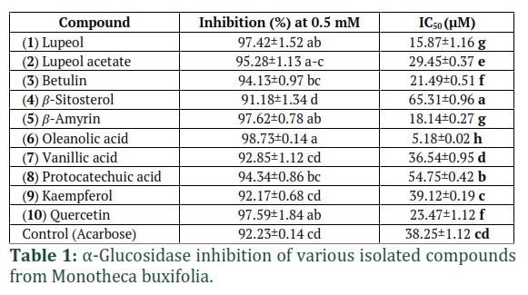
Results: All ten compounds hold α-glucosidase inhibition potential (91-99%). However, IC50 (half-maximal inhibitory concentration) values of oleanolic acid (5 µM) were 8-fold lower than that of acarbose. Moreover, inhibition potencies of lupeol (15.87 µM), β-amyrin (18.14 µM) betulin (21.49 µM), quercetin (23.47 µM), and lupeol acetate (29.45 µM) were much stronger than the inhibitory effect obtained from acarbose (38.25 µM).
Conclusion: Oleanolic acid of M. buxifolia exhibited a potent inhibitory effect against α-glucosidase, therefore, oleanolic acid may be utilized in medicinal formulations against diabetic disorders.
Keywords: Diabetes mellitus; Enzyme inhibition; Medicinal plants; Pentacyclic triterpenes
Introduction![]()
Diabetes mellitus (DM) is a chronic condition caused by hyperglycemia resulting from defects in insulin secretion and/or insulin action [1], hence leading to dysfunctioning and failure of different organs (e.g. eyes, kidneys, nerves, heart, and blood vessels) along with the development of macro- and microvascular diseases [2]. In recent times, DM is ranked as the leading cause of death [3], while its prevalence has risen from 4.7 to 8.5% from 2014 to 2019 [4] and is expected to target 5.4% of the world’s population by 2025 [5]. An inexorable rise in DM has decreased the quality of life despite the promises of a wide range of antidiabetic drugs [6]. Alternatively, the α-glucosidase inhibition may be a promising aspect to reduce the postprandial blood glucose level. The enzyme α-glucosidase secreted from intestinal chorionic epithelium help in the reduction of glycosylated hemoglobin and postprandial insulin levels by delaying carbohydrate absorption [7]. The α-glucosidase gained importance as a new class of antidiabetic drug in 1980, later it has been endorsed as the first line of treatment for lowering postprandial hyperglycemia by the Third Asia-Pacific Region Diabetes Treatment Guidelines [8].
Medicinal plants have many polyphenolic compounds that aid in prevention of fast breakdown of sugar, thereby controlling the blood sugar level by competitively inhibiting α-glucosidase activity [9]. Several natural α-glucosidase inhibitors e.g. acarbose and voglibose have been used clinically against DM, while only a limited are available commercially [10]. Therefore, more natural sources of α-glucosidase inhibitor need to be explored.
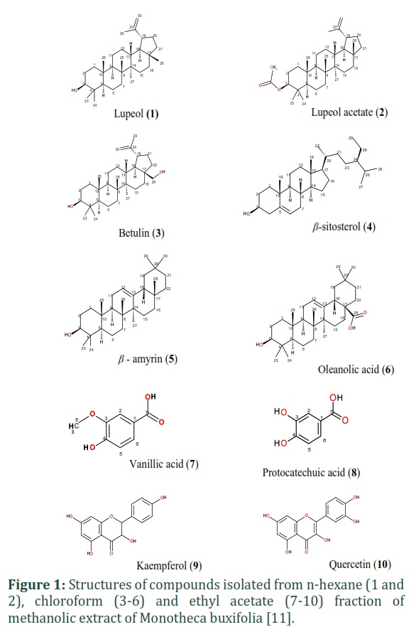
Monotheca buxifolia (Falc.) A. DC. of the family Sapotaceae is native to Pakistan and is locally known as Gwargwara is found in barren hilly areas. The plant is well-known for its medicinal values including antioxidant, digestive, hematinic, laxative, purgative, vermicidal, anthelmintic, antipyretic, etc. in South-Asia and Middle-East [11-13]. Its leaves and stem are enriched with anticancerous and antidiabetic compounds including flavonoids, phenolics, and terpenoids derivatives [11, 12]. In earlier studies, prominent α-glucosidase inhibitory activity has been documented in M. buxifolia extracts due to the presence of many important bioactive compounds and antioxidant activity [9]. Likewise, many plant-based compounds like betulinic acid, quercetin, quercitrin, α-amyrin have shown antioxidant, antiinflammatory and antidiabetic activity [14]. Znag et al., [15] reported 35.6 μM as IC50 value of oleanolic acid. Nguyen et al., [16], found IC50 values of 17 and 35 µM values for lupeol, and betulinic acid derivatives, respectively. Recently, Phan et al., [17] also explored substantial α-glucosidase inhibitory and cytotoxic activities (IC50: 20-29 µM) of lupeol derivatives containing a benzylidene chain. The present study is an extension of a previous study conducted by Javed et al., [11] where 10 compounds (viz., lupeol, lupeol acetate, betulin, β-sitosterol, β-amyrin, oleanolic acid, vanillic acid, protocatechuic acid, kaempferol, and quercetin) isolated from lipophilic hexane fraction of M. buxifolia were assessed for their antifungal activity. In the current study, all these 10 compounds were analyzed for their α-glucosidase activity.
Methods![]()
Briefly, the lipophilic hexane fraction of aerial parts (stem and leaves) of M. buxifolia was subjected to vacuum liquid chromatography over silica gel and eluted with increasing order of solvent polarity as hexane-EtOAc (0 → 10) to isolate lupeol (1) and lupeol acetate (2). The three main sub-fractions of chloroform resulted in the purification of botulin (3), botulin (4), β-amyrin (5), and oleanolic acid (6). The EtOAc fraction was chromatographed using silica gel and eluted with a solvent system of increasing polarity n-hexane, n-hexane:DCM and DCM:MeOH to attain vanillic acid (7), protocatechuic acid (8), kaempferol (9), and quercetin (10). The structures of all isolated compounds are presented in Fig. 1 [11].
The isolated compounds (lupeol, lupeol acetate, betulin, β-sitosterol, β-amyrin, oleanolic acid, vanillic acid, protocatechuic acid, kaempferol, and quercetin) were screened against α-glucosidase enzyme by a modified method described earlier [18]. Briefly, a mixture of 70 μL phosphate buffer (50 mM; pH 6.8) was mixed thoroughly with 10 μL test compound (dissolve in DMSO) and α-glucosidase enzyme (0.0234 units, 10 μL) in a 96-well plate and incubation at 37 ºC for 10 min. After incubation, each well was further added with 10 μL of 0.5 mM pNPG (p-nitrophenyl glucopyranoside). The sample-loaded 96-well plate was incubated again at 37 ºC for 10 min. Finally, 80 μL of Na2CO3 solution was added to terminate the reaction. The control and blank were prepared by adding DMSO, while acarbose was taken as a positive control. The absorbance of the samples was determined at 405 nm using a microplate reader (Synergy HT BioTek, USA). The α-glucosidase inhibitory activity was expressed as the IC50 according to the percentage inhibition.
Results
![]()
The α-glucosidase inhibitory effects of all 10 compounds and their IC50 values are presented in Table 1. All compounds exhibited substantial α-glucosidase inhibition capabilities. However, four compounds viz., oleanolic acid, β-amyrin, quercetin and lupeol exhibited significantly greater (≥ 97%) inhibitory potential as compared to acarbose. The inhibitory potential of remaining 6 compounds viz., lupeol acetate, betulin, β-sitosterol, vanillic acid, protocatechuic acid, and kaempferol was recorded in the range of 91-95%, which was statistically similar to acarbose. These compounds also exhibited variable IC50 values as compared to the control. β-sitosterol, and protocatechuic acid showed significantly greater inhibitor affinity (IC50) values of 65.31 and 54.75 µM as compared to acarbose (38.25 µM). However, IC50 values of vanillic acid and kaempferol were insignificantly different than acarbose. Moreover, the IC50 values of oleanolic acid (5 µM) followed by lupeol (15.87 µM), β-amyrin (18.14 µM) betulin (21.49 µM), quercetin (23.47 µM), and lupeol acetate (29.45 µM) were significantly lower than that of acarbose, especially oleanolic acid, with IC50value being 8-fold lower than that of acarbose.
Figures & Tables
Discussion![]()
Betulinic acid is also a lupane-type triterpenoid abundant in plants, and it has been found to be antidiabetic, anti-malarial, anti-tumor, anti-retroviral, anti-inflammatory, anti-cancer and anti-obesity activity. Betulinic acid has been recommended to be developed into an antidiabetic drug like metformin, which has both anticancer and antidiabetic properties [29]. Quercetin is plant-based polyphenol and has been regarded as a powerful antioxidant, anti-cancer, and anti-inflammatory agent, which is also active against neurodegenerative and cardiovascular diseases. The antidiabetic action of quercetin may occur through lipid peroxidation, inhibition in absorption of intestinal glucose, insulin secretion, improved glucose uptake and inhibition of α-glucosidase [30].
The gram-negative bacteria were the major cause of infections in the ICU. The commonest isolates were S.aureus, Acinetobacter, Enterobacteriaceae, and Pseudomonas Aeruginosa. The best empirical therapy should include vancomycin, Tigecycline, cefepime, and polymyxin B can be used as first-line drugs with carbapenems as second-line agents. The high frequency of multidrug resistance bacteria in ICU suggests that we need to prescribe broad-spectrum antibiotics more wisely to reduce pressure on sensitive strains. This could be beneficial for saving ICU patients and preventing the spread of resistant isolates in critical wards.
Author Contributions
The author declares that there is no conflict of interest regarding the publication of this paper.
References
- Riddle MC. Standards of medical care in diabetes. Standards of medical care in diabetes. American Diabetes Association, (2019); 42: S1-S193.
- Papatheodorou K, Banach M, Bekiari E, Rizzo M, Edmond M. Complications of diabetes. Journal of Diabetes Research, (2017); Article ID 3086167.
- Zimmet PZ, Magliano DJ, Herman WH, Shaw JE. Diabetes: a 21st century challenge. The lancet Diabetes & Endocrinology, (2014); 2: 56-64.
- Konda YP, Egi JY, Dasari S, Katepogu R, Jaiswal KK, et al. Ameliorative effects of Mentha aquatica on diabetic and nephroprotective potential activities in STZ-induced renal injury. Comparative Clinical Pathology, (2020); 29(1): 189-99.
- ReMukesh R, Namita P. Medicinal plants with antidiabetic potential-A review. American-Eurasian Journal of Agricultural & Environmental Sciences, (2017); 13: 9481-94.
- Ayele, AG, Kumar P, Engidawork E. Antihyperglycemic and hypoglycemic activities of the aqueous leaf extract of Rubus Erlangeri Engl (Rosacea) in mice. Metabolism Open, (2021); 11: Article ID 100118.
- Van de Laar FA, Lucassen PL, Akkermans RP, Van de Lisdonk EH, De Grauw WJ. Alpha-glucosidase inhibitors for people with impaired glucose tolerance or impaired fasting blood glucose. Cochrane Database of Systematic Reviews, (2006); 18: CD005061.
- Li T, Zhang XD, Song YW. A microplate-based screening method for alpha-glucosidase inhibitors. The Chinese Journal of Clinical Pharmacology, (2005); 10: 1128-1134.
- Ali JS, Saleem H, Mannan A, Zengin G, Mahomoodally MF, et al. Metabolic fingerprinting, antioxidant characterization, and enzyme-inhibitory response of Monotheca buxifolia (Falc.) A. DC. extracts. BMC Complementary Medicine and Therapies, (2020); 20(1): 313.
- Yin Z, Zhang W, Feng F, Zhang Y, Kang W. α-Glucosidase inhibitors isolated from medicinal plants. Food Science and Human Wellness, (2014); 3: 136-74.
- Javed S, Mahmood Z, Khan KM, Sarker SD, Javaid A, et al. Lupeol acetate as a potent antifungal compound against opportunistic human and phytopathogenic mold Macrophomina phaseolina. Scientific Reports, (2021); 11, Article number: 8417.
- Javed S, Shoaib A, Mehmood Z, Nawaz S, Khan KM. Phytochemical, pharmacological and GC-MS characterization of the lipophilic fraction of Monotheca buxifolia. Asian Journal of Agriculture and Biology, (2021); 3: 1-7
- Javed S, Shoaib A, Mehmood Z, Nawaz S. Hepatoprotective effect of methanolic extract of Monotheca buxifolia against isoniazid and rifampicin induced hepatotoxicity. Asian Journal of Agriculture and Biology, (2021); 4: 1-6.
- Mazura P, Susanti D, Rasadah MA. Antiinflammatory action of components from Melastoma malabathricum. Pharmaceutical Biology, (2007); 45(5): 372-375.
- Zhang BW, Xing Y, Wen C, Yu XX, Sun WL, et al. Pentacyclic triterpenes as α-glucosidase and α-amylase inhibitors: structure-activity relationships and the synergism with acarbose. Bioorganic & Medicinal Chemistry Letters, (2017); 27: 5065-70.
- Nguyen NH, Pham DD, Le TT, Nguyen TA, Huynh DL, et al. Synthesis and α-Glucosidase inhibitory activity of ursolic acid, lupeol, and betulinic acid derivatives. Chemistry of Natural Compounds, (2021); 57: 1038-41.
- Phan HV, Duong TH, Pham DD, Pham HA, Nguyen VK, et al. Design and synthesis of new lupeol derivatives and their α-glucosidase inhibitory and cytotoxic activities. Natural Product Research, (2022); 36(1): 1-7.
- Liu S, Yu Z, Zhu H, Zhang W, Chen Y. In vitro α-glucosidase inhibitory activity of isolated fractions from water extract of Qingzhuan dark tea. BMC Complementary and Alternative Medicine, (2016); 16: 378.
- Bhatia A, Singh B, Arora R, Arora S. In vitro evaluation of the α-glucosidase inhibitory potential of methanolic extracts of traditionally used antidiabetic plants. BMC Complementary and Alternative Medicine, (2019); 9: 1-9.
- Thengyai S, Thiantongin P, Sontimuang C, Ovatlarnporn C, Puttarak P. α-Glucosidase and α-amylase inhibitory activities of medicinal plants in Thai antidiabetic recipes and bioactive compounds from Vitex glabrata R. Br. stem bark. Journal of Herbal Medicine, (2020); 19: 100302.
- Lee D, Park JY, Lee S, Kang KS. In vitro studies to assess the α-glucosidase inhibitory activity and insulin secretion effect of isorhamnetin 3-o-glucoside and quercetin 3-o-glucoside isolated from Salicornia herbacea. Processes, (2021); 9: 483.
- Deng XY, Ke JJ, Zheng YY, Li DL, Zhang K, et al. Synthesis and bioactivities evaluation of oleanolic acid oxime ester derivatives as α-glucosidase and α-amylase inhibitors. Journal of Enzyme Inhibition and Medicinal Chemistry, (2022); 37: 451-461.
- Dubey VK, Patil CR, Kamble SM, Tidke PS, Patil KR, et al. Oleanolic acid prevents progression of streptozotocin induced diabetic nephropathy and protects renal microstructures in Sprague Dawley rats. Journal of Pharmacology and Pharmacotherapeutics, (2013); 4(1): 47.
- Gao D, Li Q, Li Y, Liu Z, Fan Y, Han Z, et al. Antidiabetic potential of oleanolic acid from Ligustrum lucidum Ait. Canadian Journal of Physiology and Pharmacology, (2007); 85: 1076-83.
- Teodoro T, Zhang L, Alexander T, Yue J, Vranic M, et al. Oleanolic acid enhances insulin secretion in pancreatic β-cells. FEBS Letters, (2008); 582: 1375-80.
- Malik A, Jamil U, Butt TT, Waquar S, Gan SH, et al. In silico and in vitro studies of lupeol and iso-orientin as potential antidiabetic agents in a rat model. Drug Design, Development and Therapy, (2019); 13: 1501-1513.
- Lakshmi V, Mahdi AA, Ahmad MK, Agarwal SK, Srivastava AK. Antidiabetic activity of lupeol and lupeol esters in streptozotocin-induced diabetic rats. Bangladesh Pharmaceutical Journal, (2014); 17: 138-46.
- Beserra FP, Vieira AJ, Gushiken LF, de Souza EO, Hussni MF, et al. Lupeol, a dietary triterpene, enhances wound healing in streptozotocin-induced hyperglycemic rats with modulatory effects on inflammation, oxidative stress, and angiogenesis. Oxidative Medicine and Cellular longevity, 2019; Article ID 3182627.
- Song TJ, Park CH, In KR, Kim JB, Kim JH, et al. Antidiabetic effects of betulinic acid mediated by the activation of the AMP-activated protein kinase pathway. PLoS One, (2021); 16: e0249109.
- Eid MH, Haddad S P. The antidiabetic potential of quercetin: underlying mechanisms. Current Medicinal Chemistry, (2017); 1: 24: 355-64.
This work is licensed under a Creative Commons Attribution-Non Commercial 4.0 International License. To read the copy of this license please visit: https://creativecommons.org/licenses/by-nc/4.0

