

Full Length Research Article
Construction and characterization of the ex-situ modified macroporous bacterial cellulose scaffold as a potential epidermal graft
Aylin Basaran Eroglu1*, Gokhan Coral1, Gulsen Bayrak2, Sakir Necat Yılmaz2
Adv. life sci., vol. 10, no. 3, pp. 350-355, September 2023
*– Corresponding Author: Aylin Basaran Eroglu (a.basaran.eroglu@gmail.com)
Authors' Affiliations
2. Department of Histology and Embryology, Faculty of Medicine, Mersin University, Mersin – Turkey
[Date Received: 03/06/2021; Date Revised: 30/05/2023; Date Published Online: 30/09/2023; Date Updated: 06/09/2025]
Abstract![]()
Introduction
Methods
Results
Discussion
References
Abstract
Background: Skin is a 3-dimensional (3-D) tissue that mainly consists 2 layers, comprising the epidermis and dermis. Skin tissue engineering scaffolds are used commonly as 3-D analogs of the extracellular matrix (ECM) of the skin. Bacterial cellulose (BC) has great importance in skin tissue engineering because of its resemblance to ECM and its biocompatibility. The lack of 3-D microporosity and limited biodegradation capacity have restricted its application as a scaffold for skin tissue engineering. Controlled 3-D microporosity of BC via surface modification techniques are required for potential tissue engineering applications.
Methods: Freeze-drying is an ex-situ surface modification technique for making macroporous BC scaffolds (MBCSs). This study proposed a new approach to the freeze-drying method for the arrangement of the pore size of MBCSs specifically for the human keratinocyte cell line (KER-CT). Different concentrations of MBCS (0.25%, 0.50%, and 0.75%) were prepared and the KER-CT cell viability was detected via 3-(4,5-dimethylthiazol-2-yl)-5-(3-carboxymethoxyphenyl)-2-(4-sulfophenyl)-2H-tetrazolium (MTS) assay.
Result: The results of this study indicated that the KER-CT cells were able to proliferate all of the concentrations of MBCS, and the best cell viability value was observed with 0.75% MBCS. The results were supported by FESEM and light microscopic observations.
Conclusion: While the 0.75% MBCS supported the highest level of cell proliferation, further modifications may be needed to improve cell adhesion and spreading for its effective use in epidermal tissue engineering applications.
Keywords: Bacterial cellulose; Scaffold; Keratinocyte; Skin tissue engineering
Introduction![]()
Skin is the largest and most self-renewable organ of the body in mammalians, and plays many important roles in the physiology of the body [1, 2]. Skin consists of 2 main layers, which are the dermis and epidermis. The epidermis is the first barrier of the skin and it protects the subcutaneous layer against thermal, mechanical, chemical, and pathogenic agents, and ultraviolet radiation. In large full-thickness defects of the skin, the skin is not capable of renewing itself. Hence, the skin cannot heal without resulting in the formation of scar tissue [3, 4]. Under these conditions, the best approach is to apply an artificial graft to the victims of injury. Thus, to supply the best compatibility of artificial skin as soon as possible, the basic aim of skin tissue engineering is to fabricate an easily applicable skin graft. However, there are no models of artificial skin that entirely mimic normal, healthy human skin [5, 6, 7].
The widespread attempt in tissue engineering is to prepare 3-D biocompatible scaffolds in the same shape as the target tissue. An ideal 3-D tissue engineering scaffold must have a highly porous structure and good biocompatibility. High porosity and an optimally designed pore diameter provide a proper area for cell adhesion and proliferation, so that cells attached to the scaffold are able to exchange nutrients with their micro-environment. To mimic the in vivo environment of skin, many natural and synthetic materials have been frequently investigated for tissue-engineering applications [5, 8].
Bacterial cellulose (BC) is a valuable material for skin tissue engineering due to its strong biocompatibility, purity, porosity, water retention ability, and tensile strength [9, 10, 11]. Moreover, according to the study of Czaja et al., BC may be the cheapest artificial skin scaffold ever. These features may make it excellent as a tissue engineering scaffold, despite the fact that BC has 2 challenges in this respect, comprising its biodegradation time and its pore size. The biodegradation time of BC has been estimated as approximately 10 years [9]. However, in the standard approach of tissue engineering, the ideal timing for scaffold degradation is limited to only 1 year. Hence, many researchers have endeavored to shorten the biodegradation rate of BC [12, 13].
On the other hand, the nanoporous structure of BC constitutes a much bigger problem. The size difference between the pore size and the cell could be due to block cell viability and migration. For this reason, controlled 3-D microporosity via surface modification techniques is required for potential tissue engineering applications of BC. Freeze-drying is an ex-situ surface modification technique that is used for making macroporous BC scaffolds (MBCSs) [14, 15, 16]. Poly (ethylene) glycol (PEG) is a synthetic polymer that has been used to camouflage proteins from the immune elements of human body. As a result, PEG 400 administration to BC has enhanced the biocompatibility, porosity, and surface area of BC [14, 17].
A completely differentiated epidermis, which consists of a basal, spinous, granular, and cornified layer, has not yet been able to be mimicked. The culturing of keratinocyte cells is the first step that must be taken when making an artificial epidermal layer [2, 5, 7]. To generate the epidermis on 3-D MBCS with epidermal cells, the culture media has to be supplemented with various growth factors, such as the epidermal growth factor (EGF), insulin like growth factor (IGF), and keratinocyte growth factor (KGF). These requirements necessitate the cultivation of keratinocytes in a special medium. Keratinocyte growth medium-gold (KGM-Gold) is a special medium that has fulfilled these requirements. Working with cell lines has provided some advantages for researchers, such as enhanced reproducibility, independence of donor alterations, and accessibility [18]. For this reason, the human foreskin origin keratinocyte cell line (KER-CT) was used in this work.
This study proposed a new approach to conventional methods for making a cheap and biocompatible scaffold without disrupting the purity of the BC. Moreover, the most suitable pore size via alteration of the concentrations of BC for the proliferation of the human keratinocyte cell line for skin tissue engineering applications was investigated for the first time.
Methods![]()
Production of BC
Gluconacetobacter xylinus, which produces cellulose with high efficiency, was purchased from the American Type Culture Collection (ATCC 10245, ABD). The bacteria were cultured using Hestrin-Schramm broth (HSB). The HSB contained (w/v) 2% glucose, 0.5% yeast extract, 0.5% peptone, and 0.5% Na2HPO4 0.115% citric acid at a pH of 6.0 [19]. The solution was sterilized via autoclaving. Next, 5% Gluconacetobacter xylinus suspension was cultured in the HSB for 10 days at 30°C under static conditions. After the incubation, the BC film was harvested under aseptic conditions.
Preparation of MBCS samples
The MBCS samples were prepared as defined by Gao et al., with minor modifications.
First, the BC pellicle was boiled in deionized water for 2h, followed by 0.5 M NaOH for 15 min. The pellicle was stored in 1% NaOH for 2 days. Next, the pellicle was rinsed with deionized water several times until reaching the neutral pH. The BC pellicle was frozen and lyophilized at –50°C for 1 day.
The lyophilized BC pellicle was soaked in PEG 400 for 1 day and the BC pellicle was stored in deionized water to remove the PEG 400. Finally, the BC pellicle was lyophilized for another day under the same lyophilization conditions.
For molding of the BC pellicle, the lyophilized BC pellicle was blended with deionized water using a Heidolph homogenizer (Fisher Scientific, Göteborg, Sweden) at high speed for 10 min to prepare 0.25%, 0.50%, and 0.75% (w/v) BC suspensions. The BC suspension was transferred into a 24-well plate and freeze-dried under the same conditions[14, 15]. The scaffold size was 1 cm3. The scaffolds were sterilized via autoclaving.
Scaffold characterization and cell morphology observation with FESEM
The MBCS samples were coated with an ultrathin layer of platinum using a Q150 ES ion sputter (Quorum Technologies Ltd., Laughton, East Sussex, England). The surface morphology of the MBCS samples was observed under a Supra 55 field emission scanning electron microscope (FESEM; Carl Zeiss NTS GmbH, Oberkochen, Germany).
For observation of the MBCS-KER-CT complex, the samples were dehydrated by serial acetone administrations and air-dried using an Emitech K850 dryer (Quorum Technologies Ltd.) and were sputter-coated with platinum (Quorum, Q150 ES) and then the samples were observed using the FESEM.
Cell seeding and cell viability assay
Human foreskin keratinocyte cell line (KER-CT, ATCC CRL-4048) was used in this study to investigate the optimal pore size for making an artificial epidermis prototype.
First, the scaffolds were soaked with complete growth medium (KGM-Gold, Lonza) and the cell suspension, which included 106 KER-CT cells that were inoculated on each scaffold. The scaffolds were cultured for 1, 3, 5, 7 days at 37°C in a 5% CO2 incubator. For the cell viability assay, the culture medium was aspirated and the scaffolds were transferred into a 96-well plate, and then 30 µL 3-(4,5-dimethylthiazol-2-yl)-5-(3-carboxymethoxyphenyl)-2-(4-sulfophenyl)-2H tetrazolium (MTS) solution was pipetted each well (The CellTiter 96 Aqueous One Solution Cell Proliferation Assay, MTS, Promega). The plate was incubated for 1 h at 37°C in a 5% CO2 incubator. The optical density (OD) was detected at 490 nm.
Cell imaging with Light Microscope
At the end of the culturing period, the medium was removed, and the scaffolds were washed with phosphate buffered saline. Then, freshly prepared %4 paraformaldehyde was added for fixation for 48 h. Next, 20% sucrose was added to the samples for 24 h. Following this, 30% sucrose, containing sodium azide, was added and kept at 4°C until sectioning. Sections that were 10 µm thick were placed onto adhesive slides that were coated with gelatin using a Leica CM1900 cryostat (Leica Microsystems, Wetzlar, Germany). Hematoxylin-eosin (H&E) staining was then performed. The sections were observed under an Olympus BX50 light microscope with an Olympus LC30 camera attachment (Olympus Scientific Solutions, Shinjuku, Tokyo, Japan).
Statistical analysis
The MTS experiment was conducted in replicates of six, and the pore size evaluation was performed using 20 measurements. Statistical analysis was conducted using SPSS. A p-value < 0.05 was accepted as statistically significant.
Results![]()
Preparation of MBCS and its structural pore size analysis
The pore size, which supports cell growth, differentiation, and colonization in the scaffold, varies depending on the cell type. For these reasons, this study focused on determining the optimal pore diameter for human skin keratinocyte cell growth, proliferation, and colonization.
The average pore diameter was calculated according to FESEM measurements and shown as the mean pore diameter on the graph. The average pore diameter of the scaffolds decreased significantly as the Figure 3 MBCS concentration increased from 0.25% to 0.75% (Figure 1).
Fig. 1 shows the average pore size of the different BC concentrations of MBCS. The average pore sizes of the 0.50% and 0.75% MBCS were significantly different than that of the 0.25% MBCS (P ˂ 0.05). Figs. 2a–2c. show the FESEM micrographs of the 0.25%, 0.50%, and 0.75% MBCS, respectively.
Cell proliferation and MTS assay
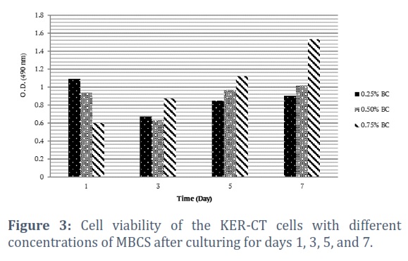
Fig.3 demonstrates the proliferation of the KER-CT cells on the MBCS on days of 1, 3, 5, and 7. The results of the MTS assay showed that the human keratinocyte cells could proliferate and grow in all of the concentrations of the MBCS. However, the 0.75% MBCS showed a significantly different increase when compared to the other concentrations of the scaffold. The number of proliferated KER-CT cells increased by approximately 2.5-fold when compared with day 1 of incubation. The MTS results of this study were supported by the FESEM and light microscopic observations in terms of cell viability, adhesion, and biocompatibility.
Observation of cell morphology on MBCS with FESEM and Light microscope
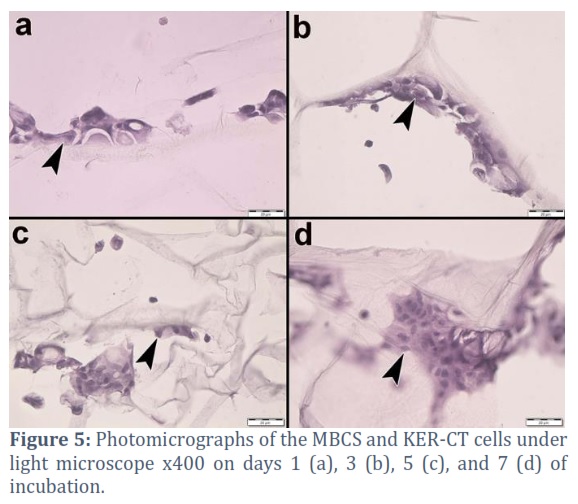
The cell morphology of the MBCS was examined via FESEM and a light microscope at the aforementioned incubation times. The results are shown in Figs. 4 and 5.
According to the FESEM and light microscope observations, the KER-CT cells attached onto the MBCS surface and micropores after day 1 of incubation, and in the progressing days of incubation, the number of colonized cells increased, as shown Fig. 5.
Figures & Tables
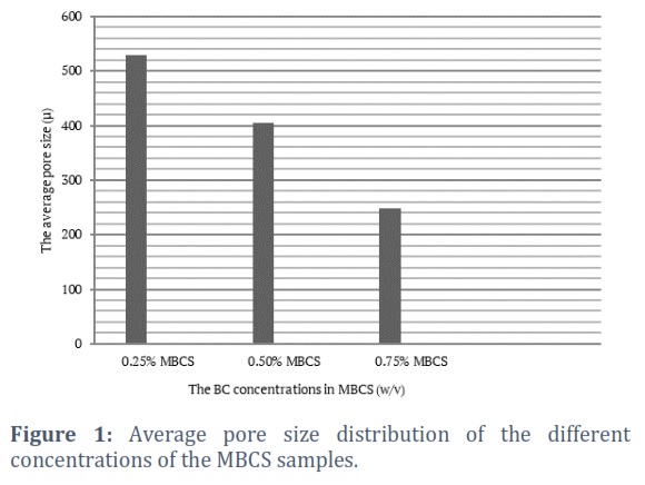
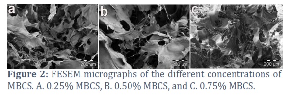
The pore diameters we obtained in the MBCS samples, was produced in this study, support the proliferation and colonization of keratinocyte cells, considering the average size of a mammalian cell. The pore size measurements via the FESEM demonstrated that the average pore sizes of the 0.50% and 0.75% MBCS were significantly different when compared to that of the 0.25% MBCS. Figs. 1 and 2 show that increasing the BC concentration in the freeze-dried MBCS samples was a simple and effective method to reduce the average pore size of the MBCS. These results align with our previous study, which also demonstrated that increasing the BC concentration is an effective method for reducing the scaffold's average pore size [20]. As shown in Fig. 1, the average pore size of the 0.25% MBCS was about 500 µm, and although the pore size formation was random, the result was in agreement with our previous study and those of Gao et al. and Xiong et al [14, 15, 20]. Our previous studies also compared that, to the 30-day in vitro degradation period of BCF and MBCS samples. The result indicated that the increased surface area and macroporous structure of MBCS can contribute to reducing the degradation process positively. However, the increased degradation rate of MBCS is still not ideal for tissue engineering applications [20].Further studies on the subject will be beneficial in terms of cumulative scientific progress.
The first report was published about keratinocyte (CRL-2309, ATCC) cell viability on unmodified BC film by Sanchavanakit et al. (2006) [21]. According to the results of their study, the keratinocytes covered the surface of an unmodified BC film and at the end of 48 h of incubation, the keratinocytes reached confluency. The phase-contrast microscopy of the study indicated that the keratinocyte cell line had good biocompatibility with the unmodified BC film. Contrary to the results of their study, the MTS results herein indicated that the confluence of the KER-CT cells was realized on day 7 of incubation. The difference in the confluence times might have been based on the dimensional difference between the BC film and the 3-D MBCS.
The previous report of Khan et al. (2018) reported much better cell viability on a microporous BC-gelatin composite scaffold with HaCaT cells. The MTT results of their study showed that the number of HaCaT cells increased by 9-fold when compared with that on day 1 of incubation [22]. The difference between their study and that herein may have arisen from the effect of the gelatin and the type of keratinocyte cell line. More detailed and comparative studies might be useful to determine the optimal keratinocyte cell line type scaffold specifically.
The experimental design, which included the FESEM observations and cell viability assay, allowed the discovery of the adhesion of the cells that held it to the surface of the scaffold and the viability capability on the scaffold surface. Moreover, the light microscopic sections allowed for an observation of the deeper layers of the MBCS. Fig. 5 demonstrates the colonization of the KER-CT cells inside the MBCS pores and on the fibers via light microscopic observations.
Freeze-drying is a traditional method that is used for making 3-D scaffolds. This study proposed a simple and applicable modification to the freeze-drying method. The method not only reduced the pore size of the BC, but also improved its biocompatibility with PEG administration.
According to the MTS assay results, the 0.75% MBCS had the most appropriate pore size for human keratinocyte cell growth and proliferation. The FESEM and light microscopic observations indicated that the concentration of 0.75% BC was the best for the human keratinocyte cell morphology and adhesion. The SEM and light microscopic observations in this study demonstrated that the KER-CT cells most often penetrated into the pores of the MBCS when compared to attaching on the surface. As a result, modified and improved 0.75% MBCS might be an epidermal graft in skin tissue engineering applications. Further and more detailed studies are suggested to determine the potential applications of MBCSs for skin tissue engineering scaffolds.
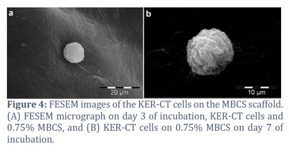
The FESEM micrographs in the current study showed that at the end of 7 days of incubation, the KER-CT cells did not exhibit the well-spread, polygonal morphology characteristic of healthy, adherent keratinocytes. The cells had a round shape and were loosely attached to the surface of the MBCS; hence, the filopodia formation was not observed (Fig. 4). On the other hand, according to the light microscopic observations, the KER-CT cells frequently attached on the inter-layer pores of the MBCS. These results were supported by the previous study of Khan et al [22]. According to the data of our study and the previous studies, MBCSs can be a good scaffold candidate for skin tissue engineering.
Conflict of Interest
The authors declare that there is no conflict of interest.
Ph.D. Aylin Basaran Eroglu, corresponding author is responsible for performing all experiments and writing the article.
Prof. Dr. Gokhan Coral, as a supervisor, he supported the creation of a microbiological and molecular biological scientific experimental structure.
Assoc. Prof. Dr. Gulsen Bayrak and Prof. Dr. Necat Yilmaz provided support on cell culture and microscopic observations.
![]()
References
- Horch RE, Kopp J, Kneser U, Beier J, Bach A D. Tissue engineering of cultured skin substitutes. Fundamentals of Tissue Engineering and Regenerative Medicine, (2009); 9: 329–343.
- Bottcher-Haberzeth S, Biedermann T, Reichmann E. Tissue Engineering of Skin. Principles of Regenerative Medicine, (2010); 36: 450–460.
- Metcalfe AD, Ferguson MWJ. Tissue engineering of replacement skin: The crossroads of biomaterials, wound healing, embryonic development, stem cells and regeneration. Journal of the Royal Society Interface, (2007); 4: 413–417.
- Bi H, Jin Y. Current progress of skin tissue engineering: Seed cells, bioscaffolds, and construction strategies. Burns & trauma, (2013); 1: 63-72
- Priya S G, Jungvid H, Kumar A. Skin tissue engineering for tissue repair and regeneration. Tissue Engineering – Part B: Reviews, (2008); 14: 105–118.
- de Oliveira Barud H G, da Silva RR, da Silva Barud H, Tercjak A, Gutierrez J, Lustri W R, de Oliveria Junior O B, Ribeiro, S. J. A multipurpose natural and renewable polymer in medical applications: Bacterial cellulose. Carbohydrate Polymers, (2016); 153: 406-420.
- Yu JR, Navarro J, Coburn J C, Mahadik B, Molnar J, et al. Current and future perspectives on skin tissue engineering: key features of biomedical research, translational assessment, and clinical application. Advanced healthcare materials, (2019); 8: 1801471.
- Groeber F, Holeiter M, Hampel M, Hinderer S, Schenke-Layland, K. Skin tissue engineering – In vivo and in vitro applications. Advanced Drug Delivery Reviews, (2011); 63: 352–366.
- Czaja WK, Young DJ, Kawecki M, Brown R M. (2007). The Future Prospects of Microbial Cellulose in Biomedical Applications, (2007); 8: 1–12.
- Fu L, Zhang J, Yang G. Present status and applications of bacterial cellulose-based materials for skin tissue repair. Carbohydrate Polymers, (2013); 92: 1432–1442.
- Jozala AF, de Lencastre-Novaes LC, Lopes AM, de Carvalho Santos-Ebinuma V, Mazzola P G, Pessoa-Jr A, Grotto D, Gerenutti M, Chaud MV. Bacterial nanocellulose production and application: a 10-year overview. Applied Microbiology and Biotechnology, (2016); 100: 2063–2072.
- Petersen N, Gatenholm P. Bacterial cellulose-based materials and medical devices: Current state and perspectives. Applied Microbiology and Biotechnology, (2011); 91: 1277–1286.
- Luo H, Xiong G, Hu D, Ren K, Yao F, Zhu Y, Gao C, Wan Y. Characterization of TEMPO-oxidized bacterial cellulose scaffolds for tissue engineering applications. Materials Chemistry and Physics, (2013); 143: 373-379.
- Gao C, Wan Y, Yang C, Dai K, Tang T, Luo H, Wang J. Preparation and characterization of bacterial cellulose sponge with hierarchical pore structure as tissue engineering scaffold. Journal of Porous Materials, (2011); 18: 139–145.
- Xiong G, Luo H, Zhu Y, Raman S, Wan Y. Creation of macropores in three-dimensional bacterial cellulose scaffold for potential cancer cell culture. Carbohydrate Polymers, (2014); 114: 553–557.
- Stumpf TR, Yang X, Zhang J, Cao X. In situ and ex situ modifications of bacterial cellulose for applications in tissue engineering. Materials Science and Engineering C, (2018); 82: 372–383.
- Cai Z, Kim J. Bacterial cellulose/poly(ethylene glycol) composite: Characterization and first evaluation of biocompatibility. Cellulose, (2010); 17: 83–91
- Tao K, Bai XZ, Zhang ZF, Shi J H, Hu XL, Tang C W, Hu DH, Han, JT. Construction of the tissue engineering seed cell (HaCaT-EGF) and analysis of its biological characteristics. Asian Pacific Journal of Tropical Medicine, (2010); 6: 893–896.
- Hestrin S, SchrammM. Synthesis of cellulose by Acetobacter xylinum and Preparation of freeze-dried cells capable of polymerizing glucose to cellulose. Biochemical Journal, (1954); 58: 345.
- Basaran Eroglu A, Coral G. Preparation and characterization of a 3-dimensional macroporous bacterial cellulose scaffold for in vitro tissue engineering applications. Digest Journal of Nanomaterials and Biostructures, (2021); 16: 1011 – 1017.
- Sanchavanakit N, Sangrungraungroj W, Kaomongkolgit R, Banaprasert T, Pavasant P, Phisalaphong M. Growth of human keratinocytes and fibroblasts on bacterial cellulose film. Biotechnology progress, (2006); 22: 1194-1199.
- Khan S, Ul-Islam M, Ikram M, Islam S U, Ullah M W, Israr M, Yang J H, Yoon S, Park J K. Preparation and structural characterization of surface modified microporous bacterial cellulose scaffolds: A potential material for skin regeneration applications in vitro and in vivo. International journal of biological macromolecules, (2018); 117: 1200-1210.
This work is licensed under a Creative Commons Attribution-Non Commercial 4.0 International License. To read the copy of this license please visit: https://creativecommons.org/licenses/by-nc/4.0