

Full Length Research Article
Predicting Leptospirosis Outcomes in Dogs with the Simplified Acute Physiology Score
Sergey Laptev*, Nikolai Pimenov, Sergej Pozyabin, Vasily Ivanyuk, Saida Marzanova, Kristina Permyakova, Marina Selina
Adv. life sci., vol. 11, no. 1, pp. 173-181, February 2024
*– Corresponding Author: Sergey Laptev (s.v.laptev@inbox.ru )
Authors' Affiliations
[Date Received: 27/07/2023; Date Revised: 27/12/2023; Date Published: 25/02/2024]
Editorial Note: You are viewing the latest version of this manuscript having minor correction in affiliation section of authors, which is different from originally published copy.
Abstract![]()
Introduction
Methods
Results
Discussion
References
Abstract
Background: Leptospirosis is a serious infectious disease affecting dogs, caused by the spirochete bacterium Leptospira. Understanding the prognosis and severity of the disease is essential for effective clinical management. This study aimed to assess the use of the PIRO and SAPS scales in predicting the outcome of leptospirosis in dogs.
Methods: The study involved 24 dogs diagnosed with leptospirosis and a control group of 22 healthy dogs. Clinical assessments were conducted, and scores on the PIRO and SAPS scales were assigned within 24 hours of admission. Statistical analyses, including correlation and regression, were employed to evaluate the relationship between scale scores and disease outcomes.
Results: In dogs with leptospirosis, SAPS scale scores exhibited a strong positive correlation with disease outcomes, indicating a robust association. Scores on the SAPS scale were associated with the severity of the disease, with specific score ranges indicative of a moderate, severe, or fatal outcome. The PIRO scale also demonstrated a substantial correlation with SAPS scores and disease prognosis. In cases where the immune system was compromised, protective mechanisms activated with a delay, increasing the risk of fatality.
Conclusion: The PIRO and SAPS scales provide valuable tools for assessing the severity and predicting the outcome of leptospirosis in dogs. These scales offer clinicians a means to promptly evaluate the risk of physiological disruptions and sepsis complications in dogs with leptospirosis, ultimately aiding in clinical decision-making and treatment planning.
Keywords: Leptospirosis; Prognosis; Physiology Score; PIRO and SAPS scales
Introduction![]()
Leptospirosis is a non-transmissible natural focal infection [1,2] that poses an epidemiological risk to both humans and animals. In dogs, leptospirosis is typically associated with representatives of the Canicola serogroup [3]. Morphologically identical leptospires have significant differences in epidemiological and epizootiological implementation [4-6]. Currently, diagnostic PCR tests have been developed to identify all known leptospira genomes [7]. Sanitary regulations are aimed at ensuring epidemiological well-being in the Russian Federation. All types of agricultural and domestic animals are vaccinated against leptospirosis in accordance with veterinary rules. As a result, veterinary services incur significant economic costs annually for the diagnosis, prevention, treatment, and containment measures related to leptospirosis [8].
The development of multiple organ dysfunction syndrome (MODS) and sepsis often arises as a result of the systemic inflammatory response syndrome (SIRS) triggered by leptospirosis infection. Neutrophilic granulocytes (NG) have a crucial function in triggering SIRS [9-12]. Leptospirosis predominantly affects purebred animals, with Dobermans and Caucasian Shepherds being particularly susceptible [13]. In these cases, MODS and hemostatic system dysfunction can lead to a fatal outcome [14-16]. To impartially evaluate and track the condition of a patient with leptospirosis, it is recommended to utilize scoring systems such as SAPS and PIRO scales [9,17,18]. The process of making decisions to predict possible complications in dogs includes assessing clinical signs of SIRS, multiple organ dysfunction syndrome (MODS), and compensatory anti-inflammatory response syndrome (CARS) with scoring systems. In the initial step of the algorithm, PIRO indicators are established [17].
SAPS is a simplified scale designed to assess physiological disturbances. This approach utilizes readily recognizable biological and clinical markers that mirror the patient's likelihood of mortality. These markers are assessed 24 hours after the patient's hospital admission, and the cumulative score helps estimate the likelihood of death.
Methods![]()
This study took place at veterinary clinics located across different regions within the Central Federal District. The data collection and initial data analysis were carried out by students from the Veterinary Academy. The research involved the evaluation of 24 dogs diagnosed with leptospirosis between 2020 and 2022, ranging in age from 8 months to 11 years. To provide a meaningful comparison, a control group of 22 healthy animals was carefully selected to match the diagnosed dogs in terms of age, gender, and breed characteristics.
All dogs included in this study were examined by veterinarians at the clinic. The target population included dogs with clinical examinations, blood profiles, biochemical records, and known outcomes, either discharged from the hospital or deceased. Only dogs with a laboratory-confirmed diagnosis were considered for inclusion in the study. At the end of the research, 21 dogs (87.5%) diagnosed with leptospirosis made a full recovery, while 3 dogs (12.5%) did not survive.
The SAPS scale is a simplified tool designed to assess physiological disturbances. This scale utilizes easily recognizable biological and clinical markers that provide an indication of the patient's risk of mortality. These markers are assessed 24 hours after the patient's admission to the hospital, and the cumulative score helps determine the likelihood of death.
The development of biomarker criteria and prognostic scales in dogs was carried out in an experimental group of animals with leptospirosis, where the risk of bacterial infections spreading was most likely according to clinical practice and veterinary research experience. In the control group, clinically healthy animals of similar weight, age, and breed were selected to match the weight, age, and breed of the diseased animals included in the experimental group. Animals in the experimental and control groups were kept under the same conditions before the onset of illness in the animals from the experimental group. All dogs in the experimental and control groups were examined by veterinarians at the clinic.
An important condition maintained during the analysis was the need to ensure comparability of indicators since only qualitatively homogeneous indicators can be compared. To achieve this, several methods of converting analytical indicators into a comparable format were used:
- Importing complex indicators for comparison into a unified structure;
- Importing comparative indicators into a unified calculation methodology;
- Replacing absolute indicators with relative ones (indicators are transformed into scores).
The use of these methods allows for the comparability of indicators within the analyzed control groups and experiments, involving animals with septic manifestations of specific types of infection or disease.
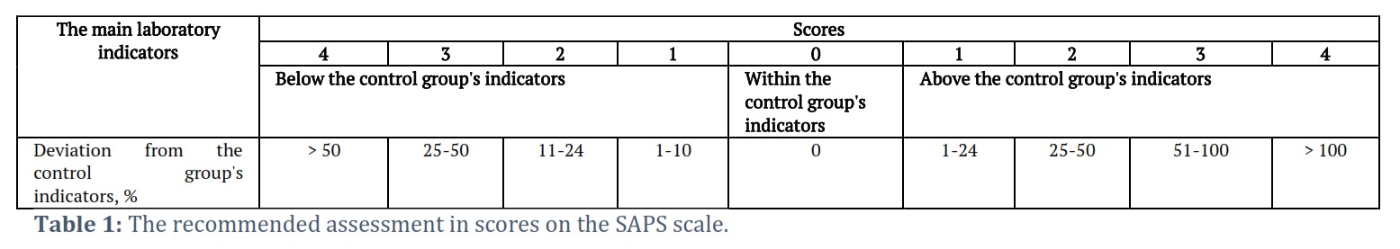
The target group includes dogs with clinical examinations, blood profiles, biochemical records, and known outcomes, either discharged from the clinic or deceased. Dogs without a laboratory-confirmed diagnosis were intentionally excluded from the study. At the end of the study, it was observed that 21 dogs (87.5%) diagnosed with leptospirosis made a successful recovery, while unfortunately, 3 dogs (12.5%) did not survive the illness. All research findings were quantified and translated into scores utilizing the Simplified Acute Physiology Score (SAPS) scale, as delineated in Table 1.
SAPS is a straightforward assessment scale utilized for evaluating physiological perturbations in patients. This scale relies on easily discernible biological and clinical parameters, effectively capturing the patient's mortality risk. These parameters are meticulously assessed precisely 24 hours after the patient's admission to the veterinary clinic. The cumulative SAPS score ascertains the patient's prognosis and likelihood of mortality.
Results![]()
Clinical and laboratory data from 24 dogs suffering from leptospirosis were thoroughly examined. These canines presented in a compromised condition, marked by elevated body temperatures within the range of 40.0 to 41.0°C. Notable clinical manifestations included petechial hemorrhaging, vomiting, diarrhea, and a discernible yellowish hue in their mucous membranes and sclera. The definitive diagnosis was confirmed through Polymerase Chain Reaction (PCR) analysis.
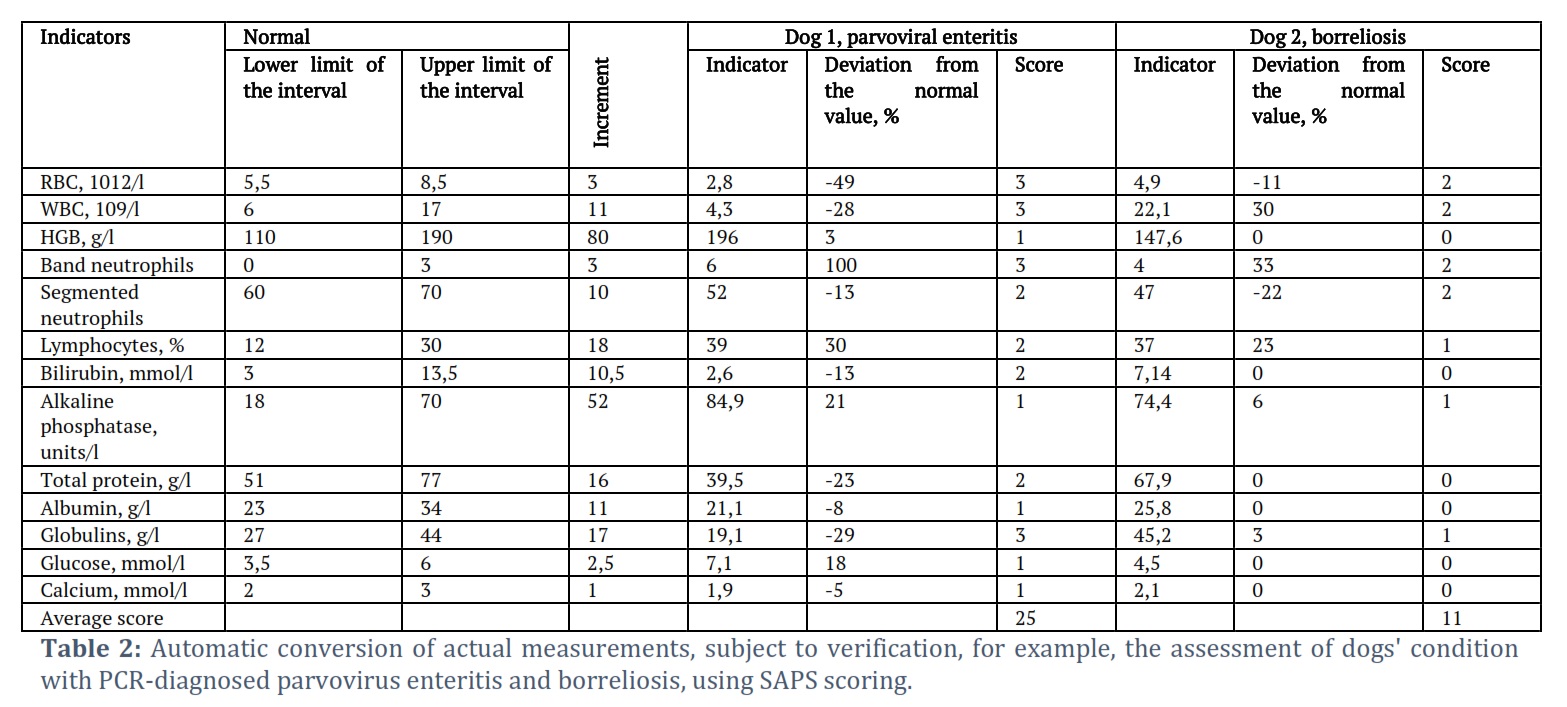
Throughout the course of the investigation, we devised an automated system for the conversion of actual measurements obtained from the subjects into SAPS scores. An illustrative demonstration of this conversion process is exemplified in Tables 2 and 3.
The level of physiological dysfunction within the animal's body is directly associated with the score it receives. A greater score corresponds to an increased susceptibility to septic complications and a higher likelihood of the animal's death.
Throughout our study, we devised a system known as the "Automated Conversion of Real-time Measurements with Verification." The health status of the animal is assessed through the SAPS scale scores. The calculation table delineates the lower and upper boundaries for each assessed reference value, with the "increment" defined as the difference between the upper and lower limits of the physiological norm. Subsequent columns calculate the percentage deviation of the parameter according to the formula, automatically determining the initial value. The degree of physiological impairment within the animal's organism is directly linked to the score obtained. A higher score indicates an increased risk of septic complications and potential fatality for the animal.
We carried out an extensive analysis of clinical and laboratory data gathered from 24 dogs diagnosed with leptospirosis. These afflicted animals presented with symptoms of depression, accompanied by elevated body temperatures ranging from 40.0 to 41.0°C. Clinical observations revealed polydipsia, anorexia, bleeding from the intradermal capillaries of the mucous membranes, as well as instances of vomiting and diarrhea. Notably, some dogs exhibited a yellowish discoloration of the mucous membranes and sclera. The definitive diagnosis was established through the application of PCR and IFA methods.
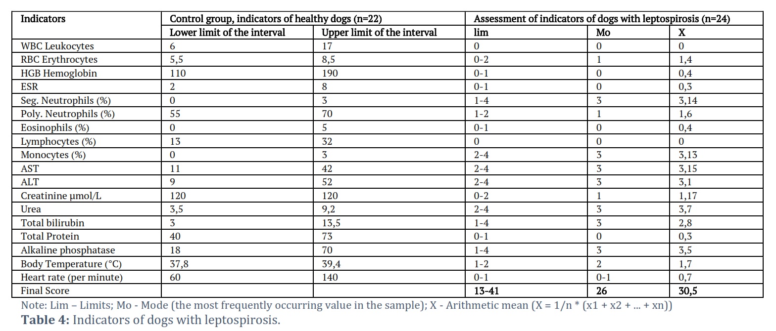
To assess the disease's severity, we performed hematological and biochemical blood analyses. Table 4 contains the SAPS scale data for dogs afflicted with leptospirosis.
In dogs with leptospirosis, complications suggestive of sepsis were associated with specific hematological changes. These changes included a mild reduction in red blood cell count, scoring between 0-2 points, and a decrease in hemoglobin levels, typically at the lower boundary of the normal range, scoring 0-1 point. These findings may indicate a gradual onset of anemia linked to the release of capillary toxins by leptospires.
Additionally, there was a slight increase in the erythrocyte sedimentation rate (0-1 point), primarily attributed to elevated levels of large-dispersed proteins. The blood also showed elevated levels of bile pigments, along with a shift in the acid-base balance towards alkalosis.
The white blood cell count remained within the normal range. Notably, there were no detectable basophils, bone marrow cells, or immature neutrophils observed in the leukocyte formula. However, there was a slight elevation in eosinophil percentages (0-1 point). Monocyte levels exhibited an increase of 2-4 points beyond the normal physiological range, while the count of rod-shaped neutrophils showed a substantial deviation from the norm, with scores ranging between 1 and 4 points. Conversely, the percentage of segmented neutrophils was reduced by 1-2 points, and the lymphocyte count remained within the normal range. It should be noted that in dogs with leptospirosis, there is liver damage, and the levels of aspartate aminotransferase (2-4 points) and alanine aminotransferase (2-4 points) increase. A sharp increase in bilirubin levels (1-4 points) was observed as the disease progressed. The levels of urea (2-4 points) and creatinine (0-2 points) also increased.
The study entailed the examination of clinical and laboratory data from 24 dogs diagnosed with leptospirosis. All of these dogs were observed to be lethargic, accompanied by a significant increase in body temperature, ranging from 40.0 to 41.0°C. Furthermore, these animals displayed jaundiced mucous membranes and sclera, suggestive of possible bile duct damage. The diagnosis was confirmed through the utilization of the polymerase chain reaction (PCR) method.
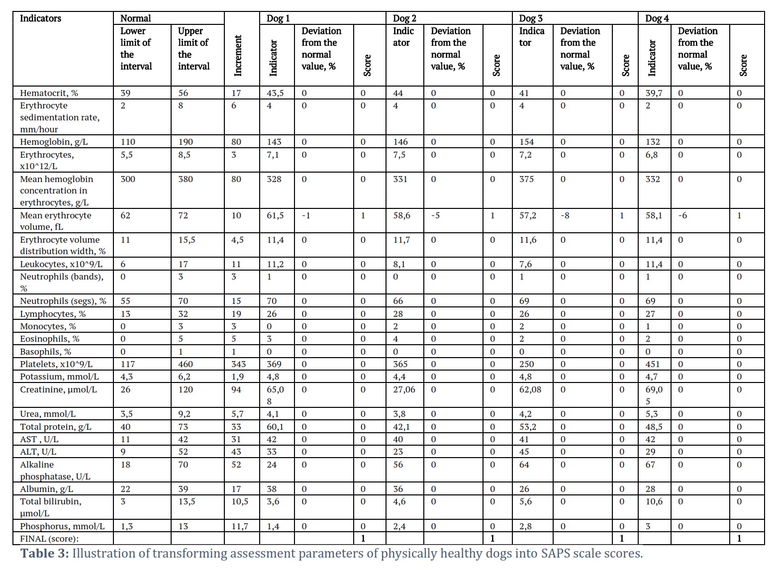
During the course of this research, a systematic approach was developed for the automatic conversion of actual measurements taken from the examined animals into corresponding scores on the Simplified Acute Physiology Score (SAPS) scale. To demonstrate this conversion process, an example is presented in Tables 2-4. The calculation table delineates the lower and upper limits of the reference range for each analyzed parameter while also establishing the "step." In the adjacent column, employing a predetermined formula, the table automatically computes the percentage variance of the parameter relative to the reference value and subsequently assigns the corresponding score accordingly.
To evaluate the overall condition of the animals, the scores obtained from individual parameters are added together. It is crucial to note that the degree of dysfunction in the animal's body is directly correlated with the cumulative score. Specifically, a higher score indicates an elevated risk of septic complications and, consequently, a heightened likelihood of mortality in the affected animal.
The leukocyte count in these dogs fell within the normal range, and no bone marrow cells, basophils, or juvenile neutrophils were detected. There was a slight increase in the percentage of eosinophils (0-1 point). Notably, the number of monocytes exceeded the physiological norm by a range of 2-4 points. Moreover, the number of rod-shaped neutrophils significantly exceeded the norm, with a range of 1-4 points, while segmented neutrophils showed a decrease of 1-2 points. These findings collectively suggest the presence of prolonged inflammatory processes in the affected animals. Lymphocyte counts remained within the normal range.
The infection route of leptospires involved their initial entry into the dog's body through damaged skin and mucous membranes. Subsequently, these leptospires circulated within the bloodstream, allowing them to infiltrate the liver, kidneys, and lungs, where they underwent multiplication. Between the 4th and 5th days of the illness, there was a notable reduction in leptospirosis agents, leading to the release of endotoxins. These endotoxins played a role in the destruction of red blood cells, contributing to the pathogenesis of the disease.
In dogs with leptospirosis, there was liver damage, leading to dysfunction of their organs, characterized by elevated levels of ALT and AST (2-4 points). This is due to the action of hemolytic toxins released in leptospirosis. Red blood cells are destroyed, anemia develops, and an excess of hemoglobin accumulates. The liver cannot process the accumulated excess hemoglobin in time. This results in an increase in bilirubin levels (1-4 points), damage to the bile ducts, and bilirubin adsorption by tissues, leading to jaundice. Due to poisoning, capillaries constrict and become obstructed by blood clots, leading to bleeding and tissue necrosis in the skin. The levels of creatinine (0-2 points) and urea (2-4 points) are also elevated, indicating severe dysfunction of the renal organs (Table 6) and leading to the development of MODS.
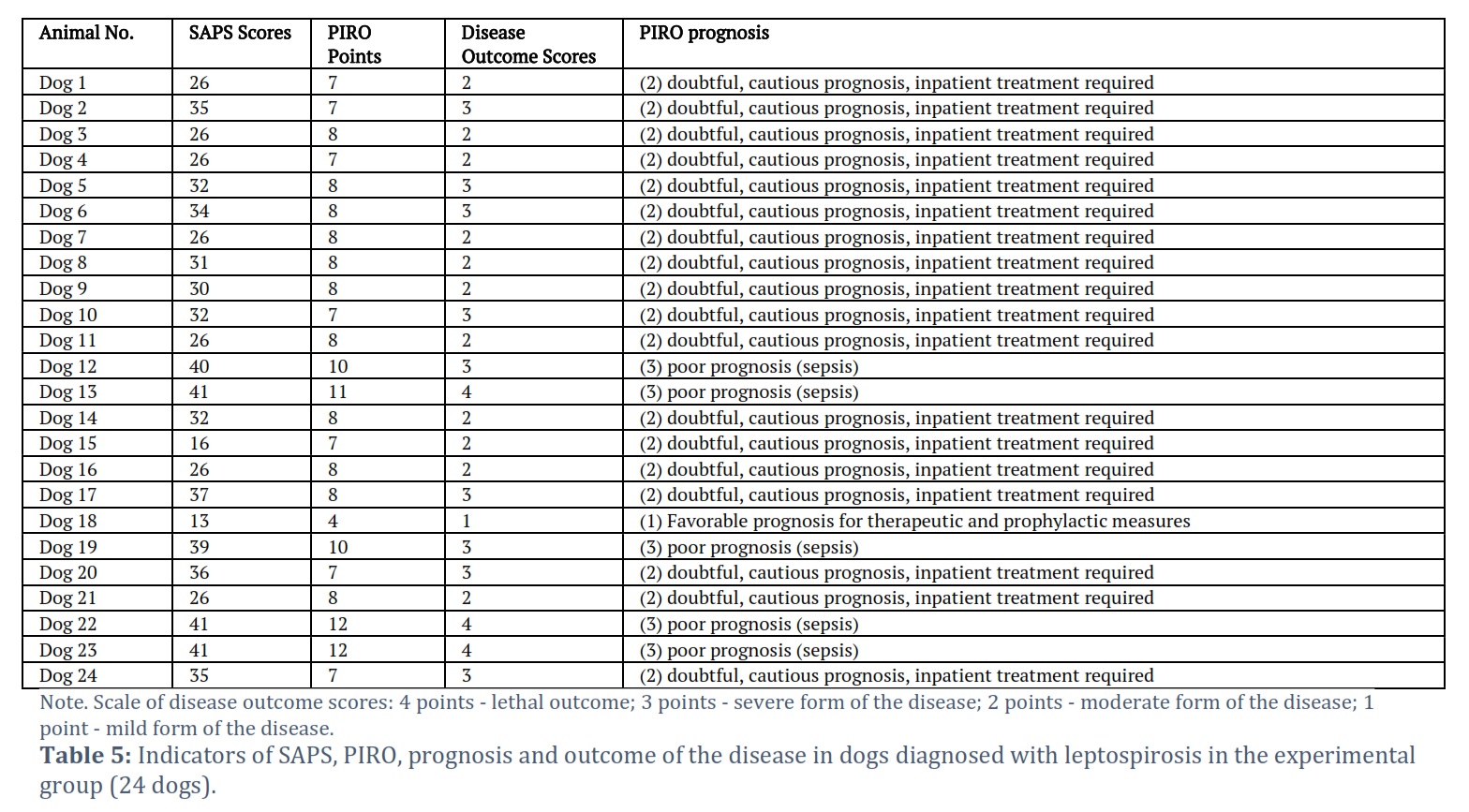
The data converted into scores on the PIRO and SAPS scales obtained from dogs diagnosed with leptospirosis are provided in Table 5.
The SAPS scale scores exhibit a strong positive correlation (r = 0.883) with the outcome of leptospirosis in dogs, indicating a high degree of association between these variables. The statistical analysis, using the Chaddock scale, classifies this relationship as robust. With 22 degrees of freedom (f) and a Student's t-test criterion of 8.824, the calculated t-value surpasses the critical t-value (2.074), signifying a highly significant statistical relationship (p = 0.000000). The paired linear regression equation is expressed as y = -0.27979 + 0.09199 * x, and the coefficient of determination (r²) is 0.780. This indicates that the factor x explains 78% of the variance in the dependent variable y. The mean approximation error, a measure of the regression model's adequacy, is 11.3%.
Additionally, SAPS scores demonstrate a substantial positive correlation (r = 0.782) with the prognosis on the PIRO scale (Table 6). Again, this relationship is characterized as strong on the Chaddock scale. With 22 degrees of freedom (f) and a Student's t-test criterion of 5.891, the t-value exceeds the critical t-value (2.074), resulting in a highly significant statistical relationship (p = 0.000008). The paired linear regression equation is represented as y = 0.59164 + 0.05060 * x, and the coefficient of determination (r²) is 0.612, meaning that the factor x accounts for 61.2% of the variance in the dependent variable y. The mean approximation error in this context is 12.0%.
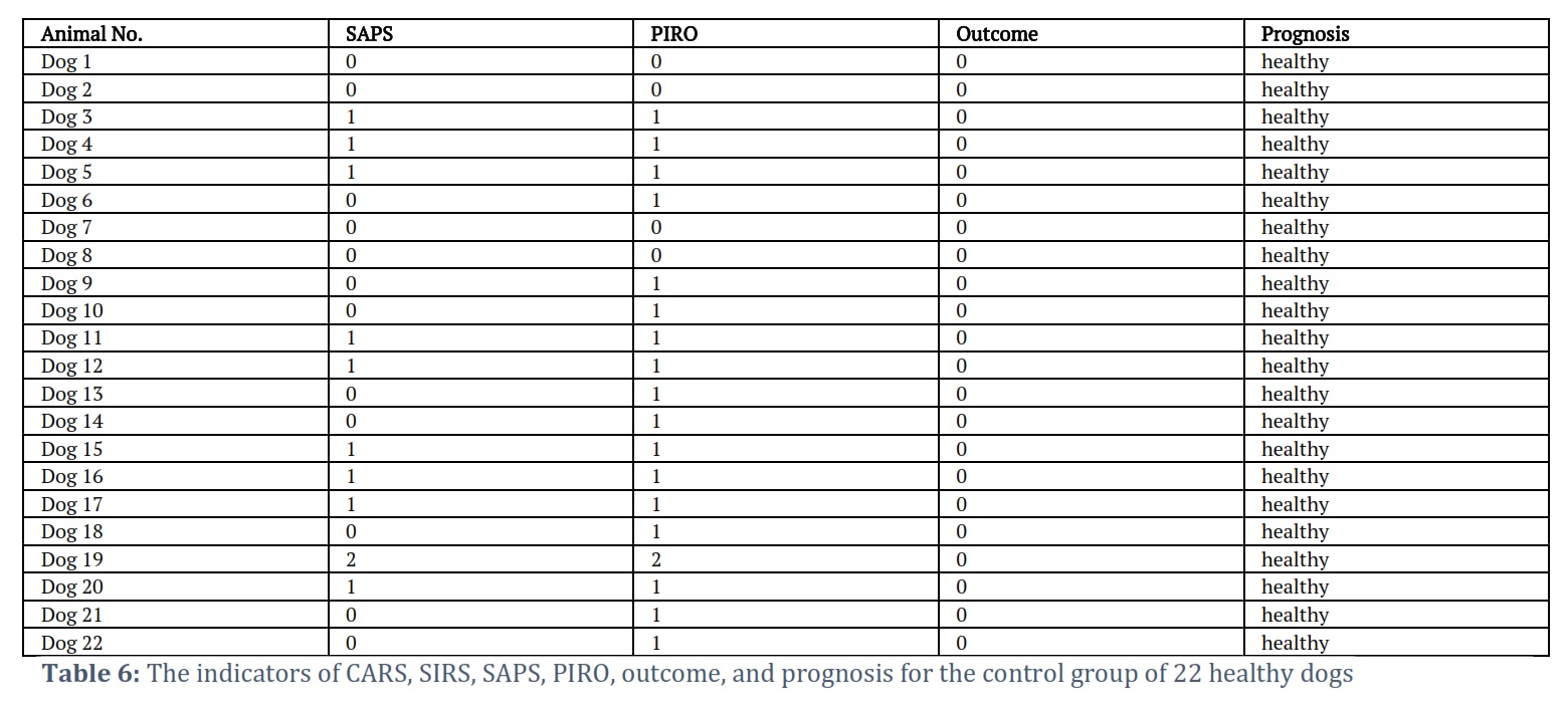
Table 6 provides an overview of the examination results for the control group, presenting scores on both the PIRO and SAPS scales. The correlation between the scores on the PIRO scale and the SAPS scale for the dogs in the experimental and control groups (Tables 5 and 6) is r = 0.972. The relationship between PIRO and SAPS scores is direct, and the strength of the relationship according to Chaddock's scale is very high. With f = 27.186, the t-criterion of Student is 2.015. t-value > t-critical, indicating that the relationship between the variables is statistically significant (p=0.000000). The regression equation is y = -2.52128 + 4.06502 * x, with an r^2 of 0.944 (the factor x explains 94.4% of the variance in the dependent variable y). The average approximation error, which serves as an indicator of the adequacy of the regression model, cannot be calculated and is considered undefined.
Figures & Tables
In cases where the animal's immune system is compromised, the protective mechanisms in dogs activate with a delay, which can ultimately lead to the dog's fatality from leptospirosis. Leptospirosis presents itself as a rapidly progressing disease, displaying acute, subacute, and chronic forms, both in typical and atypical manifestations. The fulminant form of the disease is distinguished by a sudden and sharp increase in body temperature, severe depression, and significant weakness. Some affected animals may exhibit excitement and agitation, which can escalate to uncontrolled behavior in individual cases [19]. This severe presentation highlights the rapid progression of leptospirosis in certain cases, emphasizing the critical importance of early diagnosis and intervention. The contrasting behaviors observed, from depression to agitation, underscore the variability in clinical manifestations and the challenges veterinarians face in managing such cases effectively.
During the initial hours of the disease, body temperature rises significantly, followed by a subsequent decrease to normal levels and even below 38°C. Respiratory patterns become shallow and rapid, and symptoms such as jaundice of the mucous membranes and the presence of bloody urine may be observed. Death in these cases typically occurs due to respiratory failure [20]. The development of respiratory distress underscores the systemic impact of leptospirosis, with lung involvement contributing to the disease's severity. The presence of jaundice and hematuria further reflects the multi-organ nature of the infection, complicating the clinical picture and necessitating comprehensive treatment strategies.
Puppies afflicted with acute cases exhibit a range of symptoms, including a high fever within the range of 39.5 to 41.5°C, tachycardia, loss of appetite, feelings of depression, and weakness. Their respiration becomes notably quick and shallow. By the 4th to 6th day, jaundice becomes noticeable. Additionally, these dogs encounter challenges with urination, and their urine may take on a color resembling cherry-red or brown. At the outset, dogs may exhibit diarrhea, occasionally accompanied by the presence of blood. Subsequently, this diarrhea may evolve into constipation. Laboratory examinations indicate a reduction in red blood cell count, an increase in white blood cells (leukocytosis), and elevated levels of bilirubin [21]. These hematological and biochemical abnormalities signify the systemic impact of leptospirosis on hematopoietic and hepatic functions. The decrease in red blood cells and elevated bilirubin levels reflect the severity of liver damage, while leukocytosis suggests an ongoing inflammatory response and the body's attempt to combat the infection.
Subacute cases display similar symptoms, but their onset is slower and less pronounced. Occasionally, the temperature may briefly rise above 39.5°C.
In one dog, a chronic form of leptospirosis was observed, which was marked by a gradual deterioration of the condition, anemia of the mucous membranes, necrosis, enlargement of lymph nodes in the groin and neck. There were occasional episodes of brief temperature spikes, and the urine remained discolored [22]. These atypical manifestations of leptospirosis highlight the disease's variability and the challenges it presents in diagnosis and treatment. The presence of brief increases in body temperature and persistent discolored urine in this chronic form underscores the need for vigilant monitoring and consideration of leptospirosis in cases with unusual clinical presentations.
In deceased animals, distinct post-mortem observations included prominently enlarged pale yellow lymph nodes. The liver exhibited noticeable features, characterized by enlargement, flabbiness, and a clay-like appearance upon sectioning. Kidney volumes were notably increased, and beneath the hemorrhagic capsule, the boundary between cortical and medullary layers appeared smoothed. The urinary bladder contained red-colored urine, accompanied by the presence of hemorrhagic lesions in the form of pieces and stripes on the mucous membrane.
Regarding cases of acute, rapid, and subacute leptospirosis, the overall prognosis tends to be unfavorable or uncertain, culminating in three fatal outcomes. Treatment strategies encompassed a comprehensive approach, encompassing both etiological and pathogenetic therapies to address the complex nature of the disease. However, despite these efforts, the challenging nature of leptospirosis in its severe forms often results in a grave prognosis.
When converting various indicators into scores on the PIRO and SAPS scales within 24 hours of admitting afflicted dogs to the veterinary clinic, a strong and statistically significant correlation with disease outcomes was observed for leptospirosis. SAPS scores falling within the range of 16-31 indicated a moderate form of the disease, while scores of 35-40 indicated a severe form, and scores reaching 41 or higher indicated a fatal outcome.
Leptospirosis in dogs is characterized by reduced red blood cell counts and elevated levels of ALT, AST, total bilirubin, creatinine, and urea. These changes are indicative of the progression of multiple organ failure. The SAPS scale serves as a valuable tool for promptly assessing the risk of physiological disturbances stemming from clinical signs of SIRS, MODS, and CARS. This aids in predicting sepsis complications in dogs affected by leptospirosis.
In conclusion, the study's findings on the SAPS and PIRO scales offer valuable tools for assessing and managing leptospirosis in dogs. Although the study has several limitations. The research primarily represents a veterinary perspective, and broader interdisciplinary collaboration is needed for a comprehensive understanding.
Nevertheless, future research should focus on practical application, validation, longitudinal studies, personalized treatment approaches, disease surveillance, One Health perspectives, public education, and genetic factors to further enhance our understanding and control of this disease in both canine and human populations.
Acknowledgment
The study was funded with a grant from the Russian Science Foundation No. 22-26-00091, https://rscf.ru/project/22-26-00091/.
Conflict of Interest
The authors declare that there is no conflict of interest.
All authors made equal contribution to the study.
![]() References
References
- Laptev SV, Pigina SY, Selina MV. Systematic analysis of domestic and foreign literature reflecting the peculiarities of pathogenesis in canine leptospirosis. Veterinary, Zootechnics, and Biotechnology, (2022); 1(12): 56-64
- Laptev SV, Mezentseva NI, Kamenskaya EP, Lamberova ME. Microbiology. (2012); 319. Altai State Technical University named after I.I. Polzunov, Biysk.
- Paz LN, Dias CS, Almeida DS, Balassiano IT, Medeiros MA, Costa F, Silva DN, Reis JN, Estrela-Lima A, Hamond C, Pinna MH. Multidisciplinary approach in the diagnosis of acute leptospirosis in dogs naturally infected by Leptospira interrogans serogroup Icterohaemorrhagiae: A prospective study. Comparative Immunology, Microbiology and Infectious Diseases, (2021); 77: 1-12.
- Ivanyuk VP, Mescheriakov OY, Navruzshoeva GSh. Distribution, pathogenesis, and comprehensive therapy of leptospirosis in dogs. Veterinary, Zootechnics, and Biotechnology, (2022); 11: 35-42.
- Nesterova IV, Evglevsky AA (eds). Chromatin Restructuring of Neutrophil Granulocytes in Normal and Pathology. 2017; 356. Carpicorn Publishing, Moscow.
- Volkova NI, Golubeva SV, Kulikova OL, Demidova TN, Zhavoronkova TS, Gusarova ML, Khaibrakhmanova SSh, Taymusova EN, Pashkin AV, Sochnev VV, Saushkin VV, Kozyrenko OV. Epizootological monitoring of the formation and functioning of infectious and invasive parasitic systems. Questions of Regulatory and Legal Regulation in Veterinary, (2018); 4: 59-63.
- Soboleva GL, Anan'ina YV, Nepoklonova IV. Current issues of leptospirosis in humans and animals. Russian Veterinary Journal, (2017); 8: 13-17.
- Samodelkin AG, Sochnyov VV (eds). Rapid Veterinary Response Forces. 2017; 243. BIKAR, Nizhny Novgorod.
- Pimenov NV, Laptev SV, Marzanova SN, Permyakova KY. PIRO model as a comprehensive assessment of septic complications in veterinary propaedeutics. Veterinary, Zootechnics, and Biotechnology, (2022); 4: 6-15.
- Marzanova SN, Laptev SV, Pimenov NV, Permyakova KY. Determination of the homeostasis deviation index on the model of purulent septic diseases in dogs and cats: In Biology and Biotechnology in the Service of Animal and Human Health. 2022: 60-64. National Scientific and Practical Conference of Young Scientists, Postgraduates, and Students, Moscow.
- Pimenov NV, Permyakova KY, Marzanova SN, Laptev SV. Neutrophil granulocyte reaction in the prognosis of purulent complications in dogs. Scientific Bulletin of Lugansk State Agrarian University, (2022); 2(15): 97-100.
- Marzanova SN, Laptev SV, Pimenov NV, Permyakova KY. Comparative tests for the evaluation of the phagocytic activity of animal neutrophil granulocytes: In Biology and Biotechnology in the Service of Animal and Human Health. 2022: 30-36. National Scientific and Practical Conference of Young Scientists, Postgraduates, and Students, Moscow, Russia.
- Bobrakov SI. Leptospirosis in dogs (epidemiology, pathogenesis, immunology, prevention). Dissertation abstract. 2005; 21. Altai State Agrarian University, Barnaul.
- Moiseeva DL, Gorodin VN. Clinical and pathomorphological features of hemostasis disorders in severe leptospirosis. Infectious Diseases, (2020); 18(1): 43-52.
- Delmas B, Jabot J, Chanareille P, Ferdynus C, Allyn J, Allou N, Raffray L, Gaüzere B-A, Martinet O, Vandroux D. Leptospirosis in ICU: a retrospective study of 134 consecutive admissions. Critical Care Medicine, (2018); 46(1): 93-99.
- Ludwig B, Zotzmann V, Bode C, Staudacher DL, Zschiedrich S. Lethal pulmonary hemorrhage syndrome due to leptospira infection transmitted by pet rat. IDCases, (2017); 8: 84-86.
- Pimenov NV, Laptev SV, Permyakova KYu, Ivannikova RF, Marzanova SN. Criteria in the prognosis of bacterial infections generalization in dogs with uterine inflammation. International Veterinary Bulletin, (2022); 3: 11-21
- Laptev SV, Pimenov NV, Gorbatova KS. Prognosis of septic pathologies in veterinary propaedeutics on the model of panleukopenia in cats. Veterinary, Zootechnics, and Biotechnology, (2022); 11: 52-58.
- Naviaux RK. Mitochondrial and metabolic features of salugenesis and the healing cycle. Mitochondrion, (2023); 70: 131–163.
- Oțelea MR, Fell AKM, Handra CM, Holm M, Filon FL, Mijakovski D, Minov J, Mutu A, Stephanou E, Stokholm ZA, Stoleski S, Schlünssen V. (2022). The value of fractional exhaled nitric oxide in occupational diseases – a systematic review. Journal of Occupational Medicine and Toxicology, (2022); 17: 1-12.
- Thachil J, Bates I. Approach to the Diagnosis and Classification of Blood Cell Disorders. Dacie and Lewis Practical Haematology. (2017): 497–510.
- Azócar-Aedo, L., & Monti, G. Seroprevalence of pathogenic Leptospira spp. in domestic dogs from southern Chile and risk factors associated with different environments. Preventive Veterinary Medicine, (2022); 206: 1-13.
This work is licensed under a Creative Commons Attribution-Non Commercial 4.0 International License. To read the copy of this license please visit: https://creativecommons.org/licenses/by-nc/4.0