

Full Length Research Article
Genotypic study of Trichomonas gallinae in Domestic Pigeons in Basrah Province, Iraq
Harith Abdulla Najem1*, Sarmad Awad Mozan AL-Asadi2, Isam A. Khaleefah1
Adv. life sci., vol. 11, no. 1, pp. 206-211, February 2024
*– Corresponding Author: Harith Abdulla Najem (Harith.najem@uobasrah.edu.iq)
Authors' Affiliations
2. Department of Biology, College of Education for Pure Sciences, University of Basrah, Basrah – Iraq
[Date Received: 23/05/2023; Date Revised: 07/01/2023; Date Published: 25/02/2024]
Abstract![]()
Introduction
Methods
Results
Discussion
References
Abstract
Background: The current study aimed to employ polymerase chain reaction (PCR) for validating the initial clinical diagnosis. Additionally, the research utilized wet microscopic smear sequence analysis and constructed phylogenetic trees to investigate trichomoniasis in domestic pigeons in Basrah province, Iraq.
Methods: After screening dozens of cases attending local clinics, we selected the suspected cases for additional tests and verification. Furthermore, all cases were inspected for postmortem changes and samples were collected from mouth and pharynx for microscopic examination and DNA isolation. We selected fifteen clinically suspected cases that have shown positive microscopic results. DNA samples were PCR amplified for the ITS-rDNA region, followed by sequencing and Phylogenetic analysis.
Results: Our research unveiled consistent manifestations among all positively diagnosed cases. Direct examination through wet mount smears revealed the presence of the parasite. Sequence analysis and phylogenetic investigation identified the B strain of the T. gallinae parasite in domestic pigeons within Basrah province, Iraq. This strain exhibited only two haplotypes in the region, haplotype 1 and haplotype 2. Furthermore, our study documented the prevalence of the B genotype of T. gallinae in domestic pigeons across various countries, including China, Iraq, Iran, and Saudi Arabia.
Conclusion: The verification of T. gallinae infection in domestic pigeons was achieved through a PCR technique that utilized the ITS rDNA region as a genetic marker. Furthermore, the application of haplotype network analysis provided evidence supporting the categorization of the T. gallinae parasite in domestic pigeons as belonging to the B strain.
Keywords: Trichomonas gallinae; Pigeons Canker; Sequence Analysis; Phylogenetic
Introduction![]()
Trichomonas gallinae, a flagellate protozoan characterized by four anterior flagella and an undulating membrane on one side, lacking a posterior flagellum, is responsible for avian trichomonosis. T. gallinae is a prevalent parasite in pigeons and doves [1]. The disease is commonly known as canker in pigeons and frounce in birds of prey. Flagellated protozoa of T. gallinae inhabit various organs, including the sinuses, esophagus, liver, mouth, and throat. The transmission of the disease occurs through oral secretions present in feed, water, and crop milk [2]. Key symptoms of the disease include open-mouthed breathing, drooling, repeated swallowing movements, loss of condition, and occasionally watery eyes, with nervous symptoms being infrequent. A distinctive feature is the development of canker, or caseous plaque, in the oral cavity, esophagus, and crop [3]. Additionally susceptible to infection are chickens, hawks, golden eagles, domestic and wild turkeys, and other raptors [4]. The domestic pigeon, being the primary host of T. gallinae, plays a crucial role in disease spread. While young pigeons are more prone to infection and may succumb to it, various bird species can act as carriers without exhibiting clinical signs [5]. Initial lesions in pigeon squabs appear as small spots in the mouth, particularly on the soft palate, ranging in color from white to yellow. Over 3–14 days, these lesions progress from the mouth to the esophagus, then the crop, and finally, the proventriculus [6]. Yellowish lesions with a central conical caseous spur develop into spherical elevations commonly referred to as “yellow buttons” [7]. In advanced cases, lesions may grow into substantial, solid necrotic masses, potentially obstructing the lumen. Changes in the crop manifest as a yellowish, diphtheritic membrane, gradually extending to the proven triculus. In some advanced cases, the surrounding tissue, including the skin and skull, can be affected by the spreading lesions [8].The internal transcribed spacers (ITS) of ribosomal DNA have demonstrated promise as a genetic marker for distinguishing between different Trichomonas species associated with avian trichomoniasis. This is achieved through the use of a polymerase chain reaction (PCR) technique. Trichomonas infection represents a serious health problem for pigeons in Basrah that causes financial detriment for pigeon owners. In addition, there were no previous scientific reports related to T. gallinae in domestic pigeons in Basrah. Therefore, the present study was designed to use PCR to confirm the primary clinical diagnosis and used sequence analysis and phylogenetic trees to document the type, strain, and genotype of trichomoniasis in domesticated pigeons in Basrah province, Iraq.
Methods![]()
Samples Collection
The focus of the current research was on domestic pigeons in Basrah province, Southern Iraq. This study spanned from May 2022 to October 2022. We selected Alzubir, Al- Hartha, and Abo Alkasib, regions which are famous for pigeon raising and have a significant number of pigeon breeders. After the initial screening, fifteen domestic pigeons from veterinary clinics located in in selected area ( Five birds from each clinic) were transported to the Department of Veterinary Pathology and Poultry Diseases at the Faculty of Veterinary Medicine, University of Basrah-Iraq. Six pigeons that showed severe signs of cankers were euthanized and examined individually to record a postmortem change according to [7]. The lesions were scrapped using a sterile surgical blade from the mouth and pharynx and were transferred into a sterile container containing 500 µL of 50% glycerol phosphate buffer solution and stored at -20 C for subsequent DNA isolation [9]. The current study was performed under the permission of the ethical committee in the Faculty of Veterinary Medicine, University of Basrah (Ref. No. 75/2022).
Microscopic Examination
The validation of oropharyngeal swab samples collected from suspected pigeons utilized the wet mount technique in microscopy (microscopic inspection). Swabs were blended with Phosphate Buffered Saline (PBS) and applied to sterile cotton-tipped applicators for observation under a light microscope at 40x magnification, enabling the identification of Trichomonas [10].
DNA Extraction and Polymerase Chain Reaction (PCR)
The DNA extraction from all samples was carried out using the gSYNCTM DNA Extraction kit's rapid procedure, specifically from the caseous masses in the mouth (Geneaid, Korea). The NanoDrop system was employed to determine the quantity of purified DNA. Subsequently, confirmation of all extracted DNA samples was achieved through PCR. The forward primer used was 5'-AACTAATACCAACTTCTTTT-3', while the reverse primer was 5'-TATTCGCGTAGAATAAGAAT-3'. These primers were utilized to amplify a 263 bp segment of the ITS rDNA region for each T. gallinae DNA sample using PCR. The PCR mixture included 25 μl Gotaq master mix (Promega, USA), 1.5 μl of each primer , 150 ng of DNA template, and nuclease free water up to 50 μl per each PCR reaction. The PCR was done in Simpliamp thermos cycler – Applied bio system, USA. The PCR program included 94 ̊C initial denaturation for 5 min, followed by 35 cycles at ( 94 ̊C for 30 sec, 46 ̊C for 30 sec and 72 ̊C for 30 sec), which were followed by a final 5 min extension at 72 ̊C to complete the elongation of products [11]. The PCR run was followed by gel electrophoresis of 5μl from each PCR sample on 1.5% agarose gel. After visualization of 5μl of each PCR product on 1.5% agarose gel, 30μl of the positive PCR amplicons were sent for sequencing at Macrogen company , Korea using the same forward and reverse primers of the PCR in the sequencing process.
Sequence and Bioinformatics Analysis
After visualization of 5 µL of each PCR product on agarose gel, the positive PCR samples were sent for sequencing (forward and reverse direction using the same forward and reverse primers used in the PCR amplification of the ITS rDNA region). The PCR samples were processed and shipped according to the instructions provided by the sequencing provider (Macrogen Inc., South Korea). Sequences of trichomonas were edited, aligned, and evaluated using MEGA 6.06 software. . The chromatograph files containing the genetic data were meticulously inspected for quality. The annotation and variances were arranged in clean chromatographs that show the quality of sequencing. This step aimed to ensure the quality and accuracy of genetic information. The comparison also included contrasting the detected nucleic acid sequences of the samples with the retrieved reference sequences from the gene bank database to determine variation in those ITS rDNA region sequences.
Cladogram trees were constructed using the Maximum Likelihood method and the Tamura-Nei model were employed for tree construction, allowing a nuanced analysis of taxonomic relationships through nucleotide inspection [12]. The robustness of the cladogram trees was assessed using Felsenstein’s bootstrap test. This statistical method involved resampling the dataset multiple times (2,000 times) to calculate bootstrap values. These values provided insights into the reliability and support for taxa clusters identified in the phylogenetic trees [13].
Results![]()
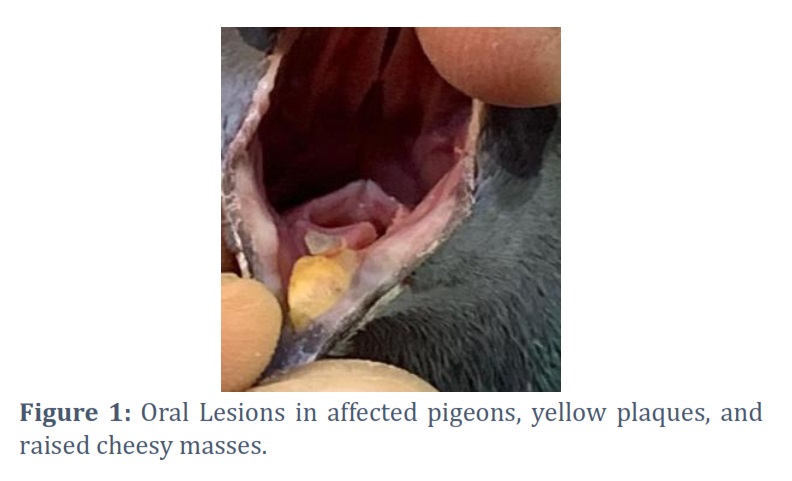
During the study period, many pigeons were examined in the targeted clinics, but only those that were clinically positive were targeted. The severity of the disease varied in between the examined pigeons. All pigeons had lesions, whereas 6 of 15 birds exhibited more severe characteristic oral lesions. Most of the affected birds showed identical typical signs such as opening mouth and complex repeated swallowing movements. The oropharyngeal cavity of the affected birds displayed a whitish to yellowish nodule of varying sizes, accompanied by ulceration and inflammation as depicted in (Figure 1).
The severity and spreading of lesions were more sever in six birds which were noticed in the esophagus, proventriculus, and crop; this led to more severe clinical signs, as specific signs of illness, including weight loss, lethargy, reluctance to eat, and greenish diarrhea. These pigeons exhibited nasal discharge and neurological symptoms such as head tremors and reduced flying ability. In addition, dehydration, dull eyes, and respiratory distress, with complex and open-mouthed breathing blocking the airways and causing the birds to pass away.
Microscopic Examination
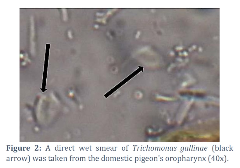
The distinctive jerky movement of the parasite was observed in the motile organism under a microscope with 40x magnification, revealing vibrant and dynamic activity. Identification of the parasite was established by discerning its unique physical attributes. The parasite exhibited a pear- or oval-shaped structure with a single undulating membrane, four anterior flagella, and a prominent axostyle at the posterior end, as illustrated in (Figure 2).
Detection of DNA by PCR
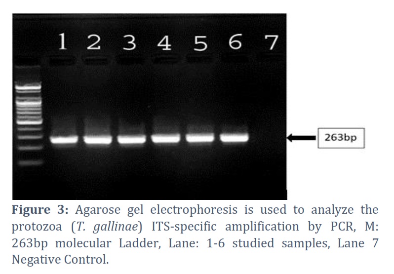
The DNA extracted from T. gallinae samples was subjected to amplification through PCR, employing both forward and reverse primers. The samples that underwent PCR analysis are depicted in Figure 3. Following analysis on an agarose gel, DNA bands of 263 base pairs in length were identified. Proper-sized DNA bands were observed in six samples.
Sequence and Bioinformatics Analysis
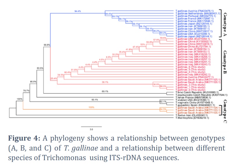
The PCR results of six samples were sequenced using ITS-rDNA-specific primers in both directions. These sequenced samples were then submitted to the GenBank database through the NCBI website, with accession numbers OP268446.1, OP268447.1, OP268448.1, OP268449.1, OP268451.1, and OP268452.1. In Figure 4, the phylogenetic analysis of the current study's samples is presented alongside sequences of Trichomonas spp., including genotypes A, B, and C of T. gallinae from different countries. All samples from the present study were grouped with genotype B of T. gallinae from China, Iran, Iraq, Austria, and Saudi Arabia.
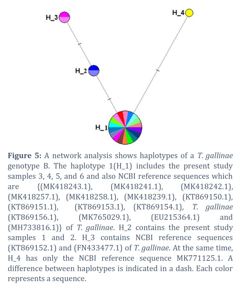
Figure 5 illustrates a haplotype network analysis of T. gallinae genotype B. Our findings revealed four haplotypes associated with T. gallinae genotype B. Specifically, present study samples 3, 4, 5, and 6 were clustered with sequences of haplotype 1 (H_1), which also included NCBI reference sequences such as T. gallinae (MK418243.1, Iraq), T. gallinae (MK418241.1, Iraq), T. gallinae (MK418242.1, Iraq), T. gallinae (MK418257.1, Iraq), T. gallinae (MK418258.1, Iraq), T. gallinae (MK418239.1, Iraq), T. gallinae (KT869150.1, Iran), T. gallinae (KT869151.1, Iran), T. gallinae (KT869153.1, Iran), T. gallinae (KT869154.1, Iran), T. gallinae (KT869156.1, Iran), T. gallinae (MK765029.1, Saudi Arabia), T. gallinae (EU215364.1, USA), and T. gallinae (MH733816.1, China). However, present study samples 1 and 2 were separated in a single haplotype (H_2), while H_3 and H_4 only contained NCBI reference sequences.
Figures & Tables
Geographically, Basrah is located in the lower south of Iraq region and a has distinct climate compared with other parts of Iraq. In addition, Basrah is a major hub for smuggling birds with other Arabian Gulf countries. Therefore, diverse types of pigeon breeds are raised in Basrah for different purposes. On occasion, [14,15,16] mentioned the intensity of infection and clinical responses differs among different breeds, species, and according to geographical locations.
Clinical symptoms and gross lesions play a pivotal role in the initial diagnosis of T. gallinae. In this study conducted in Basrah city, Iraq, pigeons infected with trichomoniasis exhibited open mouths and repeated swallowing difficulties. Gross lesions in the oropharyngeal cavity included whitish to yellowish nodules of varying sizes, accompanied by inflammation and ulceration. These lesions occasionally extended into the esophagus, crop, and proventriculus, aligning with findings from previous studies [10], which described nodules of pale to yellowish color. The varying size distribution of nodules, along with ulceration and inflammation, represented the gross lesions observed in affected birds, leading to fatal outcomes as the lesions obstructed the respiratory system. In addition, [17] described variation in the severity of lesions, which could spread to esophagus and crop in young and adult birds.
The direct wet smear analysis in our study revealed the parasite's pear- or oval-shaped form with four-front flagella and an axostyle, exhibiting lively and jerky movements. Consistent with prior research.
Also, more previous studies revealed that the parasite was recognized based on its physical traits and characteristics. It was pear- or oval-shaped, with one undulating membrane, four front flagella, and a conspicuous axostyle at the back end of the parasite [14,18,19].
Diagnosis of trichomoniasis is challenging as the oral lesions may be confused with other diseases such as, candidiasis, avian pox (pox virus) and hypovitaminosis A [20]. The molecular identification through PCR targeting the ITS-rDNA region confirmed T. gallinae infection in domestic pigeons. This PCR technique, with specific primers, has been emphasized in previous studies for identifying T. gallinae infection in various bird species, such as chickens, turkeys, feral pigeons, and common mynah [21,22].
Bioinformatic analyses based on the ITS-rDNA region were conducted to establish the relationship of our study's sequences with other Trichomonas spp. and determine the T. gallinae genotypes. Our findings affirmed that the study samples belonged to the Trichomonas genus, particularly T. gallinae. While some Trichomonas species can infect humans and animals [23]. For example, it is known that three Trichomonas species (oral T. tenax, intestinal T. hominins, and genitourinary T. vaginalis) infected humans and T. fetus species (intestinal and venereal) only infected cattle and cats. At the same time, the T. gallinae (gastrointestinal) is the only species infected birds. The T. gallinae parasite causes a common bird disease called canker (avian trichomoniasis) and has three genotypes: A, B, and C [21]. These genotypes (strains) and their virulence seemed linked with the type of birds infected [24]. In the present study, we only found the B strain of the T. gallinae parasite in domestic pigeons, and this strain only had two haplotypes in Basrah province, Iraq. Haplotype 1 had the adenine nucleotide at position 58, whereas haplotype 2 had the deletion mutation. This corresponds with previous studies that found the B genotype in domestic pigeons widespread across different countries, including China, Iraq, Iran, and Saudi Arabia [21,25]. In addition to this, based on the data used in the haplotype analyses, our results reported the B genotype could be present in four haplotypes. These haplotypes had variations at three positions (35, 58, and 72); the most significant haplotype is 1 (Figure 5). Globally others have shown variation in T. gallinae genotypes presence in birds such as genotype D [26] in Hungary, genotypes (A) in the United States [27]. In conclusion, the confirmation of T. gallinae infection in domestic pigeons using a PCR technique based on the ITS rDNA region as a genetic marker. In addition, the haplotype network analysis proved the T. gallinae parasite in domestic pigeons belonging to the B strain.
Acknowledgement
The authors would like to acknowledge the help of staff members at the Department of Pathology and Poultry Diseases, Faculty of Veterinary Medicine, University of Basrah, Iraq.
Conflict of Interest
We have read and understood ALS policy on declaration of interests and declare that we have no competing interests.
Harith Abdulla Najem: contributed to the design of the research and Field diagnosis of the disease.
Sarmad Awad Mozan AL-Asadi: genetic analysis of the results and the manuscript's writing.
Isam Azeez Khaleefah: molecular detection.
![]() References
References
- Nematollahi A, Ebrahimi M, Ahmadi A, Himan M. Prevalence of Haemoproteus columbae and Trichomonas gallinae in pigeons (Columba domestica) in Isfahan, Iran. Journal of Parasitic Diseases, (2012); 36(1):141-142.
- Saikia M, Bhattacharjee K, Sarmah PC, Deka DK, Buragohain LM, Tamuly S. Molecular detection and characterization of Trichomonas gallinae isolated from pigeon and chicken of Assam, India. Journal of Entomology and Zoology Studies,(2023); 11(1): 101-105
- Amin A, Nöbauer K, Patzl M, Berger E, Hess M, Bilic I. Cysteine peptidases, secreted by Trichomonas gallinae, are involved in the cytopathogenic effects on a permanent chicken liver cell culture. PLoS One, (2012);7(5):e37417.
- Urban EH, Mannan RW. The potential role of oral pH in the persistence of Trichomonas gallinae in Cooper's Hawks (Accipiter cooperii). Journal of Wildlife Diseases, (2014); 50(1):50-55.
- Bunbury N, Jones CG, Greenwood AG, Bell DJ. Epidemiology and conservation implications of Trichomonas gallinae infection in the endangered Mauritian pink pigeon. Biological Conservation, (2008); 141(1): 153-161.
- Begum N, Mamun MA, Rahman SA, Bari AS. Epidemiology and pathology of Trichomonas gallinae in the common pigeon (Columba livia). Journal of the Bangladesh Agricultural University, (2008); 6(2): 301-306.
- Abd-El-Motelib TY, GALAL BE. Some studies on Trichomonas gallinae infection in pigeons. Assiut Veterinary Medical Journal, (1993); 30(59): 277-288.
- Saikia M, Bhattacharjee K, Sarmah PC, Deka DK, Upadhyaya TN, Konch P. Prevalence and Pathology of Trichomonas gallinae in Domestic Pigeon (Columba livia domestica) of Assam, India. Indian Journal of Animal Research, (2021); 55(1): 84-89
- Mohamed HM, Saad AS, Khalifa MM, Abdel-Maogood SZ, Awadalla SM, Mousa WM. Detection and molecular characterization of Trichomonas gallinae recovered from domestic pigeons in Egypt. Parasitology Research, (2023); 122(1): 257-263
- Elbahy, N., Gomaa, A., AbouLaila, M., ElKhatam, A., & Anis, A. Trichomonas gallinae, Prevalence and Histopathology in Domestic Pigeons in Sadat District, Egypt. Journal of Current Veterinary Research, (2023); 5(1): 223-230.
- Qiu SB, Lv MN, He X, Weng YB, Zou SS, Wang XQ, Lin RQ. PCR identification and phylogenetic analysis of Trichomonas gallinae from domestic pigeons in Guangzhou, China. Korean journal of parasitology, (2017); 55(3): 333-336.
- Tamura K, Peterson D, Peterson N, Stecher G, Nei M, Kumar S. MEGA5: molecular evolutionary genetics analysis using maximum likelihood, evolutionary distance, and maximum parsimony methods. Molecular biology and evolution, (2011); 28(10): 2731-2739.
- Felsenstein J. Confidence limits on phylogenies: an approach using the bootstrap. evolution, (1985); 39(4): 783-791.
- Al-Hasnawy MH, Rabee AH. A review on trichomonas species infection in humans and animals in Iraq. Iraqi Journal of Veterinary Sciences, (2023); 37(2): 305-313.
- hamza Hussein H. Trichomonasis detection in pigeons (Columba livia domestica) in Diyala province. Diyala Journal for Veterinary Sciences, (2023);1(1): 85-97.
- Jaafar A. Avian Trichomoniasis Prevalence in Domesticated Pigeons in Thi-Qar Province, Iraq. University of Thi-Qar Journal of agricultural research, (2023); 12(2): 89-108.
- Echenique JV, Soares MP, Bruni M, Farias NA, Moretti VD, Bandarra PM, Albano AP, Schild AL. Oral trichomoniasis in raptors in Southern Brazil. Pesquisa Veterinária Brasileira, (2020); 39(12): 983-988.
- El-Khatam AO, AbouLaila MR, Ibrahim M, AbdEl-Gaber MM. Trichomonas gallinae: Prevalence and molecular characterization from pigeons in Minoufiya governorate. Egypt. Experimental parasitology, (2016); 170: 161-167.
- Fadhil LT, Faraj AA, AL-Amery AM. Trichomonas gallinae identification and histopathological study in pigeon (Columba livia domestica) in Baghdad, Iraq. The Iraqi Journal of Veterinary Medicine, (2020); 44(E0): 57-63.
- Marx M, Reiner G, Willems H, Rocha G, Hillerich K, Masello JF, Mayr SL, Moussa S, Dunn JC, Thomas RC, Goodman SJ. High prevalence of Trichomonas gallinae in wild columbids across western and southern Europe. Parasites & Vectors,(2017);10( 242):1-11
- Chou S, Hadano S, Kojima A, Yorisaki M, Yasuda M, Ike K, Tokiwa T. Genetic characterization of Trichomonas gallinae (Rivolta, 1878) in companion birds in Japan and the genotypical relationship in the Asia region. Journal of Microbiology, Immunology and Infection, (2022); 55(3) :527-534.
- Arabkhazaeli F, Madani SA, Ghorbani A. Parasitological and molecular survey of scattered parasitism by trichomonads in some avian species in Iran. Avian Pathology, (2020); 49(1): 47-55.
- Saleem MH, Khan MS, Chaudry AS, Samad HA. Prevalence of trichomoniasis in domestic and wild pigeons and its effects on hematological parameters. Pakistan Veterinary Journal, (2008); 28(2): 89-91.
- Fadhil LT, Faraj AA. Survey of Trichomonas gallinae isolates in pigeons by microscopy and PCR. Veterinary Research, (2019); 23(4): 321-329.
- Alrefaei AF. Molecular detection and genetic characterization of Trichomonas gallinae in falcons in Saudi Arabia. PLoS One, (2020); 15(10): e0241411.
- Tuska-Szalay B, Sipos G, Takács N, Kontschán J, Sándor AD, Péter Á, Berta K, Kerek Á, Jerzsele Á, Votýpka J, Hornok S. Molecular epidemiological study of Trichomonas gallinae focusing on central and southeastern Europe. Frontiers in Veterinary Science, (2022); 9: 1050561.
- Mousa AJ, Hamid NR. Trichomonas gallinae prevalence and genetic characteristics from racing pigeons in Thi-Qar Province, Iraq. Advances in Animal and Veterinary Sciences, (2023); 11(10): 1690-1696.
This work is licensed under a Creative Commons Attribution-Non Commercial 4.0 International License. To read the copy of this license please visit: https://creativecommons.org/licenses/by-nc/4.0