

Full Length Research Article
Phytochemical Screening of leaves of Polyalthia sclerophylla using Classical Methods and GC-Mass Spectroscopy: Cytotoxicity and Antibacterial Activities
Zahraa Sabbar Omran1, Hend Ahmed Abbas2, Farqad A. Albaidhani3, Huda M. Almusawi4, Mustafa Mudhafar5*, Mustafa M. Karhib6, H. A. Alsailawi1
Adv. life sci., vol. 11, no. 1, pp. 212-219, February 2024
*– Corresponding Author: Mustafa Mudhafar (almosawy2014@gmail.com)
Authors' Affiliations
2. Department of Chemistry and Biochemistry, College of Medicine, Al-Nahrain University, Baghdad – Iraq
3. Department of Prosthetics and Orthotics Engineering, College of Engineering, University of Kerbala, Karbala – Iraq
4. Department of Biology, College of Education for pure Sciences, University of Karbala, Karbala – Iraq
5. Department of Medical Physics, Faculty of Applied Medical Sciences, University of Kerbala, Karbala – Iraq
6. Department of Medical Laboratory Techniques, Al Mustaqbal University College, 51001 Hillah, Babylon – Iraq
[Date Received: 23/01/2023; Date Revised: 25/10/2023; Date Published: 25/02/2024]
Editorial Note on Version of Record
31 May 2025: This article has been corrected. See https://doi.org/10.62940/als.v13i0.4288 for more information.
Abstract![]()
Introduction
Methods
Results
Discussion
References
Abstract
Background: Polyalthia sclerophylla (P. sclerophylla) is a member of the Annonaceae family, with a wide distribution in tropical and subtropical regions. In traditional Chinese medicine, various members of this genus have been employed as medicinal plants to address refractory ailments. The current study aims to extract the leaves of Polyalthia sclerophylla (TLPS) and determine their chemical contents using GC-MS and standard phytochemical techniques.
Methods: The antibacterial and cytotoxicity activities of TLPS were used to evaluate its bio-medical characteristics. In that sequence, three solvents were used to extract TLPS to produce three samples: MTLPS, DTLPS, and HTLPS.
Results: Phytochemical analysis indicated terpenoids and glycosides in all prepared models, but no alkaloids were observed. The GC-MS data showed twenty-one chemical compounds. Pyridine, 2-Undecanol, 2-methyl-2-oxiranyl- and cyclobutanone were observed with higher percentages, while the 2(5H)-Furanone, 5-methyl, 1,1,3-Trimethylcyclopentane and 1,6-Heptadiene were observed with lower rates. The cytotoxicity study of TLPS was performed using Alamar blue assay using MG-63 cells. However, results show no detrimental influence at any dosage as cell availability increased. MTLPS, DTLPS and HTLPS were treated with six bacteria pathogens, and all of them showed significant effects against these bacteria to inhibit the growth of the bacteria. The extract of methanol (MLPS) was more effective in inhibiting the development of the bacteria compared with the DTLPS and HTLPS.
Conclusion: The current work has shown that the chemical composition of crude TPLS varies, leading to notable chemical, biological, and medicinal features, and their non-toxic impact.
Keywords: Polyalthia sclerophylla; GS-Mass; Antibacterial; Cytotoxicity activity
Introduction![]()
The Polyalthia genus is part of the Annonaceae family, which comprises approximately 130 genera and 2300-2500 species. Polyalthia, which has over 120 species, is a tropical and subtropical plant species native to South Asian nations such as Malaysia, Thailand, and Indonesia [1-4]. Polyalthia plants are well-known for their excellent medicinal and biological efficiency. They exhibit anti-cancer, anti-inflammatory, anti-oxidant, anti-plasmodial, anti-bacterial, and anti-DENV2 characteristics. Polyalthia genus stems, stem sections, roots, barks, leaves, and twigs have all been recognized via research. Traditional medicine uses Polyalthia plants to cure many ailments, including diabetes, dysmenorrhea, fever, helminthiasis, hypertension, stomach aches, pharyngeal neurosis, and skin issues [5-11]. Alkaloids, carbohydrates, terpenoids, tannins, saponins, phenolic compounds, flavonoids, and tannins are among the phytochemicals identified in the Polyalthia species. Because of their large chemical groups, these plants have unique biological activities that contribute to their efficiency. This study assesses one of these genera based on its chemical composition and biological activity [12-14]. The leaves of Polyalthia sclerophylla (TLPS) have been selected for various reasons. No studies have been reported on its chemical structures, antibacterial activity, or cytotoxicity. Nevertheless, there is a likelihood of getting comparable or unique phytochemicals with antimicrobial action from LPS leaves. Second, LPS is accessible locally and is deemed economical (low-cost). Third, because there was little information regarding LPS, the current study would explore creating a database for the researcher. This study investigated the biological activities of TLPS extracted using methanol, hexane, and dichloromethane, assessing their antibacterial activity against six bacteria. A toxicity investigation was conducted on the MG-63 human cell line. The methanol extract contained carbohydrates, glycosides, terpenoids, tannins, and steroids, indicating its significant influence on bacteria.
Methods![]()
Preparation of TLPS
In Perak, Malaysia, TLPS leaves were collected and cleansed with distilled water to remove any fungal contaminants or dust. TLPS was sun-dried for seven days before being chopped into small pieces and powdered for future use.
Extraction of TLPS
The Soxhlet technique extracted potential chemicals from TLPS via three solvent systems: hexane, dichloromethane (DCM), and methanol (MeOH). 50 g of TLPS were removed to obtain three produced cures were labelled as MTLPS, DTLPS, and HTLPS, which were then kept at four °C for potential research [15].
The Phytochemical analysis of TLPS
MTLPS, DTLPS, and HTLPS were cured using normal chemical procedures to determine their chemical groupings.
Test for alkaloids
Two stages were employed to detect the presence of alkaloids. The preliminary tests come first, followed by the confirmation testing. Mix 10 ml of diluted TMLPS, TDLPS, and THLPS to summarise the preliminary examination in HCl and filter the liquid. The filtered solution was used to treat Mayer’s and Dragendroff’s reagents. The second test involved combining 1 g of MTLPS, DTLPS, and HTLPS with 40% Ca(OH)2 solution until alkaline was visible on litmus paper, followed by two chloroform extractions. A Chloroform extract revealed the presence of thin layer plates. The chromatogram was created using ethyl acetate, n-hexane 1:4 solvent system, with a Dragendorff reagent, then sprayed in the chromatogram solution. The existence of alkaloids in the background can be recognised by monitoring the yellow and orange colours [16, 17].
Test for Flavonoids
There are several ways to detect Flavonoids in medicinal plants. Three techniques were utilised in this study to determine the percentage of Flavonoids in TPLS.
Determine the Flavonoids in TLPS
The mixture of MTLPS, DTLPS, and HTLPS with 5 mL of ethyl acetate, then put in the steam bath for heated for 3 minutes and filtered with high-quality filter paper. After adding 1 ml of ammonium dilute, the filtered solution was shaken. The presence of flavonoids may be identified by observing the yellow colour [18].
Test for Carbohydrates
The carbohydrates in TLPS were detected using Molisch’s assay and Fehling’s reagent. It was discovered that reducing sugar was present in PS leaves by dissolving MTLPS, DTLPS, and HTLPS in distilled water, adding Fehling’s reagent, and witnessing the colour shift to brick red.
Detect of Phenolic compound in TLPS
The mixture was prepared by taken 1 g of MTLPS, DTLPS, and HTLPS and solved in 100 ml of distilled water, then added a drop of Fe2(SO4)3. The presence of a phenolic group was detected as dark violet [19].
Test of Salkowski to Detect the Terpenoids in TLPS
Initially, 2 ml of CHCl3 was mixed with 10 ml of MTLPS, DTLPS, and HTLPS, followed by adding the H2SO4. Terpenoids are present when the mixture’s colour turns reddish-brown [20].
Test for Saponins (Froth test)
A test tube was filled with MTLPS, DTLPS, and HTLPS, then combined with 10 ml of distilled water and shaken vigorously for 1 minute. For more than 30 minutes, the tube was connected at an angle, and saponins were discovered by inspecting the surface honeycomb [21, 21].
Detect of Glycosides
The Keller-Killani assay was used to detect glycosides in LPS by dissolving 1 g of MTLPS, DTLPS, and HTLPS in DW, adding sulphuric acid and ferric chloride, and observing the formation of reddish and reddish-brown layers [23].
Test of Ferric chloride to detect Tannins
Before filtering, 1g of mixed MTLPS, DTLPS, and HTLPS was heated in a conical flask with 50 DW for 20 minutes. The samples were carefully soaked in 0.1% FeCl3. Black-Blue and green brownish were seen to determine the availability of tannins in the samples [24].
Test for Steroids (Lieberman’s test)
Acetic anhydride was mixed with 10 ml each of MTLPS, DTLPS, and HTLPS before being cooled in an ice bath. Several drops of sulphuric acid have been incorporated into the mixes Combinations of steroids can be identified by a shift in colour to blue or green from violet [25].
The test of Lead acetate
The mixture of MTLPS, DTLPS, and HTLPS (10 ml) was heated using a hot plate. The mixture was then treated with 1 ml of lead acetate (10%). The yellow precipitate format indicates the presence of flavonoids [26].
Reaction with Sodium hydroxide
The hot plate was used to heat 10 ml of MTLPS, DTLPS, and HTLPS. The mixture was then treated with diluted sodium hydroxide. The yellow precipitate format indicates the presence of flavonoids [27].
Detect of volatile compounds using GC-MS
To determine the volatile compounds in the TLPS extracts was analysed using a gas-chromatography-mass spectroscopy (GC-MS) analyser.
The toxicity of TLPS
The cytotoxicity of TLPS on the MG-63 human cell line was assessed using the Alamar Blue assay. For 24 hours, the TLPS powder was incubated in a complete medium containing roughly 250 mg/mL, and then extracts were produced for cell viability test and sterilized using a 0.2 m syringe. The pure extracts were diluted with medium to achieve different weight-to-volume ratios of 50, 100, 150, 200, and 250 mg/mL. The extracts were applied to the healthy MG-63 cell monolayer, and the cells were grown in incubator carbon dioxide for 24 hours at 37°C. The Alamar Blue assay was utilized to assess cell viability, which involves staining and incubating the culture for four hours.
Antibacterial activities of TPLS
The potential of MTLPS, DTLPS, and HTLPS samples is tested using six species of bacteria. The bacteria consist of six species: three gram-negative and three gram-positive. Include Yersinia pestis (Y. pestis), Pseudomonas aeruginosa (P. aeruginosa), Escherichia coli (E. coli), along with three Gram-positive including Staphylococcus aureus (S. aureus), Streptococcus pneumonia (S. pneumonia) and Streptococcus pyogenes (S. pyogenes).
Agar Preparation
40 g of the nutrient broth agar was dissolved in 2000 ml of DW and sterilized using an autoclave at 121 °C for 25 minutes. Then, it was cooled to 60 °C, and 26ml of cooled media was poured onto the plate and allowed to solidify before being kept in dark conditions at four °C for future experiments. A diffusion approach was used to evaluate the ability of TPLS as an antibacterial agent using MTLPS, DTLPS, and HTLPS. To test the antibacterial capabilities, the inhibition zone of bacteria was studied. 10 mL of bacterial culture was used to observe the culture of both bacterium strains. For 24 hours, the MLPS, DLPS, and HLPS were cultured at 37°C in a 100mL bacterial culture dispersed over nutrient agar plates. The inhibitory zone was then calculated.
Results![]()
Chemical compositions of TLPS
TLPS were extracted using three solvents to produce MTLPS, DTLPS, and HTLPS; then, it was cured to determine the chemical arrangements by employing phytochemical screening using GC-MS spectroscopy and traditional techniques.
Phytochemical screening
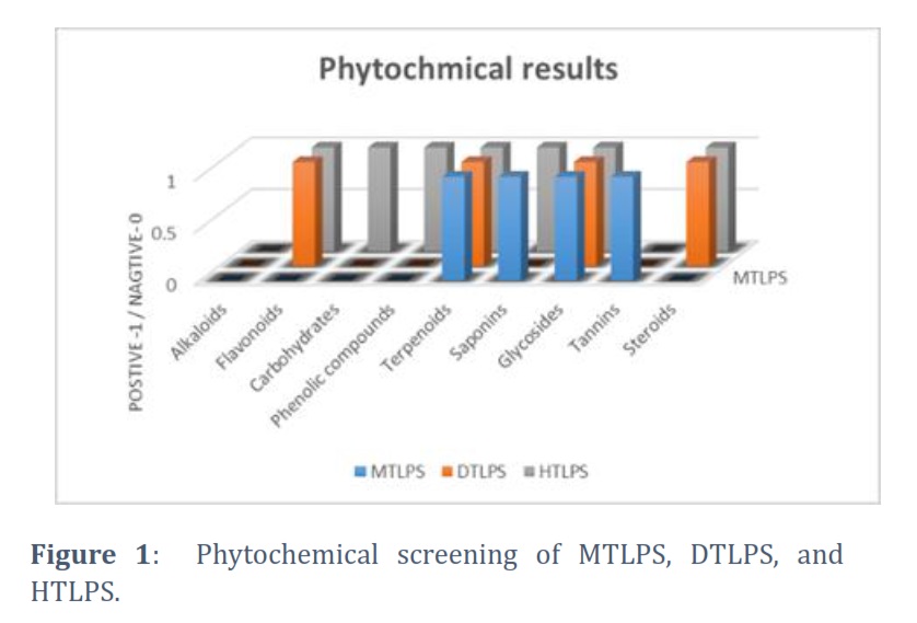
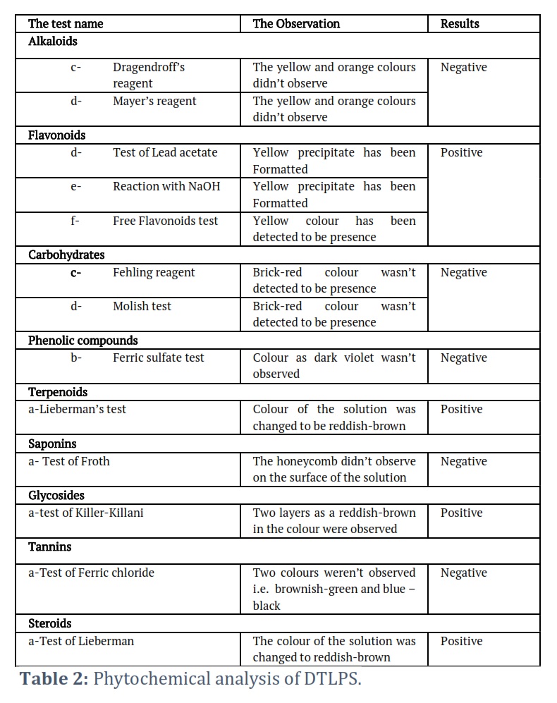
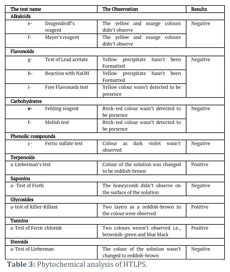
Table 1 presents the phytochemical analysis of MLPS, revealing the existence of carbohydrates, flavonoids, glycosides, phenolic compounds, terpenoids, tannins, and steroids, whereas there were no alkaloids or saponins. Table 2 demonstrates that glycosides, flavonoids, and terpenoids were found in the phytochemical screening of DTLPS, while carbohydrates, alkaloids, tannins, saponins, and phenolic chemicals weren’t detected to be present. TLPS were extracted using hexane to detect the possible presence of the non-polar compounds in it. Table 3 shows the phytochemical results of HTLPS. Four of nine chemical groups evaluated in the current study were detected to be present, i.e., glycosides, terpenoids, tannins, and saponins.
Figure 1 shows the presence of phytochemicals groups in the prepared samples, indicating the presence of terpenoids and glycosides in all samples, but no alkaloids were observed.
GC-MS of TLPS
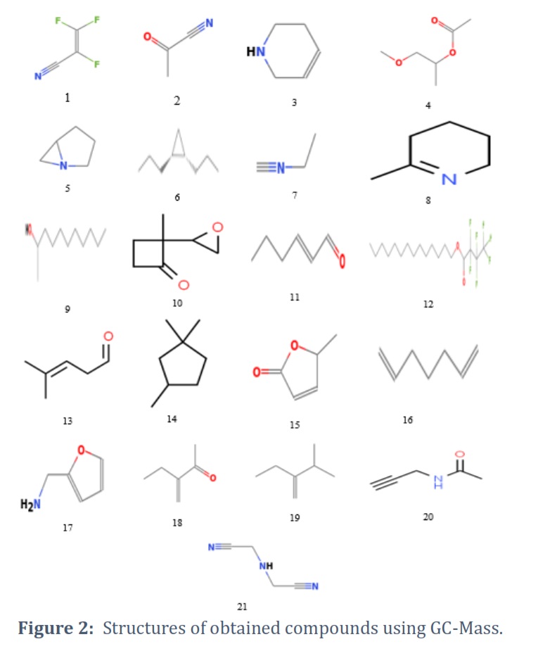
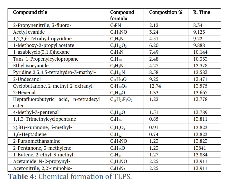
MeOH was utilized to extract TLPS, and the photochemical screening portion revealed that MTLPS had more chemical groups than DTLPS and MTLPS. GC-MS with MTLPS was used to identify the chemical substances. This section’s data identified 21 compounds in the TPLS. As shown in the table4, cyclobutanone,2-methyl-2-oxirany (10), 2-Undecanol(9), and Pyridine,4, 2,3,4,5-tetrahydro-3-methyl (8) were discovered in higher percentages, whereas 2(5H)-Furanone, 5-methyl-(15), 1,1,3-Trimethylcyclopentane(3), and 1,6-Heptadiene(16), were found in lower percentages. Figure 2 shows the chemical structures of the twenty-one chemical compounds obtained from GC-MS spectroscopy of the leaves of Polyalthia sclerophylla.
Cytotoxicity study of TLPS
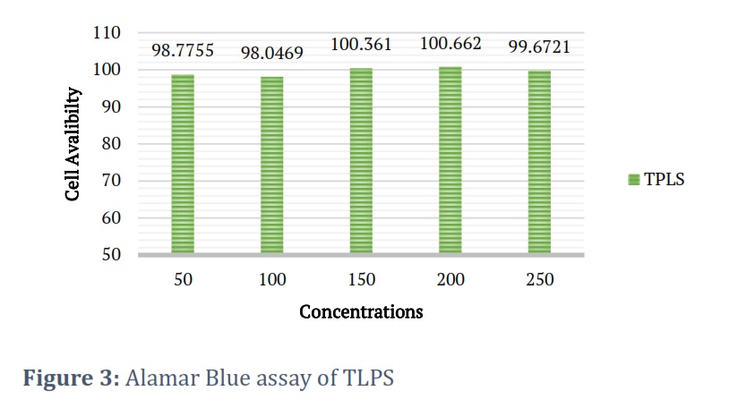
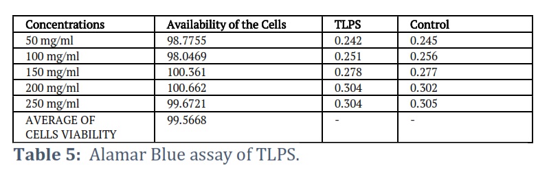
A cytotoxicity research for MTLPS was conducted in this section to determine whether or not this plant is hazardous. One of the most significant tests for medicinal materials is their poisonous impact, because of the influence on the final product that may be utilised therapeutically. The M-63 human cell line was exposed to five different concentrations of MLPS. Table 5 shows that all concentrations of MTLPS had no harmful impact. Previous research on the Polyalthia genus has revealed that its species have a nontoxic impact [35]. The current study’s findings revealed that TLPS had no harmful impact.
Figure 3 shows the availability of M-63 human cell line in the different five concentrations of leaves of Polyalthia sclerophylla. The results that obtained were observed there were no toxic effect of the extract on the MG-63 human cell and the availability of the cells were 98%.
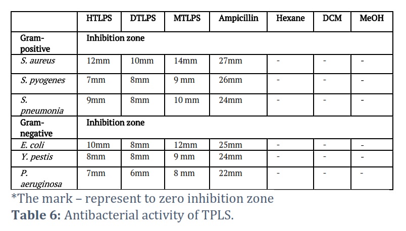
Antibacterial activities of leaves of Polyalthia sclerophylla
TLPS was tested for its antibacterial activity against six bacterial species, including gram-negative and gram-positive. As shows in table 6 MTPLS, DTLPS, and HTLPS were found to be effective against gram-negative bacteria. Substantial suppression of bacterial growth was seen at 8, 9, and 12mm (a), 6, 7, and 8mm (b), and 7, 8, and 10mm (c), respectively. The study discovered that crude extracts of MPLS, DLPS, and HLPS successfully suppressed the growth of gram-positive bacteria, S. pneumonia, S. pyogenes, and S. aureus, with growth rates of 10, 9, and 14mm (a), 8, 8, and 10mm (b), and 9, 7 and 12mm (c), respectively. The solvent utilised affects bacterial growth suppression, with methanolic extract having a higher effect on both bacteria. Grammes showed a smaller effect than dichloromethane extract.
Figures & Tables
Previous research has employed MeOH as a solvent for phytochemical screening, i.e., Kujur et al. [28] investigation on Stevia rebaudiana leaves, which discovered phenolic compounds, saponins, tannins, and steroids, but no alkaloids were detected. Kaur et al. [29] discovered carbohydrates, glycosides, flavonoids, saponins, and tannins in Caesalpinia sappan leaves and no alkaloids. These results are consistent with our present research, identifying the accessible chemical groups in plant leaves responsible for their actions. The presence of these chemicals has been approved, which has resulted in MTPLS actions.
In several investigations, DCM was formerly utilized as a solvent to extract medicinal plant leaves and assess chemical contents. Many chemical group substances have been recognized as being present. DCM extract of Adenanthera pavonina L., (DEAP) was observed to be available for terpenoids, steroids, flavonoids, alkaloids, and tannins; in contrast, extract of Euodia ridleyi (DERE) was observed to be available for terpenoids, flavonoids, tannins, alkaloids, and steroids, and while phenolic compounds and tannins didn’t detect in DERE and saponins and carbohydrates weren’t detected in DEAP [30, 31]. Both of these experiments had essentially identical results.
Previous studies on extracting medicinal plants using hexane revealed compounds; flavonoids, steroids, tannins and saponins, [32, 33]. Our findings corresponded with their findings.
To extract TLPS, three solvents are currently used: MeOH, DCM, and Hexane. The data show no alkaloids were detected in any extracts, while terpenoids and glycosides are present in MTLPS, DTLPS, and HTLPS. Figure 1 shows the absence of steroids and flavonoids in HTLPS and present in MTLPS and DTLPS. Hexane is a non-polar solvent. However, chemical groups require polar solvents to dissolve [34].
GC-MS analysis is used in the study to evaluate the chemical structures of twenty-one compounds, as depicted in Figure 2. Three compounds had a more significant proportion of functional groups: compounds 8, 8,9, and 10, highlighting the TLPS’s potential in biological sectors. These compounds have a high concentration of productive groups such as NH2, (C(=O)OH), OH, and CH3, indicating their potential for use in various biological applications.
Two critical parameters must be addressed in this section based on the data received. The efficacy of crude extracts in inhibiting bacterial growth is influenced by various factors, including the solvent used in extracting medicinal plants and the compounds present in the plants. Three solvents were examined, each with varying effects on bacterial growth. MTPLS revealed more chemical groups in crude, suggesting more significant potential for bacteria, while DTLPS had fewer chemical groups and less antibacterial activity, indicating a different impact on bacterial growth. The second point concerns TLPS, which has been shown to have a beneficial impact on bacteria. The Polyalthia genus is known for its antibacterial properties against various types of bacteria [36, 37]. Furthermore, TLPS has shown high anti-HIV activity; nevertheless, The TLPS was isolated and evaluated for antibacterial activity against all species of bacteria investigated in the current investigation.
The current study used three solvents to extract TLPS, analyse its chemical constituents, and investigate its biomedical and biological properties. In GC-MS spectroscopy, the methanolic extract of TLPS revealed 21 components. Three of the 21 compounds were found in high percentages, compound number, 8,9, and 10. While three were found in low percentages, compounds 3, 3,15, and 16, The researchers discovered that three crude extracts had substantial antibacterial efficacy against all bacterium species, indicating they can be used as antibacterial agents. MTLPS had a more significant impact than the other extracts. These crudes are allowed for biomedical safety as the cytotoxicity testing showed no hazardous effects on MG-63 cells. Our research will serve as a valuable resource for future TLPS research.
Conflict of Interest
The authors declare that there is no conflict of interest.
All authors contributed equally in conducting and reporting this research.
![]() References
References
- Firdous SM, Ahmed SN, Hossain SM, Ganguli S, Fayed MA. Polyalthia longifolia: phytochemistry, ethnomedicinal importance, nutritive value, and pharmacological activities review. Medicinal Chemistry Research, (2022);31(8):1252-64.
- Dashora A, Rathore K, Raj S, Sharma K. Synthesis of silver nanoparticles employing Polyalthia longifolia leaf extract and their in vitro antifungal activity against phytopathogen. Biochemistry and Biophysics Reports, (2022); 31(9):101320.
- Kolaprath MK, Benny L, Varghese A. A facile, green synthesis of carbon quantum dots from Polyalthia longifolia and its application for the selective detection of cadmium. Dyes and Pigments, (2023); 210(2):111048.
- Annapureddy PK, Kishore N. Non-isothermal pyrolysis of Polyalthia longifolia using thermogravimetric analyzer: Kinetics and thermodynamics. Journal of Renewable and Sustainable Energy, (2023); 15(5):053101.
- Ghous M, Dogar Na, Hanif A, Jabbar M. Phytochemical Analysis And Anti-Oxidant Potential Of Ethanolic Extract Of Polyalthia Longifolia Leaves. Pakistan Journal of Science, (2023);75(02):434-8.
- Zhangxin Y, Xinming S, Yuan Y, Jun Y, Shuo Y, Xiaobao L. Anti-inflammatory Clerodane Diterpenoids from Polyalthia longifolia. Chinese Journal of Organic Chemistry, (2023);43(2):751.
- Siddappa RY, Aditya Rao SJ, Usha BM, Verma B, Mahadevappa P. Anti-proliferative Activity of Labdane Diterpenes Isolated from Polyalthia cerasoides and their Molecular Interaction Studies. Current Drug Discovery Technologies, (2022);19(5):78-85.
- Zareen S, Adnan M, Khan SN, Alotaibi A. Anti-plasmodial potential of selected medicinal plants and a compound Atropine isolated from Eucalyptus obliqua. Open Chemistry, (2023);21(1):20220281.
- Maulana I, Fasya D, Ginting B. Biosynthesis of Cu nanoparticles using Polyalthia longifolia roots extracts for antibacterial, antioxidant and cytotoxicity applications. Materials Technology, 2022;37(13):2517-21.
- Lo IW, Liao GY, Lee JC, Chang CI, Wu YC, Chen YY, Liu SP, Su HJ, Liu CI, Kuo CY, Lin ZY. Novel Aporphine-and Proaporphine–Clerodane Hybrids Identified from the Barks of Taiwanese Polyalthia longifolia (Sonn.) Thwaites var. pendula with Strong Anti-DENV2 Activity. Pharmaceuticals, (2022);15(10):1218.
- Mudhafar M, Zainol I. Medical values, antimicrobial, and anti fungal activities of Polyalthia genus. International Journal of Pharmaceutical Research, (2019) ;11(1): 90–96
- Firdous SM, Ahmed SN, Hossain SM, Ganguli S, Fayed MA. Polyalthia longifolia: phytochemistry, ethnomedicinal importance, nutritive value, and pharmacological activities review. Medicinal Chemistry Research, (2022); 31(8):1252-64.
- Shinde PK, Kokate RH, Gawade GS. Physicochemical, phytochemical, biological and chromatographic evaluation of Polyalthia longifolia plant leaves-A review. Research Journal of Science and Technology, (2023);15(1):41-8.
- Alsailawi HA, Mudhafar M, Hanan AH, Ayat SS, Dhahi SJ, Ruaa KM, Raheem HA. Phytochemical screening and antibacterial activities of antiaris toxicaria stem, Polyalthia rumphii leaves and Polyalthia bullata stem extracts. InAIP Conference Proceedings, (2023); 2845 (1). AIP Publishing.
- Ghous M, Dogar NA, Hanif A, Jabbar M. PHYTOCHEMICAL ANALYSIS AND ANTI-OXIDANT POTENTIAL OF ETHANOLIC EXTRACT OF POLYALTHIA LONGIFOLIA LEAVES. Pakistan Journal of Science, (2023);75(02):434-8.
- Bhatt B, Chaurasia H, Singh R, Kaushik S. Phytochemical Profile and in vitro Sun-Protective Activity of Polyalthia longifolia (Sonn.) Thwaites Bark Extracts. Trop J Nat Prod Res, (2022);6(8):1174-7.
- Dashora A, Rathore K, Raj S, Sharma K. Synthesis of silver nanoparticles employing Polyalthia longifolia leaf extract and their in vitro antifungal activity against phytopathogen. Biochemistry and Biophysics Reports, (2023);1:101320.
- Mudhafar M, Alsailawi HA, Zorah M, Karhib MM, Zainol I, Kadhim FK. Biogenic Synthesis and Characterization of AgNPs Using CEPS: Cytotoxicity and Antibacterial Activites. Journal of Advanced Research in Fluid Mechanics and Thermal Sciences, (2023);106(1):65-75.
- Baqir S. Exploration of Antimicrobial Activities of an Ethnobotanically important Tree Terminalia arjuna of Family Combretaceae. GU Journal of Phytosciences, (2022);2(4):228-34.
- Parusnath M, Naidoo Y, Singh M, Rihan H, Dewir YH. Phytochemical Composition of Combretum molle (R. Br. ex G. Don.) Engl. & Diels Leaf and Stem Extracts. Plants, (2022);12(8):1702.
- Iyekowa O, Uwumarongie OH, Emmanuel KC, Omoagbotse PI, Okhions SO, Izuegbunam CL, Oligie MH, Eduwuirofo LO, Iyekekpolor RM, Okafor VC, Michael OT. Phytoconstituents, Acute Toxicity and In-Vivo Anti-anxiety Activity of Chloroform and Ethylacetate Extracts of Datura Stramonium in Balb/C Mice. Journal of Science and Technology Research, (2023);5(1):158-70.
- Shree TJ, Poompavai S, Begum SM, Gowrisree V, Hemalatha S. Cancer-fighting phytochemicals: another look. J Nanomedine Biotherapeutic Discov. (2019);8:162:1-7.
- Mudhafar M, Zainol I, Alsailawic HA, Karhib MM, Zorah M, Alnagdi FH, Hamzah MS. Bioactive chemical constituents of three crude extracts of Polyalthia Sclerophylla using GC-MS and phytochemical screening and their antibacterial and cytotoxicity activities. Eurasian Chemical Communications, (2023);5:675-90.
- Shaikh JR, Patil MK. Qualitative tests for preliminary phytochemical screening: An overview. International Journal of Chemical Studies, (2020);8(2):603-8.
- Siddappa RY, Aditya Rao SJ, Usha BM, Verma B, Mahadevappa P. Anti-proliferative Activity of Labdane Diterpenes Isolated from Polyalthia cerasoides and their Molecular Interaction Studies. Current Drug Discovery Technologies, (2022);19(5):78-85.
- Ahmed M, Ji M, Qin P, Gu Z, Liu Y, Sikandar A, Iqbal MF, Javeed A. Phytochemical screening, total phenolic and flavonoids contents and antioxidant activities of Citrullus colocynthis L. and Cannabis sativa L. Appl. Ecol. Environ. Res, (2019);17(3):6961-79.
- Hossain MA, AL-Raqmi KA, Al-Mijizy ZH, Weli AM, Al-Riyami Q. Study of total phenol, flavonoids contents and phytochemical screening of various leaves crude extracts of locally grown Thymus vulgaris. Asian Pacific journal of tropical biomedicine, (2013);3(9):705-10.
- Kujur RS, Singh V, Ram M, Yadava HN, Singh KK, Kumari S, Roy BK. Antidiabetic activity and phytochemical screening of crude extract of Stevia rebaudiana in alloxan-induced diabetic rats. Pharmacognosy research, (2010);2(4):258.
- Kaur H, Amini MH, Prabhakar PK, Singh A, Suttee A. Phytochemical screening and antimicrobial activity of Caesalpinia sappan L. leaves. International Journal of Pharmacognosy and Phytochemical Research, (2016);8(6):1040-5.
- Maskam NA, NH MR, HH MH. Phytochemicals screening and antioxidant activity of three different solvent extracts of Euodia redleyi leaves. Mod. Agric. Sci. Technol, (2017);3(5):18-21.
- Arzumand Ara M, Saleh-e-In M, Ahmed NU, Ahmed M, Abul M. Phytochemical Screening, Analgesic, Antimicrobial and Anti-oxidant Activities of Bark Extracts of Adenanthera pavonina L.(Fabaceae). Advances in Natural and Applied Sciences, (2010);4(3):352-60.
- Mudhafar M, Zainol I, Alsailawi HA, Zorah M, Karhib MM. Preparation and characterization of FsHA/FsCol beads: Cell attachment and cytotoxicity studies, Heliyon. (2023);9(5).
- Dhawan D, Gupta J. Research article comparison of different solvents for phytochemical extraction potential from datura metel plant leaves. International Journal of Biological Chemistry 2017);11(1):17-22.
- Hemagirri M, Sasidharan S. In vitro antiaging activity of polyphenol rich Polyalthia longifolia (Annonaceae) leaf extract in Saccharomyces cerevisiae BY611 yeast cells. Journal of ethnopharmacology, 2022;290):115110.
- Zorah M, Mudhafar M, Majhool AA, Abbood SF, Alsailawi HA, Karhib MM, Mustapa IR. Review of the Green Composite: Importance of Biopolymers, Uses and Challenges. Journal of Advanced Research in Fluid Mechanics and Thermal Sciences, (2023);111(1):194-216.
- Vinay, K. Antibacterial activity of crude extracts of Spirulina platensis and its structural elucidation of bioactive compound. Journal of Medicinal Plants Research, (2011);5(32), 7043-7048.
- Alja’afreh IY, Alaatabi RM, Hussain Aldoghachi FE, Mudhafar M, Almashhadani HA, Kadhim MM, Hassan Shari F. Study the antioxidant of Matricaria chamomilla (Chamomile) powder: In vitro and vivo. Revis Bionatura, (2023); 8 (1) 63.
This work is licensed under a Creative Commons Attribution-Non Commercial 4.0 International License. To read the copy of this license please visit: https://creativecommons.org/licenses/by-nc/4.0