

Short Communication
Pathological and Biochemical Methods for Detection of Uranium Pollution in the Diyala-bridge Region, Iraq
Hashim M. Obaid1*, Bushra I. Al. Kaisi2
Adv. life sci., vol. 11, no. 1, pp. 263-267, February 2024
*– Corresponding Author: Hashim. M. Obaid (dr_hashimalshumay@moen.gov.iq)
Authors' Affiliations
2. Department of pathology and poultry, College of Veterinary Medicine, University of Baghdad, Baghdad – Iraq
[Date Received: 16/10/2022; Date Revised: 27/12/2023; Date Published: 25/02/2024]
Editorial Note on Version of Record
27 May 2025: This article has been corrected. See https://doi.org/10.62940/als.v13i0.4281 for more information.
Abstract![]()
Introduction
Methods
Results
Discussion
References
Abstract
Background: The present study comprised two distinct experiments. The first experiment involved a field survey conducted in the Diyala Bridge area of Baghdad, Iraq, spanning from December 2021 to May 2022. Soil, milk, and blood samples were collected from locally grazed cattle to ascertain uranium concentrations. In the second experiment, rats were subjected to poisoning with Uranyl acetate. Upon completion of the experiment, samples of liver, kidneys, and blood were collected for histopathological and biochemical analyses.
Methods: Twenty-four albino male rats were divided equally and randomly into two groups i.e., control group and intoxicated group. Uranyl acetate was given daily for 120 days by 0.05 mg/kg to the latter group. At the end of the experiment liver and kidney sample were taken for histopathological examination and stained by Masson-trichrome stain.
Results: The finding of eosin and hematoxylin stain and biochemical assay for CuZn-SOD U/mg and catalase K/mg protein were showed significantly elevated while liver tissue showed necrosis atrophy atherosclerosis cirrhosis and carcinoma characterized by irregular cell mitotic figure extensive fibrous tissue, tissue kidney presented interstitial hemorrhage and multiple granuloma.
Conclusion: Intoxicant by Uranyl acetate showed significant elevating for CuZn-SOD U/mg and catalase K/mg protein, in contrast liver tissue showed necrosis atrophy atherosclerosis cirrhosis and carcinoma, tissue kidney presented interstitial hemorrhage and multiple granulomas.
Keywords: Uranyl acetate; Granuloma; Xenobiotic; Granuloma
Introduction![]()
Pollution has been a global problem for a long time yet just began to be critical after the biotransformation of chemical material like dioxin radioactive pollutant, etc. [1]. Uranium occurs naturally in very small amounts in rocks, soils, water bodies, plants and animals. The history and use of uranium reflect advances in mining, chemistry and physics, and in our understanding of radioactive substances and their potential applications [2]. Earth’s crust contains Uranium as a natural component, and it exists naturally in oxidative state [3]. Uranium reach human via food like table salt vegetables cereals which transfer from soil and pollutant soil to plant and human or animals’ product mostly (milk, Eggs, meat and fish meat) [4, 5]. Uranium forms are soluble complexes with bicarbonate, citrate or proteins in the plasma when immediately absorbed from tissue and bone [6]. And is directly harmful to the lungs and kidneys [7]and its compounds cause changes in the kidney, lung, liver, nerves, circulation, reproductive system, hemopoietic, DNA deformation and decrease bone development and its known carcinogenic and genotoxic agents [8, 9]. Uranyl nitrate molecular formula UO2 (NO3), 6H2O with molecular weight 502.13 is a water soluble salt it’s a radioactive and oxidizing compound composed of yellow bipyramidal rhombic crystals it easily absorbed by human pulmonary, cutaneous and GIT routes [10&11].
One of the major sites of Uranium pollution in Iraq is AL-Tuwaitha nuclear research site which is located [18] km South west Baghdad and in the Diyala Bridge areas (AL-Arifi and Altaamim Distract) and at Gulf War in 1981 was destroyed and mostly Nowruz reactor in AL-Tuwaitha nuclear research [12].
Methods![]()
Sample collection
Collection of the soil samples
Randomly 100 soil samples were collected from the lands of Diyala Bridge area like (AL-Tuwaitha, AL-Arifi and Altaamim Distract) Soil samples about 500gm were collected by cutting out the soil (50-100)cm depth then the samples saved in glass containers for laboratory test. Soil samples in laboratory were grinding in mortar and Sieve in fine mesh about 75 m then the soil powder after homogenized about 1 gm mixed with 0.2 gm. of methylacellouse then pressed using a piston into a pellet with dimension of 1 cm. diameter and 1.5 mm thickness these pellet to make a pair were covered with CR-39 debtor on both sides [13, 14].
Cattle blood collection
Hundred blood samples were collected from both sexes randomly from local grazing cattle in Diyala Bridge area and blood about 10 CC collected from cattle jugular vein, labeled and saved in heparinized tube in cold box for Uranium detection [13, 14].
Collection milk sample
Collection from female cattle 100 milk samples were collected daily from the cattle above and area two drops of milk by drying milk and left square piece of CR-39 detector at room temperature for Uranium detection sample save in glass container [13, 14]. Natural Uranium stander samples were prepared according to (IAEA) International Atomic Energy Agency [13, 14]. The experiment analysis above was done in the Ministry of Science and Technology in Baghdad, Iraq.
Experimental animals
The doses of uranyl acetate (0.05) mg/kg body weight were determined. of uranyl acetate according to [15] and is administration orally. Twenty – four albino male rats aged (120) days and body weight (200-250) gm. were housed in sterilized polypropylene cages (120*120*30) Cm3 in the animal house of the department of Pathology, Collage of Veterinary Medicine, Baghdad University, Iraq, in twelve hours dark light cycle with (21 0C)ambient Celsius degree temperature. All rats were given normal rat pellet and drinking water ad libitum [16].All animals were under identical management protocol and guide line of Institutional Animal Ethics Committee.
Intoxication of rats
Twenty – four albino male rates were divided randomly and equally into two groups:-1st group (12) animals act as control group (C) received normal rat diet and water ad libitum while 2end group (12) albino male rats daily and orally administration ( 0.05 )mg/kg B.W. of Uranyl acetate for 120 days. Uranyl acetate dihydrate UO2 (CH3COO)2H2O (Purity99%) was obtained from AccuStandard Inc (New Haven, Connecticut, USA). All rats scarified under ethics protocol under ether anesthesia at 120 days for biochemical and pathological examination.
Blood collection
Cardiac puncture technique on fasting blood were collected (3 ml) and then blood left (15) minutes to stand and coagulate in refrigerator then for (15) minutes at (500) rpm in centrifuge. Serum was separated and kept at (-20) CO till assay.
Biochemical assay
All below chemicals (purity ≥99%) were obtain from Sigma chemical Co.(St. Louis, Missouri, USA) and were of analytical grade or of the highest grade available and were done in laboratory of Science and Technology Ministry, Baghdad, Iraq.
a. CuZn-SOD (Iu/mg) activity [17] was measured by inhibition of nitro blue tetrazolium (NBT) reduction due to O2 generation by the xanthine / xanthine oxidase system and marked by spectrophotometer at (560nm).
b. Catalase (CAT) activity (k/mg) [18]. The enzymatic decomposition of H2O2 followed by decrease in absorbance at (240nm).
c. Serum creatinine (mg/dl) [19].
Histopathological change
At days (120) all animals groups were sacrificed and liver and kidney were carefully dissected from abdominal cavity then immediately fixed in 10% neutral buffered formalin for routine histopathological technique [20]. All tissue stained with Masson trichrome stain for fibrous connective tissue detection and eosin and hematoxylin stain.
Statistical analysis
All the groups’ data were statistically read by SPSS program, version 17software (2010).Including one way ANOVA for comparisons among groups. P value of less than ≤0.05 were considered statistically significance, all data were expressed as means ±standard error (SE) [21].
Results![]()
Determination of Uranium soil (mg/kg), blood and milk (mg/L)
Uranium concentration in milk (0.43±4.22) mg/L, in blood (0.55±4.22) Mg/L and in soil (6.23±0.54) mg/kg samples were showed significant increased (P≤0.05) when compare to minimum acceptable Uranium concentration in milk (1.2±0.00) Mg/L, in blood (1.8±0.00) Mg/L and in soil (2.8±0.00) mg/kg
Biochemical assay
In current study the level of SOD, CAT and protein in 2nd group which administrated (0.05) mg/kg B.W showed significant decreased p≤0.05 in CuZn-SOD U/mg protein (255.0 ±16.2) when compared with control group (370.0±12.0 )U/mg protein.
Catalase k/mg protein in 2nd group decreased significantly (p≤0.05)(4.50±0.3) when compared with control group(8.60±0.4) k/mg.
Serum concentration of creatinine (mg/dL) indicated significant increased p≤0.05 in Uranium group when compared to control group.
Histopathological Examination
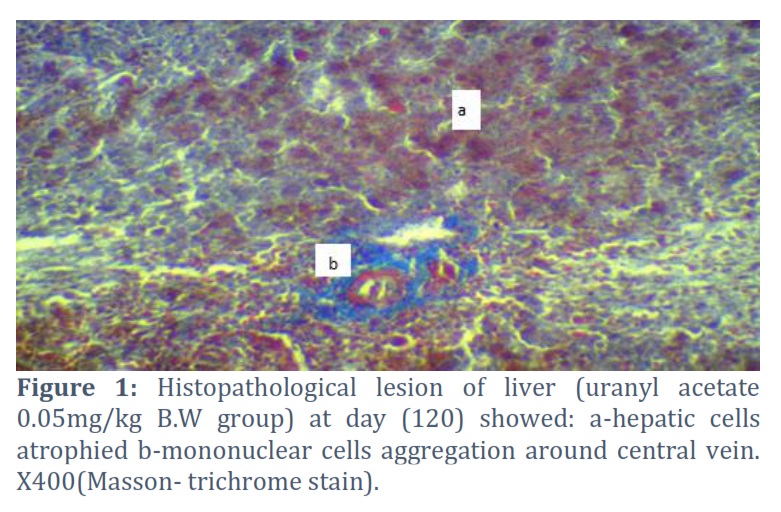
No important histopathological lesions were observed in livers and kidney of 1st group (control group), while 2nd group at (120) days livers showed hepatic cells atrophied with increased width of sinusoids figure (1).
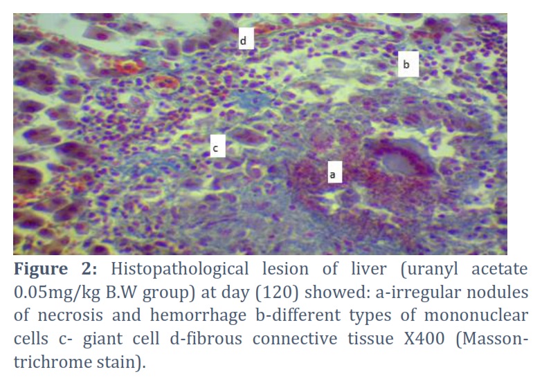
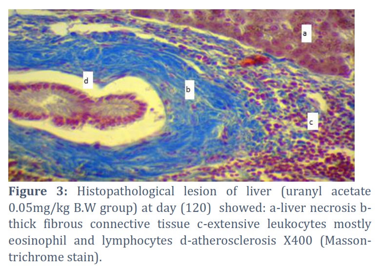
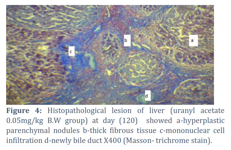
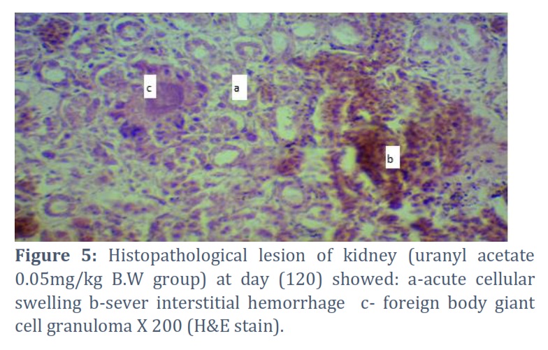
Granulomatous areas showed increase in the fibrous connective tissue with blue coloration, other liver cells consist a huge amount of leukocytes mostly eosinophil’s and lymphocytes, collapsed of all arteries with sever atherosclerosis figure (3). Liver cirrhosis characterized by hyperplastic parenchymal nodules separated by fibrous bands with distortion of the normal liver architecture with moderate infiltration of mononuclear cells with newly bile duct fig(4). Kidney section in 2nd group presented sever acute cellular swelling, sever interstitial hemorrhagic with granuloma formation consist of (necrotic center surrounded by foreign body giant cells) Fig (5).
Figures & Tables
Our results showed significant increase in Uranium concentration in soil, milk and blood of cattle when compared to minimum accepted uranium concentration. Uranium accumulation in Diyala bridge area like (AL-Tuwaitha), AL-Arifi and Altaamim) District due to war (1981) and terrorism [22] also gulf war resulting in presence radioactive of Uranium in soil surface and the invade the deeper layer of ground [23]. The bioaccumulation of soil uranium in the above areas that act as potentially long-term source of food contamination because Uranium half-life(4.5 x 10⁹) years [24], when ingested randomly by cattle which grazing in these contaminated areas accumulate in milk, blood, meat and different tissue like kidney, bone, brain and mostly liver [25]. The current study showed significant decreased in CuZn-SOD and catalase with in creatinine which induction of oxidative stress in tissue and may be due to lipid peroxidation causing increase cell fluidity and cell death due to oxidative stress and free radical [26]. Liver is often show by toxic effects of xenobiotic due to its ability to accumulate xenobiotic by active transport [27]. Uranium has ability to accumulate in hepatocytes and cause toxicity [28 and 29], by induce oxidative stress in mitochondria or lysosome [30]. In rats hepatocytes uranium is potential oxidative marker including reactive oxygen species (ROS) formation and generation excessive (ROS) in liver. Liver cirrhosis is the global lesion occurs due to liver damage from toxin which causes losses of liver function in human and animals [31]. Formation of pseudolobules without central vein surrounding by fibrous connective tissue (cirrhosis) to maintain regeneration of hepatocyte due to Uranium mutagenicity effect of uranium genetic mutation causing liver carcinoma and such effects can include genetic mutations generation of (ROS) lead to liver carcinoma [32]. Chronic toxicity due to Uranium exposure experimental due to excreted via kidneys especially in proximal convoluted tubules causing renal tubular epithelial degeneration, necrosis with inflammatory cells infiltration and increased serum creatinine due to brush cell injury and alteration in cells and lysosome membrane with injury to mitochondria leading to impaired energy and altered calcium ion hemostasis, uranium induction cellular oxidative stress [33], by increased lipid peroxidation and depletion of antioxidant enzyme[34], causing vacuolation mesangial and epithelial cells of glomeruli, dilated renal tubules, interstitial fibrosis with mononuclear cells infiltration[35], with fibroblastic proliferative by produce protein like collagen I, IV and fibronectin-growth- factor β (TGF-β) and platelet- derived growth factor (PDGF) [36].
Author Contributions
Hashim M. Obaid: Research article, funding the acquisition and experiment design.
Bushra. I. al. Kaisi: Preparing materials, explain the finding, statistical analysis, review and editing.
The authors declare that there is no conflict of interest.
![]() References
References
- Humadi A, Sabeeh I, Al-Kaisei I, Al-Ezzy I. Toxicopathological and Biochemical impacts of 2, 3, 7, 8 Tetrachlorodibenzo-P-Dioxin (TCDD) on liver of Albino male rats. International Journal of Pharmaceutical Research, (2021); 1(13): 2536-2543
- Balaram A, Rani A, Rathore D. Uranium in groundwater in parts of India and world: A comprehensive review of sources, impact to the environment and human health, analytical techniques, and mitigation technologies. Geosystems and Geoenvironment, (2022); 1(2):100043
- Brugge D, Jamie L, deLemos L, Beth O. Exposure pathways and health effects associated with chemical and radiological toxicity of natural uranium: a review. Reviews on environmental health, (2005); 20(3): 177-194.
- AL-Tameemi H. Radiological and chemical hazards of public exposure to uranium-235 at Al-Twaitha nuclear research site. Iraqi Journal Biotechnol, (2010); 9(2): 226-38.
- Priest D. Toxicity of depleted uranium. The Lancet, (2001); 357(9252): 244-246.
- Cooper R, Stradling N, Smith H. The behaviour of uranium-233 oxide and uranyl-233 nitrate in rats. International Journal of Radiation Biology and Related Studies in Physics, Chemistry and Medicine (1982); 41(4): 421-433.
- Patocka J. Human health and environmental uranium. Military Medical Science Letters, (2014); 83.3: 120-131.
- Craft S, Abu-Qare W, Flaherty M, Garofolo C, Rincavage L, Abou-Donia B. Depleted and natural uranium: chemistry and toxicological effect. Journal of Toxicology and Environmental Health, (2004); 7(4) : 297_317
- Hartmann M, Frederick A, Halil A. Overview of toxicity data and risk assessment methods for evaluating the chemical effects of depleted uranium compounds. Human and Ecological Risk Assessment, (2000); 6.5: 851-874.
- Kozlova D, Matyukha V, Dedov N. Mechanism and kinetics of thermal decomposition of uranyl nitrate hexahydrate under the nonisothermal conditions. Radiochemistry, (2007); 49.2: 130-134.
- Heffernan E, Lodwick C, Spitz H, Neton J, Soldano M. Solubility of airborne uranium compounds at the Fernald Environmental Management Project. Health Physics, (2001); 80(3): 255-262.
- Al-Shammari M. Environmental pollutions associated to conflicts in Iraq and related health problems. Reviews on environmental health, (2016); 31(2): 245-250.
- Aziz. A, Majed M, & Tawfiq F. Evaluation of Uranium Concentration in the Blood of Cancer Patients in Salah Al-Din Governorate. Tikrit Journal of Pure Science, (2021); 26(2): 94-97.
- Hassan A. Determination of uranium in fishes samples from selected regions in Iraq using neutron activation technique for nuclear track detectors. AL-Qadisiyah Journal of pure Science, (2017); 22(2): 47-59.
- Domingo L, Ortega A, Paternain L, Corbella J. Evaluation of the perinatal and postnatal effects of uranium in mice upon oral administration. Archives of Environmental Health: An International Journal, (1989); 44(6): 395-398.
- Humadi A, Bushra I, Taghreed H. Acrylonitrile testicular seminoma in beagle male dogs (Pathological and Hormonal assay). Plant Archives, (2020); 20(1): 1903-1909
- Ciftci O, Sadettin T, Ahmet G. Protective effect of curcumin on immune system and body weight gain on rats intoxicated with 2, 3, 7, 8-Tetrachlorodibenzo-p-dioxin (TCDD). Immunopharmacology and Immunotoxicology, (2010); 32(1): 99-104.
- Damiano S, Longobardi C, Andretta E, Prisco F, Piegari G, Squillacioti C, Ciarcia R. Antioxidative effects of curcumin on the hepatotoxicity induced by Ochratoxin A in rats. Antioxidants, (2021); 10(1): 125.
- Henry J, Cannon D, Winkelman W. Clinical chemistry. Principles and techniques,2nd Edition, Harper and Row, Hagerstown, (1974); 525-527
- Humadi A, Bushra I, Marah S. Pathological and hormonal effects of 2, 3, 7, 8- Tetrachlorodibenzo-p-dioxin (TCDD) Versus antioxidant activity of Curcumin in Sprague dawley male rats. Veterinary Practitioner, (2020); 21(2): 439-443
- Duncan B. “Multiple range and multiple F tests.” Biometrics, (1955); 11(1): 1-42.
- Brown J. Reconstructing the environment in Iraq. Environmental health perspectives, (2004); 112(8): A464-A464.
- Katsoyiannis A, Hug J, Ammann A, Zikoudi A, Hatziliontos C. Arsenic speciation and uranium concentrations in drinking water supply wells in Northern Greece: correlations with redox indicative parameters and implications for groundwater treatment.” Science of the Total Environment, (2007); 383(1-3): 128-140.
- Claiborne P. Handbook of methods for oxygen radical research. Florida: CRC Press, Boca Raton, (1985); 283-284.
- Jeambrun M, Pourcelot L, Mercat C, Boulet B, Loyen J, Cagnat X, Gauthier-Lafaye F. Study on transfers of uranium, thorium and decay products from grain, water and soil to chicken meat and egg contents. Journal of Environmental Monitoring, (2012); 14(8): 2170-2180.
- Dublineau I, Grison S, Baudelin C, Dudoignon N, Souidi M, Marquette C, Gourmelon P. Absorption of uranium through the entire gastrointestinal tract of the rat. International journal of radiation biology, (2005); 81(6): 473-482.
- Racine R, Gueguen Y, Gourmelon P, Veyssiere G, Souidi M. Modifications of the expression of genes involved in cerebral cholesterol metabolism in the rat following chronic ingestion of depleted uranium. Journal of molecular neuroscience, (2009); 38(2): 159-165.
- Mohammed A, Eyad A. The biochemical and histological effects of uranyl acetate in male New Zealand rabbits. Al-Qadisiyah Journal of Veterinary Medicine Sciences, (2015); 14(2): 25-30
- Arfsten P, Kenneth R, Glenn R. Review of the effects of uranium and depleted uranium exposure on reproduction and fetal development. Toxicology and industrial health, (2001); 17(5-10): 180-191.
- Fukuda S, Ikeda M, Nakamura M, Yan X, Xie Y. Acute toxicity of subcutaneously administered depleted uranium and the effects of CBMIDA in the simulated wounds of rats. Health physics, (2009); 96(4): 483-492.
- Asrani K, Larson J, Yawn B, Therneau M, Kim R. Underestimation of liver-related mortality in the United States. Gastroenterology, (2013); 145(2): 375-382.
- Wanless R, Eisuke N, Morris S. Regression of human cirrhosis: morphologic features and the genesis of incomplete septal cirrhosis. Archives of pathology & laboratory medicine, (2000); 124(11): 1599-1607.
- Shim S, Park H, Ahn J, Han L, Jin R, Li H, Shim K. Testosterone‐independent down‐regulation of Oct2 in the kidney medulla from a uranyl nitrate‐induced rat model of acute renal failure: Effects on distribution of a model organic cation, tetraethylammonium. Journal of pharmaceutical sciences, (2009); 98(2): 739-747.
- Sun F, Fujigaki Y, Fujimoto T, Goto T, Yonemura K, Hishida A. Relation of distal nephron changes to proximal tubular damage in uranyl acetate-induced acute renal failure in rats. American journal of nephrology, (2002); 5-6(22): 405-416.
- Hamid J. Uranyl nitrate induce histological changes in the kidney. International Journal of Engineering and Technology, (2012); 12(4): 9-13.
- Banday A, Priyamvada S, Farooq N, Yusufi K, Khan F. Effect of uranyl nitrate on enzymes of carbohydrate metabolism and brush border membrane in different kidney tissues. Food and chemical toxicology, (2008); 46(6): 2080-2088.
This work is licensed under a Creative Commons Attribution-Non Commercial 4.0 International License. To read the copy of this license please visit: https://creativecommons.org/licenses/by-nc/4.0