

Full Length Research Article
Isolation and Purification of Glycyrrhizic acid and Determination of its Biological Activity Against Microbial Pathogens
Ayat Abdaljabar Al-Kubaise, Ayyad W. Al-Shahwany*
Adv. life sci., vol. 11, no. 2, pp. 346-353, May 2024
*– Corresponding Author: Ayyad W. Al-Shahwany (ayyad.alshahwany@sc.uobaghdad.edu.iq)
Authors' Affiliations
[Date Received: 14/06/2023; Date Revised: 12/02/2024; Date Available Online: 18/04/2024]
Editorial Note on Version of Record
31 May 2025: This article has been corrected. See https://doi.org/10.62940/als.v13i0.4280 for more information.
Abstract![]()
Introduction
Methods
Results
Discussion
References
Abstract
Background: The World Health Organization estimates that 80% of the world’s population uses plant extracts or their active components in traditional therapies. Recently, there has been a lot of interest in the quest for new antioxidant and antibacterial plant-based medications. Additionally, Glycyrrhiza glabra is a commonly used food flavoring and sweetening ingredient.
Methods: This work was conducted to isolate and purify licorice root’s Glycyrrhizic acid (GA) and investigate its antimicrobial properties against S. aureus and K. pneumoniae were isolated from urine, blood, wounds, ear water, sewage, soil, and water, totaling 12 isolates of each kind, Analytical method for the measurement of Glycyrrhizic acid from the root extract that has been suitably supported by a mass spectroscopic analysis.
Results: The results showed the effects of GA with different concentrations on S. aureus and. K. pneumoniae. Obviously, the MIC ranged from 18.75 to 37.5 % for S. aureus isolates, and 4.68 to 75 for K. pneumoniae isolates. The results indicated that GA reduced the metabolic activity of cells in biofilm on S. aureus and K. pneumoniae with an an inhibition percentage
Conclusion: GA may have an effect on cell membrane permeability, biofilm formation, and efflux activity, which inhibits bacterial growth.
Keywords: Glycyrrhizin acid; Staphylococcus aureus; Klebsiella pneumoniae; Antibiotic; Biofilm
Introduction![]()
The World Health Organization estimates that 80% of the world’s population uses plant extracts or their active components in traditional therapies, since ancient times, plant sources of therapeutic chemicals have continued to play important role in preserving human health, Modern clinical medications with a greater than 50% natural product component [1]. in recent years, there has been a surge in interest in the study of plant-based natural medicines for the development of novel antioxidant and antimicrobial agents. In addition, the pharmaceutical and food industries are increasingly asking for plants that are currently used in conventional medicine, the licorice root, Glycyrrhiza glabra is a member of the Fabaceae family and is frequently used in food as a sweetener and flavor, it has been proposed for use in various clinical applications [2]. Although clinical and experimental studies indicate that licorice root has a number of other useful pharmacological properties, including anti-inflammatory, antiviral, antimicrobial, anti-oxidative, anti-cancer activities, immune modulator, hepatic protective, and cardio protective effects, it is primarily used as a traditional medicine to treat peptic ulcer, hepatitis, and pulmonary and skin diseases, There are numerous substances found in licorice that have been isolated, including triterpene saponins, flavonoids, isoflavonoids, and chalcones, while glycyrrhizic acid (GA) is typically thought to be the most physiologically active substance [3]. The therapeutic benefits of licorice are due to two of its constituents, glycone and aglycone, GA is a significant molecule that gives licorice its pharmacological and biological activities, GA is a triterpenoid disaccharide glycoside that has been shown to have anti-inflammatory, anti-diabetic, anti-allergic, and many other properties, GA is a yellow to orange powder with a molecular weight of 822.93 g/mol, the current investigation’s goal was to separate GA from licorice roots, the physicochemical properties were used to validate the extracted GA (High-performance liquid chromatography) [4]. One molecule of the triterpenoid aglycone glycyrrhizic acid, which appears in 18-α or 18-β stereoisomeric forms, is conjugated to two molecules of glucuronic acid to generate the saponin complex known as GA, Commercial licorice products are frequently made from licorice root extracts GA, sometimes referred to as glycyrrhizin or glycyrrhizinate, is thought to be the main active component in licorice root extract and makes up 10% to 25% of the root extraction [5]. Licorice is used medicinally to treat respiratory conditions like dry cough and asthma, licorice has been found to induce apoptosis in melanoma and gastric cancer cells, and GA also have anti carcinogenic, antidote, and antioxidant properties [6]. Alkaloids, glycosides, carbohydrates, starches, phenolic compounds, flavonoids, proteins, pectin, mucilage, saponins, lipids, tannins, sterols, and steroids were found in the G. glabra root during the phytochemical screening process, It shown pharmacological activities such as memory improvement, antidepressant, antibacterial, anticancer, antioxidant, and many more [7]. High-performance liquid chromatography, often known as HPLC or high-pressure liquid chromatography, is a particular type of column chromatography that is frequently used in biochemistry and analysis to separate chemicals [8]. The primary components of HPLC are a stationary phase (column holding packing material), a pump to drive the mobile phase(s) through the column, and a detector to show the molecules’ retention times. Retention time is influenced by the interactions of the stationary phase, the molecules under investigation, and the utilized solvent(s) [9]. The analysis sample is added in a tiny amount to the mobile phase stream and is slowed down by particular chemical or physical interactions with the stationary phase, the kind of analyte and the makeup of the stationary and mobile phases both affect the amount of retardation; retention time refers to the time at which a particular analyte elutes (comes out of the end of the column); popular solvents employed include any miscible mixtures of water or organic liquids (methanol and acetonitrile are the most frequently used) , Gradient elution is a separation method applied to alter the composition of the mobile phase during the analysis [10]. Multiple techniques have shown that GA exhibits strong inhibitory effects on both Gram-positive and Gram-negative bacterial activity; the majority of GA’s pharmacological activities also involve antimicrobial activity, which prevents bacterial infection by lowering gene expression, impeding bacterial development, and lessening the synthesis of microbial toxins [11].
As a commensal bacterium in humans, Staphylococcus aureus can also be harmful, S. aureus is predominantly an opportunistic infectious agent and is responsible for a number of suppurative illnesses, food poisoning, pneumonia, and sepsis [12]. Is one of the typical flora on the skin surface, the mucosal surfaces of the respiratory tract, the upper part of the digestive system, and the urogenital tract, Staphylococcus is a Gram-positive bacteria genus that belongs to the Bacillus order’s Staphylococcaceae family [13]. One of Staphylococcus aureus defense mechanisms is its ability to build biofilms. Bacteria submerged in biofilms are sometimes challenging to eliminate using conventional antibiotic regimens and are intrinsically resistant to host immune responses [14].
Additionally, Klebsiella pneumoniae contributes significantly to the transfer of environmental bacteria’s antimicrobial resistance genes to clinically significant bacteria [15]. A Gram-negative encapsulated bacterium called K. pneumoniae thrives in water, soil, and animal mucous membranes. K. pneumoniae typically colonizes the oropharynx and gastrointestinal tract in humans. From there, it can quickly spread to other tissues and the bloodstream to cause infections like bacteremia, septicemia, surgical site infections, urinary tract infections, ventilator-associated pneumonia, and pneumonias that are hospital acquired. Additionally, it helps explain why opportunistic infections are so common in those with immune system problems like diabetes or bladder neuropathy [16, 17]. Another characteristic of K. pneumoniae that is well known is its capacity to create biofilms, which are bacterial populations embedded in an extracellular matrix, proteins, exopolysaccharides, DNA, and lipopeptides make up this matrix [18]. The World Health Organization (WHO) identified antibiotic resistance as one of the three global health crises, citing a variety of mechanisms of antimicrobial resistance that may adversely affect therapeutic outcomes [19]. This study aimed to identify, and isolation of GA and investigation the effect of GA extract on S. aureus and K. pneumoniae and evaluate the biofilm effect.
Methods![]()
Collection of Plant Samples
The roots of G. glabra were purchased from the local herbal merchandise, in Baghdad, Iraq and the plant has been diagnosed in the herbarium of the College of Science – Department of Biology. They were air-dried, ground to powder and stored overnight at 4°C.
Preparation of GA Extract
Extraction using a water bath apparatus at 50°C for 4 hours, 250 g of crushed G. glabra was weighed and added to 500 ml of 30% ethanol. After cooling with continuous slow mixing, the mixture was filtered in a rotary evaporator at 60°C until a thick solution was obtained; the solution was then dried in a vacuum evaporator [20]. After that, a powder was obtained and macerated in acetone and diluted nitric acid for two hours, after filtering the contents, 20 ml more of acetone was added to the marc and gently warmed, filtrate was produced when the contents were filtered; A suitable amount of diluted ammonia solution was added to this filtrate in order to finish the precipitation of ammonium glycyrrhizinate, the precipitate was gathered, rinsed with acetone for five milliliters, dried, and collected [4]. The resulting deposit was dissolved in DMSO to prepare the doses.
HPLC Analysis of GA
A method for estimating the amount of GA in an extract once a mass spectrometric analysis has adequately supported it. However, numerous chromatographic estimation techniques have been published in which an isocratic elution is followed by a UV detector monitoring the eluent at 254 nm. A C18 column is employed with an aqueous phase containing either an acid modifier like acetic acid or phosphoric acid or a mobile phase made of either methanol or acetonitrile, an organic component [21].
Preparation of Different Concentrations of Glycyrrhizic Acid
Three grams of the dried extract were mixed with five milliliters of DMSO (dimethyl sulfoxide) to make stock solutions. Following that, various diluent concentrations (300, 150, 75, 37.5, 18.75, 9.37, and 4.68 mg mL-1) were used.
Selection of the Isolates
Twelve isolates of S. aureus and K .pneumoniae from urine, blood, wounds, ear water, sewage, soil, and water were used in this study. By utilizing the Kirby- Bauer disc diffusion method, the isolates revealed enhanced resistance to widely used antibiotics. According to Bergey’s Manual of Systematic Bacteriology, the bacteria were identified using common microbiological techniques (Gram staining, colony morphology, catalase test, cytochrome oxidase reaction, motility, and other biochemical tests)[22].
Antibiotic Susceptibility Test
4-5 colonies of bacterial isolates were taken from an overnight culture plate and suspended in 5ml of sterile normal saline until the turbidity was about similar to the McFarland No. 0.5 turbidity standard [23]. Ten minutes later, using sterile forceps, Mueller Hinton plates were covered with antimicrobial discs Amoxicillin, Ampicillin, Cefotaxime, Methicillin, and Tetracycline. For 18 to 24 hours, the plates were incubated at 37°C. After incubation, the plates were checked to see if there was an area surrounding the antimicrobial discs that inhibited bacterial growth, using a a metric ruler and the Clinical and Laboratory Standards Institute’s standard inhibition zone as a reference, the diameter of the zone of inhibition was measured in millimeters [24].
Determination of minimum inhibitory concentration (MIC)
MIC of GA extracts was estimated by microdilution method in sterile 96-well microtiter plates according to the procedure described previously, different plant extracts concentrations (300, 150, 75, 37.5, 18.75, 9.37, 4.68 and 2.34 μg/ml) (W/V) were prepared to contain bacterial cells comparable to McFarland standard no. 0.5 in a final volume of 200 μl, after 24 h at 37°C, the MIC of each sample was determined. The MIC is regarded as the lowest antimicrobial concentration that, after a 24-hour incubation period, will prevent a bacterium from showing detectable growth [25].
Biofilm Formation Assays by using Tissue Culture Plate (TCP) Method
Hassan et al., described this quantitative test. Regarded as the most effective technique for detecting biofilms [26] . In 10 ml of trypticase soy broth with 1% glucose w/v, isolated organisms from freshly prepared agar plates were inoculated; For 24 hours, broths were incubated at 37°C. Following a 1:100 dilution with fresh media, the culture was injected into each well of a sterile 96-well flat-bottom polystyrene tissue culture plate, for 24 hours, the plate was incubated at 37°C, following incubation; each well’s contents were gently tapped out. Once, sterilized distilled water was used to clean the wells; This eliminated microorganisms that were floating around, (0.1%) w/v crystal violet was used to stain the bacterial biofilm adhered to the wells. Using distilled water, extra stain was removed, and the plates were kept to dry. Using a micro-ELISA auto reader at a wavelength of 630 nm, the optical density (OD) of stained adherent biofilm was measured. The experiment was performed in triplicate and repeated three times [27].
The Effect of GA on Biofilm Formation
Biofilm formation assays were performed using 96_ well microtiter plate, based on the protocol with some modification; TSB + 1% w/v glucose was added to the cultured S. aureus and K. pneumoniae after it had been cultivated in trypticase soy broth for an overnight period, each microtiter plate well was filled with 100 μl of medium and 100 μl of glycyrrhizic acid, each concentration for every Glycyrrhizic acid tested was assayed triplicate, the plate then incubated at 37Cº for 24 hrs.; Shaking the dish over a garbage tray loaded with sterile distilled water eliminated the plank tonic bacteria, then, each well received 0.1% w/v crystal violet solution, and the plate was left to stain for 10 minutes at room temperature, the plate was then placed in a water pan to drain the crystal violet solution; The plate was then inverted, placed on paper towels, and allowed to dry by air, then, the dye was solubilized by treating the stained wells with 95% v/v ethanol for 10 min at room temperature[28]. Each well’s bacterial solution was well mixed before having its optical density measured at 630 nm in a microplate reader [29, 8]. The following formula was used to determine how much biofilm development was inhibited-mediately reduced [30].
% of biofilm inhibition = O.D. of control _ O.D. of treatment/O.D. of control × 100
Statistical Analysis
One-way analysis (ANOVA) tests were used for data analysis in SPSS version 25, the significant difference between means was compared using the least significant difference (LSD), and differences were deemed significant when P≤ 0.05 [31].
Results![]()
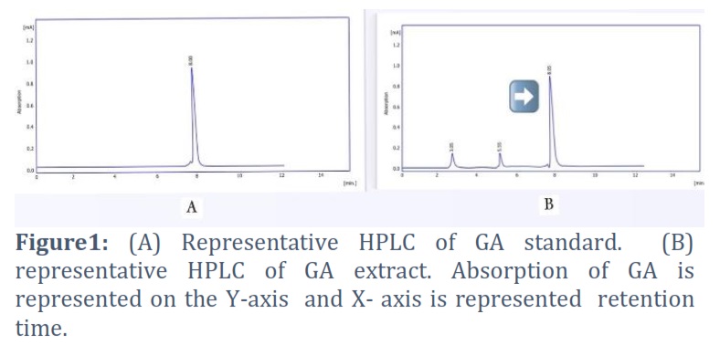
Identification of GA by HPLC
For the identification of the main bioactive of licorice like GA, an HPLC method was achieved by comparison of retention time of GA extract with standard. The result in figure 1 (A) showed the peak of standard and retention time (8.00),area (100%) , and in figure1 (B) showed peak of GA extract. The result showed the appearance GA compound in licorice extract (90%) of the total area and concentration (101 mg/gm). The concentration of GA extract calculated by the following formula:
C sample = Cstandard * A sample /Astandard *D.F/wtgm
C: concentration, A: area, D.F : dilution factor, wt: weight
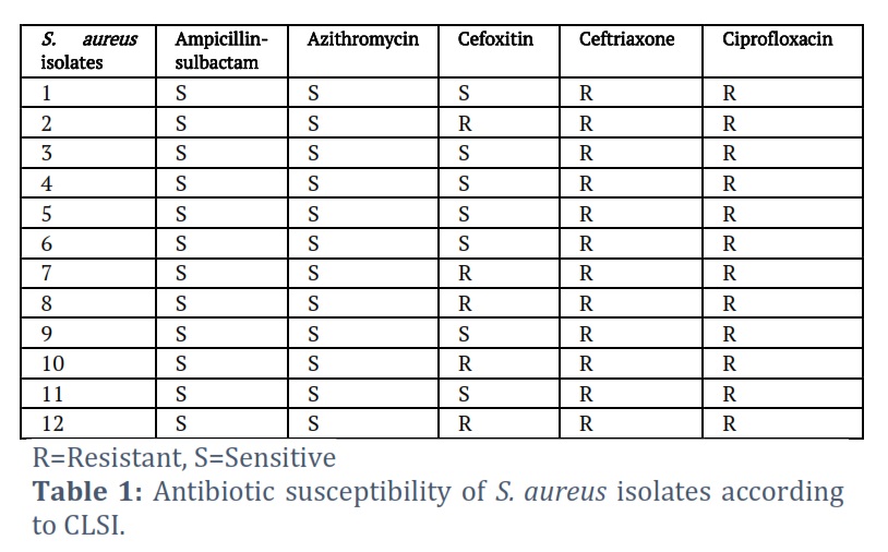
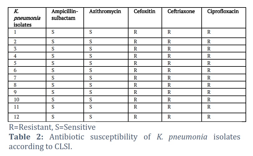
Staphylococcus aureus and K. pneumoniae isolates’ antibiotic susceptibility
Bacteria are more resilient to certain antimicrobial treatments when they are in biofilm form. In accordance with CLSI [35] recommendations, results of susceptibility tests for isolates of S. aureus and K. pneumoniae to five different antibiotics are summarized in Tables 1 and 2. Due to their method of action, which prevents the production of cell walls and results in the release of bacterial cell DNA into the environment, these antibiotics were used in this investigation [36]. Table1 and Table 2 indicate that all isolates were resistant to Ciprofloxacin and Ceftriaxone, all isolates of K. pneumoniae, and 5 isolates of S. aureus (S2, S7, S8, S10, S12) resistant to Cefoxitin and other isolate sensitive. All isolate sensitive to Ampicillin-sulbactam and Azithromycin.
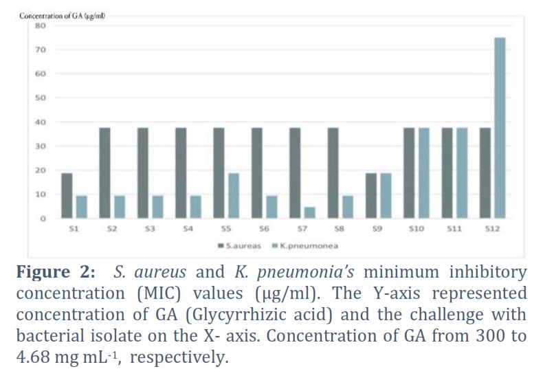
Minimum Inhibitory Concentration (MIC)
The minimum inhibitory concentration (MIC) value is essential for assessing an antibacterial agent’s effectiveness. A low MIC value could mean that a bioactive substance is highly effective or that microorganisms are unable to become resistant to it. Figure 2 represents the effects of GA concentrations with varying MIC values on S. aureus and K. pneumoniae. Obviously, For S. aureus isolates, the MIC concentration ranged from 18.75 to 37.5%, with the S1, S9 isolate having the most effect and other isolates having the lowest effect. And 4.68 to 75 for K. pneumoniae isolate, the highest effect was observed on K. pneumoniae in S7, and the lowest effect was observed in S12.
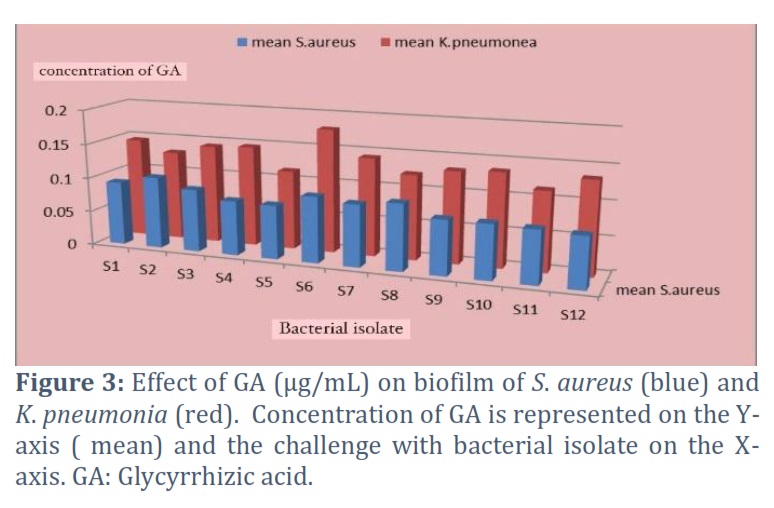
Effect of GA on Biofilm Formation
Along with MIC studies, the effects of GA on existing bacterial biofilms were also examined. These included S. aureus and K. pneumoniae. Due to the fact that biofilms naturally resist antibacterial therapy, based on the MIC data, the treatment concentrations were chosen. The results indicated that of GA extract reduced metabolic activity of cell in biofilm on S. aureus and K. pneumoniae showing an an inhibition percentage, in comparison to the S. aureus and K. pneumoniae biofilm control groups that weren’t treated with GA for 24 hours, there was a substantial decrease in absorbance A630. Figure 3 below shown that their significant differences between bacterial isolates and GA extract inhibit the formation of biofilm, the highest effect of GA in S2 isolate inhibited 66% and lowest effect in S4 isolate inhibited 34% of S. aureus biofilm. Moreover, the highest effect of GA in S6 isolate inhibited 80% and lowest effect in S12 isolate inhibited 54% of K. pneumoniae biofilm.
Figures & Tables
The results are in line with Rohinishree and Negi (2016), whereby they used 80% of GA as an external standard to detect GA in licorice extract using HPLC and a C18 column [32]. Liquiritin, liquiritigenin, and isoliquiritigenin are three flavonoids, and glycyrrhizin, 18-glycyrrhetinic acid, 18-glycyrrhetinic acid, and 18-glycyrrhetinic acid methyl ester are four triterpenoids, all from licorice [33]. For the purpose of identifying the active ingredients in licorice extract, such as glycyrrhizin or glycyrrhizic acid, an upgraded high-performance liquid chromatographic technology with a photodiode-array detection system is used [34].
These findings concur with Gamal M. Hamad et al., findings. According to MIC results, ethanol extract of licorice root was effective against K. pneumonia, Bacillus cereus, Staphylococcus aureus, and Salmonella spp., [37]. In this investigation, we showed that GA had antibacterial properties against K. pneumoniae and S. aureus isolates. These compounds exhibited strong antibacterial activities. Similar observation was reported by Kentaro Oyama et al., GA displayed potent antibacterial action against a number of S. aureus strains, and this activity may have been influenced by the blockage of a number of metabolic pathways for carbohydrates and amino acids [38]. Investigations were conducted on the antibacterial activity of (GA) against S. aureus and its impact on the production of S. aureus alpha-haemolysin (Hla) [39]. De Leon et al.’s subsequent research with Bacillus subtilis, another Gram-positive bacterium, demonstrated that triterpenoids disrupt cellular membranes as part of their mode of action [40]. Triterpenoic acids show many pharmacological effects, among them an anti-inflammatory or an antitumor activity [41]. Glycyrrhizic acid, one of the proposed chemo preventive drugs, was used to inhibit arylamine N-acetyltransferase (NAT) activity in Klebsiella pneumoniae [42]. B. pneumonia, Pseudomonas fluorescens ATCC12296 and EMCC1221 shown sensitivity to licorice ethanol extract [43]. They observed that using the agar diffusion method, licorice extracts (Glycyrrhiza glabra) showed a variety of antibacterial properties against both Gram-positive and Gram-negative bacteria. A microdilution technique and enzyme-linked immunosorbent test (ELISA) were used to determine the antibacterial activity of Glycyrrhiza glabra root extract against gram-positive and gram-negative bacteria, including Salmonella enteritidis, Escherichia coli, Bacillus cereus, and Staphylococcus aureus [44]. The treatment of MRSA biofilms with GA for 24 h caused a significant reduction in absorbance in comparison to the untreated MRSA biofilm control groups, according to Alan J. Wevaer et al., GA itself had no significant effect on biofilm viability, but it seems to be an effective biofilm disruptor, In addition, CFUs were considerably lower after GRA treatment, and these findings suggested that GRA therapy might improve biofilm dispersal [45]. Gram-positive bacteria are the only ones that triterpenes’ antibacterial action has been shown to affect [40,46,47] including biofilm inhibition and reduction [48,49]. It inhibits the growth and generation of bacterial biofilms as well as the coaggregation of P. gingivalis and plaque bacteria, GA is one of a good candidate for a natural agent that reduces the onset and progression of periodontal disease. Additionally, the same effect was confirmed with supragingival plaque bacteria [50]. Additionally, Pseudomonas aeruginosa bacteria are prevented from growing by glycyrrhizic acid due to its effects on cell membrane permeability, biofilm formation, and efflux activity [51].
Conflict of Interest
The authors declare that there is no conflict of interest.
All authors contributed equally to the manuscript.
![]() References
References
- Kirbağ S, Zengin F, Kursat M. Antimicrobial activities of extracts of some plants. Pakistan Journal of Botany, (2009); 41(4): 2067- 2070.
- Peng C, Zhu Y, Yan F, Su Y, Zhu Y, Zhang Z, Peng D. The difference of origin and extraction method significantly affects the intrinsic quality of licorice A new method for quality evaluation of homologous materials of medicine and food. Food Chemistry, (2021); 340: 127907.
- Alwan A, Nesrullah Z, Faraj E. Study the effect of ethanolic extract of Glycyrrhiza glabra on pathogenic bacteria. International Journal of Current Microbiology and Applied Sciences, (2015); 4(5): 473- 484.
- Chauhan S, Gulati N, Nagaich U. Glycyrrhizic acid: extraction, screening and evaluation of anti–inflammatory property. Ars Pharmaceutica (Internet), (2018); 59(2):61-67.
- Isbrucker R, Burdock G. Risk and safety assessment on the consumption of Licorice root (Glycyrrhiza sp.) its extract and powder as a food ingredient with emphasis on the pharmacology and toxicology of glycyrrhizin. Regulatory Toxicology and Pharmacology, (2006); 46(3): 167 -192.
- Fiore C, Eisenhut M, Ragazzi E, Zanchin G, Armanini D. A history of the therapeutic use of liquorice in Europe. Journal of ethnopharmacology, (2005); 99(3): 317- 324.
- Al-Snafi A E. Glycyrrhiza glabra: A phytochemical and pharmacological review. IOSR Journal of Pharmacy, (2018); 8(6): 1 -17.
- Martin M, Guiochon G. Effects of high pressure in liquid chromatography. Journal of Chromatography A, (2005); 1090 (1 -2):16- 38.
- Xiang Y, Liu Y, Lee ML. Ultrahigh pressure liquid chromatography using elevated temperature. Journal of Chromatography A, (2006); 1104 (1- 2): 198 -202.
- Abidi SL. High-performance liquid chromatography of phosphatidic acids and related polar lipids. Journal of Chromatography A, (1991); 587(2):193 -203.
- Nascimento M, de Araújo DR. Exploring the pharmacological potential of glycyrrhizic acid: From therapeutic applications to trends in nanomedicine. Future Pharmacology, (2022); 2(1): 1- 15.
- Oyama K, Kawada Matsuo M, Oogai Y, Hayashi T, Nakamura N , Komatsuzawa H. Antibacterial effects of glycyrrhetinic acid and its derivatives on Staphylococcus aureus. PloS one, (2016) ; 11(11) e0165831.
- El-Jakee J, Nagwa AS, Bakry M, Zouelfakar SA, Elgabry E, El-Said WG. Characteristics of Staphylococcus aureus strains isolated from human and animal sources. American-Eurasian Journal of Agricultural & Environmental Sciences, (2008); 4(2): 221 -229.
- Croes S, Deurenberg R , Boumans M , Beisser P , Neef C , Stobberingh E . Staphylococcus aureus biofilm formation at the physiologic glucose concentration depends on the S. aureus lineage. BMC microbiology, (2009); 9(1): 1- 9.
- Wyres KL and Holt KE. Klebsiella pneumoniae as a key trafficker of drug resistance genes from environmental to clinically important bacteria. Current opinion in microbiology, (2018); 45: 131- 139.
- Seifi K, Kazemian H, Heidari H, Rezagholizadeh F, Saee Y Shirvani F, Houri H. Evaluation of biofilm formation among Klebsiella pneumoniae isolates and molecular characterization by ERIC-PCR. Jundishapur journal of microbiology, (2016); 9(1) : e30682.
- Ashurst JV, Dawson A. Klebsiella Pneumonia. In: StatPearls. StatPearls Publishing, Treasure Island (FL); 2023. PMID: 30085546.
- Donlan R M. Biofilms: microbial life on surfaces. Emerging infectious diseases, (2002); 8(9): 881.
- Miller WR, Munita JM , Arias CA. Mechanisms of antibiotic resistance in enterococci. Expert review of anti-infective therapy, (2014); 12(10):1221 -1236.
- Tian M, Yan H, Row K H. Extraction of glycyrrhizic acid and glabridin from licorice. International journal of molecular sciences, (2008); 9(4): 571 -577.
- De AK, Datta S, Mukherjee A. Quantitative analysis of Glycyrrhizic acid from a polyherbal preparation using liquid chromatographic technique. Journal of advanced pharmaceutical technology & research, (2012); 3(4): 210.
- Bardaweel SK. D-amino Acids: Prospects for new therapeutic agents. Journal of Medical and Bioengineering, (2014); 3(3): 195-198.
- Morello JA, Mizer E, Granato P A. Laboratory manual and workbook in microbiology , (2006) .
- Hoelzer K, Cummings KJ, Warnick LD, Schukken YH, Siler JD, et al. Agar disk diffusion and automated microbroth dilution produce similar antimicrobial susceptibility testing results for Salmonella serotypes Newport Typhimurium and 4 5 12: I but differ in economic cost. Foodborne pathogens and disease, (2011); 8(12): 1281- 1288.
- Wiegand I, Hilpert K, Hancock RE. Agar and broth dilution methods to determine the minimal inhibitory concentration (MIC) of antimicrobial substances. Nature protocols, (2008); 3(2): 163 -175.
- Hassan A, Usman J, Kaleem F, Omair M, Khalid A, Iqbal M. Evaluation of different detection methods of biofilm formation in the clinical isolates. Brazilian journal of infectious diseases, (2011); 15: 305- 311.
- Tawfeeq HK. The effect of D and L-amino Acids on Biofilm Formation in Different Microorganisms. Iraqi Journal of Science, (2016); 570- 575.
- Goh S, Fernandez A, Ang S, Lau W, Ng D, Cheah E. Effects of different amino acids on biofilm growth, swimming motility and twitching motility in Escherichia coli BL21. Journal of Biology and Life Science, (2013); 4(2): 103- 115.
- Eftekhar F, Speert DP. Biofilm formation by persistent and non-persistent isolates of Staphylococcus epidermidis from a neonatal intensive care unit. Journal of Hospital Infection, (2009); 71(2): 112 -116.
- Namasivayam S, Roy E. Anti-biofilm effect of medicinal plant extracts against clinical isolate of biofilm of Escherichia coli. International Journal of Pharmacy and Pharmaceutical Sciences, (2013); 5(2): 486- 489.
- Mason RL, Gunst RF, Hess JL. Statistical design and analysis of experiments: with applications to engineering and science John Wiley & Sons, (2003) .
- Rohinishree Y S, Negi P S. Effect of licorice extract on cell viability biofilm formation and exotoxin production by Staphylococcus aureus . Journal of food science and technology, (2016); 53 : 1092 – 1100
- Wang YC, Yang YS. Simultaneous quantification of flavonoids and triterpenoids in licorice using HPLC. Journal of Chromatography B, (2007); 850 (1 2): 392 – 399.
- Tsai TH, Chen CF. Determination of three active principles in licorice extract by reversed-phase high-performance liquid chromatography. Journal of Chromatography A, (1991); 542: 521- 525.
- Cetti RJ, Venn S, Woodhouse CR. The risks of long‐term nitrofurantoin prophylaxis in patients with recurrent urinary tract infection: a recent medico‐legal case. BJU international, (2009); 103(5): 567- 569.
- Seguin JC, Walker R D, Caron JP, Kloos WE, George C G, et al. Methicillin-resistant Staphylococcus aureus outbreak in a veterinary teaching hospital: potential human-to-animal transmission. Journal of clinical microbiology, (1999); 37(5): 1459 -1463.
- Hamad G, Elaziz A, Hassan S, Shalaby M , Mohdaly A. Chemical composition antioxidant antimicrobial and anticancer activities of licorice (Glycyrrhiza glabra L.) root and its application in functional yoghurt. Journal of Food and Nutrition Research, (2020) ; 8(12): 707- 715.
- Oyama K, Kawada-Matsuo M, Oogai Y, Hayashi T, Nakamura N , Komatsuzawa H. Antibacterial effects of glycyrrhetinic acid and its derivatives on Staphylococcus aureus . PloS one, (2016); 11(11): e0165831
- Li HE, Qiu JZ, Yang ZQ, Dong J, Wang JF, et al. Glycyrrhetinic acid protects mice from Staphylococcus aureus pneumonia. Fitoterapia, (2012); 83(1): 241 -248
- de León L, Beltrán B, Moujir L. Antimicrobial activity of 6-oxophenolic triterpenoids Mode of action against Bacillus subtilis. Planta medica, (2005); 71(04): 313- 319
- Csuk R, Schwarz S, Siewert B, Kluge R , Ströhl D. Synthesis and antitumor activity of ring a modified glycyrrhetinic acid derivatives. European journal of medicinal chemistry, (2011); 46(11): 5356- 5369
- Lo HH, Yen YS, Hsieh SE, Chung JG. Glycyrrhizic acid inhibits arylamine N‐acetyltransferase activity in Klebsiella pneumoniae in vitro. In Journal of Applied Toxicology: An International Forum Devoted to Research and Methods Emphasizing Direct Clinical Industrial and Environmental Applications, (1997); 17(6): 385 -390.
- Zadeh JB, Kor ZM , Goftar MK. Licorice (Glycyrrhiza glabra Linn) as a valuable medicinal plant. International journal of Advanced Biological and Biomedical Research, (2013); 1(10): 1281- 1288
- Karami Z, Mirzaei H, Emam-Djomeh Z, Mahoonak A R , Khomeiri M. Effect of harvest time on antioxidant activity of Glycyrrhiza glabra root extract and evaluation of its antibacterial activity. International Food Research Journal, (2013); 20(5): 2951.
- Weaver Jr A J, Borgogna T R, O’Shea Stone G, Peters T R, Copié V, Voyich J , Teintze M. 18β-Glycyrrhetinic Acid Induces Metabolic Changes and Reduces Staphylococcus aureus Bacterial Cell-to-Cell Interactions. Antibiotics, (2022); 11(6): 781
- Verstraeten S, Catteau L, Boukricha L, Quetin-Leclercq J , Mingeot Leclercq MP. Effect of Ursolic and Oleanolic Acids on Lipid Membranes: Studies on MRSA and Models of Membranes. Antibiotics, (2021); 10(11): 1381.
- Chung PY, Navaratnam P, Chung LY. Synergistic antimicrobial activity between pentacyclic triterpenoids and antibiotics against Staphylococcus aureus strains. Annals of clinical microbiology and antimicrobials, (2011); 10: 1- 6.
- Evaristo FFV, Albuquerque MRJR, dos Santos HS, Bandeira PN, Avila FDN, da Silva BR , Teixeir EH. Antimicrobial effect of the triterpene 3β, 6β, 16β-trihydroxylup-20 (29)-ene on planktonic cells and biofilms from Gram positive and Gram negative bacteria. BioMed Research International, (2014); ( 2014) : 7 pages.
- Yu J, Lee D, Lee S. Destabilizing effect of glycyrrhetinic acid on pre-formed biofilms of Streptococcus mutans. Journal of Korean Academy of Oral Health, (2016); 40(1):38- 42.
- Dewake N, Ma X, Sato K, Nakatsu S, Yoshimura K, et al. Β‐Glycyrrhetinic acid inhibits the bacterial growth and biofilm formation by supragingival plaque commensals. Microbiology and Immunology, (2021); 65(9): 343- 351.
- Chakotiya AS, Tanwar A, Narula A, Sharma RK. Alternative to antibiotics against Pseudomonas aeruginosa: Effects of Glycyrrhiza glabra on membrane permeability and inhibition of efflux activity and biofilm formation in Pseudomonas aeruginosa and its in vitro time-kill activity. Microbial pathogenesis, (2016); 98: 98- 105.
This work is licensed under a Creative Commons Attribution-Non Commercial 4.0 International License. To read the copy of this license please visit: https://creativecommons.org/licenses/by-nc/4.0