

Full Length Research Article
Hematological effects for rabbits immunized by Corynebacterium pseudotuberculosis sonicated antigen
Basil R. F. Razook1, Mariam Hamdi Abdulkareem2*, Ansam Khalid Mohammed2
Adv. life sci., vol. 11, no. 2, pp. 375-379, May 2024
*– Corresponding Author: Mariam Hamdi Abdulkareem (marym.h@covm.uobaghdad.edu.iq)
Authors' Affiliations
2. Department of Microbiology, College of Veterinary Medicine, University of Baghdad – Iraq
[Date Received: 31/07/2023; Date Revised: 16/03/2024; Date Available Online: 18/04/2024]
Editorial Note on Version of Record
31 May 2025: This article has been corrected. See https://doi.org/10.62940/als.v13i0.4270 for more information.
Abstract![]()
Introduction
Methods
Results
Discussion
References
Abstract
Background: Analyzing Hematological parameters is usually used to monitor various circumstances such as infection, inflammation and anemia. For that we studied blood parameters which are related to immunized laboratory animals (rabbits) after immunizing these animals with Corynebacterium pseudotuberculosis then used an adjuvant of Pseudomonas aeruginosa to stimulate the immune response cells after 2 to 3 weeks of immunization. Different concentrations were used to examine the effect on the animals’ blood parameters changing.
Methods: Corynebacterium pseudotuberculosis bacterium was used at current study for evaluating the effect of immunizing laboratory rabbits with two different immune stimulators working together as adjuvant on the blood picture with the aid of 12 breed rabbits from different genders, dividing them at the base of inoculation with pre–killed and sonicated cells of bacteria (antigens) onto 4 categories as follows: the group-1 members of rabbits were injected with the antigen, group-2 and group-3 included rabbits injected with the two inoculations at various concentrations; final group, group-4 was the control group.
Results: Thus, white blood cells known to be our main line of defenses act firstly in our body towards different infecting microorganisms, their count always was increased through the immunization correlated with other measured parameters. From the results we can see that the lymphocytes percentage had no alteration between the infected and the immunized compared to control group, while the percentage of MID that refers to the monocytes was at the same range in both infected and the immunized control rabbit’s groups. Granulocyte cells percentage, which is including heterophilic, basophilic and eosinophilic cells, was significantly decreased (P<0.05) compared with control at all groups.
Conclusion: We conclude that C. pseudotuberculosis might function as a potent immunogen to notice the complete blood picture variations.
Keywords: Corynebacterium pseudotuberculosis; Pseudomonas aeruginosa; Sonicated antigen; Blood parameters; Blood picture; Adjuvant
Introduction![]()
Corynebacterium pseudotuberculosis is a gram-negative bacterium which has a vigorous exotoxin that is called phospholipase D (PLD) acts as an essential virulence factor in developing caseous lymphadenitis (CLA) in animals [1]. PLD was described for the first time in 1940 from this bacterium, then it had been detected PLD in every C. pseudotuberculosis studied isolate [2], either in its type I or Type II from all mammalians studied species [3]. Research done assured that the CLA at its initial stage might facilitate multiplication of C. pseudotuberculosis within the macrophages themselves [4,5]. However, Phospholipase D used to play a major role in infecting through aiding in escaping for the organism at the hydrolysis process inside macrophages, affecting the the inner phospholipid layer for the macrophage cell membrane [6].
Infectious diseases cause blood alteration leading to pathological conditions associated with these alterations [7]. The immune system of the host varies at its responses that different components reveal different inflammatory reactions, pathogenesis, and pathogens; increased neutrophilic activity resulted from acute bacterial infections, monocytosis and lymphocytosis are resulted from chronic bacterial infections [8, 9]. CLA incubation period ranges between three and twenty weeks with the incubation periods are shorter [10] thus, few animals might express signs clinically as the showing changeable heart or respiratory conditions, fever and minimization nutritional consumptions. The CLA, however, noticed changes but minor ones at the hematological rates in some kinds of animals that were challenged with C. pseudotuberculosis [11,12]. This bacterial genus cell walls have a complex structure act as a virulence factor [13]; That it lipids are containing 2-branched 3-hydroxy of fatty acid under the name mycolic acid (MA) [14] as a result, C. pseudotuberculosis buildings units giving it virulence, and helping this bacteria at its survival acting as facultative intracellular parasite [15]. Besides, Mycolic acid contained in this bacterium expresses cytotoxic effects that might stimulate immune response [16]. It was proved (in-vitro) that phagocytic activity of white blood cells significantly degenerated during C. pseudotuberculosis infection [17] and it could resist cellular digestion by phagocytes [18].
Methods![]()
Experimental Animals
Rabbits were used as a model for the demonstration of the doses that twelve rabbits were divided into 4 groups, each healthy animal was of nearly 1Kg weight, and was placed in a proper plastic cage at a good, air-conditioned room, they were left for adaptation for 2 weeks at the animal housing unit. Chaw pellets, herbs and tap water were the feeding materials used for nutrition.
Experimental bacteria and their source
Sheep infected lymph nodes were the Corynebacterium pseudotuberculosis source, while Pseudomonas aeruginosa was isolated from Human lymph node (swelling one).
Experimental Procedure
Different gender rabbits at body weight of 1000±30gm were used in the current experiment divided into four divisions, they were inoculated with the pre–killed and sonicated cells of bacteria (antigens) that group-1 included 4 rabbits, they were injected with 1 mg/ml antigen, group-2, included 4 rabbits and were injected with both (1 gm/ml of C. pseudotuberculosis and 1 gm/ml Pseudomonas aeruginosa) antigens; group-3 included 4 rabbits injected with antigen of both (1 gm/ml of C. pseudotuberculosis and 0.5 gm/ml Pseudomonas aeruginosa). Final group, group-4 included 4 rabbits as control group injected with Phosphate buffer saline (phosphate-buffered saline (PBS)). After 14 days of the immunization, all of the experimental first 3 groups were injected with booster doses of same sonicated bacterial antigen from Cor. Pseudotuberculosis to enhance immune response. Finally, animals were sacrificed, and the hematological changes were examined. At the laboratories of the department of Microbiology/ College of Veterinary Medicine/ University of Baghdad, both types of the bacteria (C. pseudotuberculosis and P. aeruginosa) were isolated, biochemically identified and molecularly ensured. Preparing the antigens submitted to the method of Motiva and his co-workers [19].
Results![]()
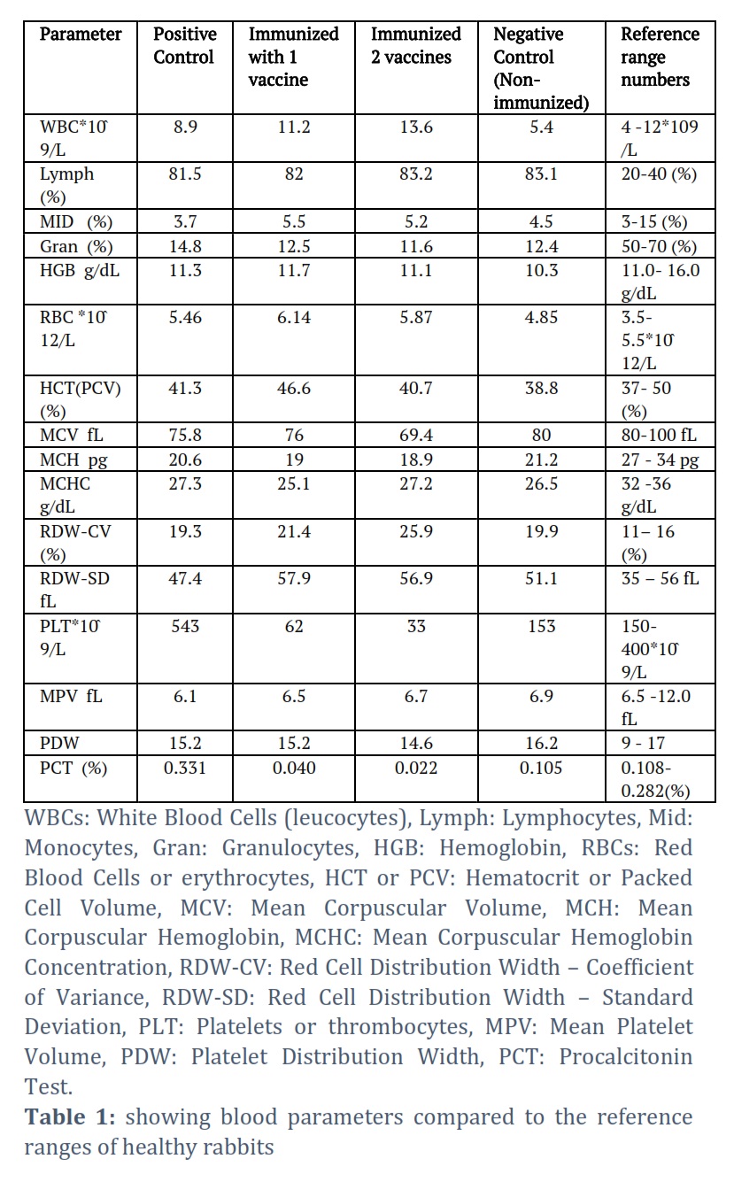
Hematological parameters are used to provide information on the body’s health conditions (such as infection, inflammation and anemia). Thus, our investigation was to research the effect of enhanced immunization on various hematological parameters in rabbits as shown in table 1 below.
Figures & Tables
During infection of tissue, many inflammatory mediators were released producing a local inflammatory response resulting in accumulation of serum proteins and phagocytic cells at the site such as phagocytic cells which include eosinophils and neutrophils, they have the ability to engulf and terminate the bacteria [20].
Many studies admitted that immunization with sonicated antigens might stimulate efficient immune response at the host cells [21]. Bone marrow is responsible of producing the granulocytes which subsequently are released into peripheral blood then migrate to the tissues in order to complete their action for the body as responding to specific stimulation which requires production and mobilization. Animals’ plasma which suffers leukopheresis have Leukopoietic factors, while animals which exposed to endotoxin mobilizing factors, they became responsible for releasing the granulocytes into the peripheral circulation. At the time, leukotoxin (which is a polypeptide from inflammatory exudates) was found to be responsible for increasing capillary permeability and inducing local granulocytes migration. When the antigen-antibody was reacting, eosinophils rottenly mobilized at the site of their presence leading to increment in the number of the eosinophils at that blood stream. Inflammatory conditions are always associated with the presence of neutrophils which could be available with large numbers in tissues infected with pyogenic germs. Additionally, the inflammatory reaction might be (in low chance) initiated by the granules of the basophils which contain the heparin (responsible of inhibiting the clotting mechanism) and histamine. Lymphocytes in the animal body are constantly in a state of circulation and recirculation, this made the lymphocytic cells unknown at the body, hence, unknown factors regulating blood lymphocytes levels also occurred; Mechanisms of proliferation and differentiation for the cellular events took place after g exposure to antigen, with the contribution of lymphocytes (as presenting essential metabolites for the proliferating cells). Macrophages, which developed from monocytes, have the capability to enter inflammation sites in which fungi and protozoa are present in order to do phagocytosis in tissues [22]. The four mentioned factors could influence the levels of blood cells that circulate at the blood vessels, which affect total and differential white blood cells count as what was found by [23,24]. A report [25] assumed that the CLA at its initial stage might facilitate multiplication of C. pseudotuberculosis within the macrophages themselves [8], However, phospholipase-D might have a positive influence in infections through playing part in escaping of germs from hydrolysis process inside the macrophages, affecting the the inner layer of the phospholipid for the macrophage’s cell membrane [26] for the previous reason we found our results are logically accepted for their variation between infected and immunized groups.
Our results exemplified that normal complete blood count (CBC) in the fourth control group were matching the normal readings rates while the positive first groups exhibited slight raising in the RBCs & lymphocytes which are in tune with findings of Abdulkareem and his team [27] whom mentioned that there was a severe leucocytes infiltration in the histopathological sections for the internal organs of rabbits that injected with different antigens.
The increase noticed in RBC count and Hb might be from good health and no presence of hepatotoxicity and dysfunction, that led to the thought that haem and globin molecules synthesized and sufficient liver iron storage, this agrees with the results of previous report [28], thus, it goes with studies mentioned that the increase in WBC indicate an inflammation, beside a slight descending in RBC [29].
One of the cases is that HGB value is high the chronic diseases and it is obvious from our results that the animals did not suffer from chronic diseases or anemia either. However, the minor rise in the values might be because of dehydration. Platelet count and its indices (MPV & PDW) considered parameters for maintenance CBC checking, they considered major growth factors sources such as platelet-derived growth factor (PDGF), vascular endothelial growth factor (VEGF), and transforming growth factor-β (TGF-β) giving the platelets the importance in achieving various pathways such as repair, inflammation, regeneration of tissues and angiogenesis [30]; this agrees with our results which were showing that the PTC was normal at the negative control group and there was a raise in the levels of the PLT, MPV, PDW and PCT values, this could be attributed to the inflammations, tissues regenerating processes or repairing at the body tissues. Moreover, it was assumed that the size and the volume of the platelets altered, that in minor inflammation disorders, values of MPV increases; oppositely, at major inflammation disorders there would be decrease in MPV levels resulting from consumption for the big platelets in the inflammatory site [31], thus, from our results it is clear that there are no major inflammations occurred at the rabbits tissues, with slight calcification at the tissues of the infected group, might be due to the harmful effects resulted from C. pseudotuberculosis PLD or exotoxin especially in the cells of endothelium for vascular system leading to disrupt of hemopoietic pathways or damage caused at the bone marrow [32, 33].
Analyzing changes in blood due to the hematology evaluation after response to C. pseudotuberculosis in rabbits revealed an experimental challenge that immune stimulators could elevate a pattern in which sonicated bacterial antigens inoculation caused significant difference indicating cellular immune response, progressing the infection from acute to chronic.
Conflict of Interest
The authors declare that there is no conflict of interestregarding the publication of this paper.
All authors equally contributed to the current study.
![]() References
References
- Baird GJ, Fontaine MC. Corynebacterium pseudotuberculosis and its role in ovine caseous lymphadenitis. Journal of Comparative Pathology, (2007); 37:179–210.
- Carne HR. The toxin of Corynebacterium bovis. Journal of Pathology and Bacteriology, (2007); 51:199–212.
- Songer JG, Beckenbach K, Marshall MM, Olson GB, Kelley L. Biochemical andgenetic characterization of Corynebacterium pseudotuberculosis. American Journal of Veterinary Research, (1988); 49:221–6.
- Hard GC. Comparative toxic effect of the surface lipid of Corynebacterium ovis on peritoneal macrophages. Infection and Immunity, (1975); 12:1439–49.
- Paton MW. The Epidemiology and Control of Caseous Lymphadenitis inAustralian Sheep Flocks.PhD thesis, (2010); Murdoch University.
- Titball RW. Bacterial phospholipases C. Microbiology and Molecular Biology Reviews, (2012); 57:347–66.13.
- Abdullah FFJ, Latif NAA, Chung ELT, Aimi S, Sarah MZS, Haron AW, Lila MAM, Zakaria Z and Norsidin MJ. Changes in the reproductive hormones of non-pregnant does infected intradermally with Corynebacterium pseudotuberculosis in chronic form. International Journal of Livestock Research, (2015); 5(7): 33-40.
- Othman AM, Abdullah FFJ, Nordi M, Rina NA, Ilyasu Y, Zamri-Saad M, Wahid AH, Saharee AA, Mohd-Azmi ML. Biochemical and serum electrolyte changes in non-pregnant boer does inoculated with Corynebacterium pseudotuberculosis via various routes. IOSR Journal of Agriculture and Veterinary Science, (2014); 7(10):5–8.
- Zeru F, Kahsay AG. Caseous lymphadenitis in goats from Borena range Land South Ethiopia slaughtered at luna export abattoir. Journal of. Veterinary Medicine and Animal Health, (2014); 6(6): 168-173.
- Abdullah FFJ, Osman AY, Adamu L, Zakaria Z, Abdullah R, Saad MZ and Saharee AA. Acute phase protein profiles in calves following infection with whole cell, lipopolysaccharides, and outer membrane protein extracted from Pasteurella multocida Type B:2. Asian Journal of Animal and Veterinary Advances, (2013); 8(4): 655-662.
- Junior JP, Oliveira A, Alves F, Silva L, Rabelo S and Mota R. Corynebacterium pseudotuberculosis experimental infection of goats mamary gland. Arquivos do Instituto Biológico, (2006); 73(4): 395-400.
- Latif NAA, Abdullah FFJ, Othman AM, Rina A, Chung ELT., Zamri-Saad M, Saharee AA, Haron AW and Lila MAM. Isolation and detection of Corynebacterium pseudotuberculosis in the reproductive organs and associated lymph nodes of non-pregnant does experimentally inoculated through intradermal route in chronic form. Veterinary World, (2015); 8(7): 924.
- Collins MD, Burton RA and Jones D. Corynebacterium amycolatum sp. Nov. A new mycolic acid-less Corynebacterium species from human skin. FEMS Microbiology Letters, (1988); 49(3): 349-352.
- Marrakchi H, Lanéelle MA and Daffé M. Mycolic acids: Structures, biosynthesis, and beyond. Chemical Biology, (2014); 21(1): 67-85.
- Vasco A. Corynebacterium pseudotuberculosis: Immunological responses in animal models and zoonotic potential. Journal of Clinical and Cellular Immunology, (2012); 1(S4): 5.
- Abdulkareem, MH, Razook BRF. and AL-Rubaiey MGA. Histopathological Changes in few Digestive Organs Affected by Corynebacterium pseudotuberculosis. Biochemical and Cellular Archives, (2022); 22(2): 3743-3750.
- Ozkanlar Y, Aktas M, Kaynar O, Ozkanlar S and Kireccl E. Bovine respiratory disease in naturally infected calves: Clinical signs, blood gases and cytokine response. Revue de Medecine Veterrinaire, (2012); 163: 123-130.
- Pazatsen R, Shun S, Abdullah FFJ, Saad MZ and Haron A. Clinical Response and Pathological Changes Associated with Corynebacterium pseudotuberculosis Infection in Mice. UPM Serdang, Malaysia. 7th Proceedings of the Seminar in Veterinary Sciences, 27 February – 02 March (2012).
- Motiva I, Denchen V and Linda K. Humoral and mediated immunity in mice after immunization with live oral vaccine of Salmonella typhimurium: auxotrophic mutant with two attenuating markers. Vaccine.Microbiology, (1992); (10):61-66.
- Jayapal V. Fundamentals of Medical Immunology. 1st ed. Jaypea Brothers, Medical Publishers LTD, (2007); New Delhi: 180-189.
- Scavone P, Miyoshi PA, Rail a, Chabalgoity A, langella P, Azerdo V and Zunino P. Intranasal immunization with recombinant Lactococcus lactis displaying either anchored or secreted forms of Proteus mirabilis – Mrp A fimbriae protein confers specific immune response and induces a significant reduction of kidney bacterial colonization in mice. Microbes and Infection, (2007); 9:821-828.
- Coles EH. Veterinary Clinical Pathology. 2nded. W.B.Saunders Company. (1974); Philadelphia. USA:70-83.
- Al-Samarrae EA. Evaluation of Proteus vulgaris fimbriae antigen by delayed type hypersensitivity (DTH)-skin test in rabbits. Iraqi Journal of Veterinary Medicine, (2011); 35(1): 100-106.
- Rzook BRF, Al-Samarrae IAA, Al-Rubaie HMA. The Effect of Proteus vulgaris Sonicate fimbriae antigens in some blood parameters and humoral immune response. Baghdad Science Journal, (2015); 12(2):301-306.
- Mahmood Z, Jesse F, Saharee A, Jasni S, Yusoff R. Wahid H. Assessment of blood changes post-challenge with Corynebacterium pseudotuberculosis and its exotoxin (phospholipase D): A comprehensive study in goat. Veterinary World, (2015); 8(9):1105.
- Mahmood ZK, Jin ZAM, Jesse FF, Saharee AA, Sabri J, Yusoff R, Haron AW. Relationship between the Corynebacterium pseudotuberculosis phospholipase D inoculation and the fertility characteristics of crossbred Boer bucks. Livestock Science, (2016);191:12–21.
- Abdulkareem MH, Razook BRF, Abdul Ameer AH and Alrubaye B. Histopathological Changes of Rabbits’ Vital Organs Associated with Corynebacterium Pseudotuberculosis Infection. International Journal of Special Education, (2022); 37(3):13243-13255.
- Abdulkareem MH, Razook BRF, Taher DD. Changes in Liver Function Enzymes of Rabbits Immunized by Corynebacterium pseudotubeculosis and Pseudomonas aeruginosa Antigens. Indian Journal of Ecology, (2022); 49 special issue (19):230-233.
- Abdel-Wareth AAA, Hammad S, Ahmed H. Effects of Khaya senegalensis Leaves on Performance, CacassTraits, Hematological and Biochemical Parameters in Raabbits. Experimental and Clinical Sciences Journal, (2014); 2014;13:502-512 – ISSN 1611-2156
- Turgut A, Sak ME, Ozler A. Alterations of peripheral blood cells in tubal ectopic pregnancy. Ginekologia Polska,(2013); 84:193-196.
- Gasparyen AY, Ayvazyen L, Mikhalidis D. MPV: A link between thrombosis and inflammation. Current Pharmaceutical Design, (2011); 17(1): 47-58.
- Osman AY, Abdullah FFJ, Saharee AA, Haron AW, Sabri I, and Abdullah R. Haematological and biochemical alterations in mice following experimental infection with whole cell and exotoxin (PLD) extracted from C. Pseudotuberculosis. Advances in Animal and Veterinary Sciences, (2012); 11(24): 4660-4667.
- Russell KE, Grindem CB. Secondary thrombocytopenia. In: Schalm’s Veterinary Hematology. Lippincott Williams and Wilkins, Philadelphia, (2000); 487-495.
This work is licensed under a Creative Commons Attribution-Non Commercial 4.0 International License. To read the copy of this license please visit: https://creativecommons.org/licenses/by-nc/4.0