

Full Length Research Article
Histopathological Detection of the Protective Role of Hydroxytyrosol Against the adverse Effect of Azithromycin in Rats
Ghusoon Abdul Kareem Neamah1*, Ekhlas Abid Hamza Alalwany1, Fadak Bahaa Rabee1, Adnan Mansour Jasim1, Ahmed Samir Abukhomra2
Adv. life sci., vol. 11, no. 2, pp. 386-391, May 2024
*– Corresponding Author: Ghusoon Abdul Kareem Neamah (Ghusoon.alneamah@vet.uoqasim.edu.iq)
Authors' Affiliations
2. Anesthesia Department, Hilla University College, Babylon – Iraq
[Date Received: 05/08/2023; Date Revised: 19/11/2023; Date Available Online: 18/04/2024]
Abstract![]()
Introduction
Methods
Results
Discussion
References
Abstract
Background: Adverse medication reactions affecting one or more organ systems can manifest as side effects of antibiotics. There is a possibility that some antibiotics will have adverse effects that are fatal. The present study aims to examine the ability of hydroxytyrosol (HT) to protect the liver, heart , and improvement of kidney function against the adverse effects of azithromycin.
Methods: Thirty adult male rats were randomly divided into three equal groups of 10 animals each. The first group, designated negative control (NC), was the daily oral administration of normal saline. The second group (T1) was subject to daily oral administration of azithromycin (30 mg/kg). The last third group (T2) was administered azithromycin (30 mg/kg) with hydroxytyrosol (50 mg/kg). After two weeks the animals were sacrificed for serum collection, histopathological specimens, and drug docking evaluation using Auto Dock Vina.
Result: The present study showed that score binding in 18gs Glutathione S-transferase was preferable in hydroxytyrosol with (-5.95 Kcal/mol), while vitamin E was in (-7.80 Kcal/mol) and (-6.16 Kcal/mol) for glutathione. On the other aspect, antioxidant coenzyme Q10 showed less binding with 18gs Glutathione S-transferase to recorded binding at (-4.9) with Root Mean Square Deviation (RMSD) (29.3, 30.3, 23.9 Kcal/mol) and (28.6) Kcal/mol, respectively. The histopathological result for T1 showed several histological effects in the liver, heart, and kidney, while in T2 group showed improvement in tissue architecture.
Conclusion: The study concluded that hydroxytyrosol binding glutathione S-transferase could predict potent antioxidants near glutathione, might be responsible for scavenger free radicals, and protect the tissue against the toxic effects of azithromycin.
Keywords: Histopathology; Rat; Hydroxytyrosol; Azithromycin; Glutathione S-transferase
Introduction![]()
A phenolic phytochemical having in vitro antioxidant effects is hydroxytyrosol. It is a naturally occurring substance that may be extracted from olive oil and leaves. Extra virgin olive oil serves as the primary dietary source of hydroxy aerosol. The pharmaceutical and food industries see hydroxytyrosol as a high-quality food supplement due to its bioavailability, chemical characteristics, simple formulation, and absence of toxicity [1]. According to [2], hydroxytyrosol and oleuropein, two components of olive oil, may prevent LDL cholesterol, which is a major cause of atherosclerosis, stroke, and heart attacks. In a dose-dependent manner, hydroxytyrosol is rapidly absorbed from the GIT, primarily by passive transport, with an efficiency ranging from 75% to 100% [3]. Additionally, HT enhanced the activity of immune system cells by shielding neutrophils from oxidation caused by hydrogen peroxide thanks to its strong antioxidant capacity[4]. Additionally, HT successfully shields the DNA of peripheral monocytes and blood mononuclear cells in Alzheimer's disease patients.
Drugs containing nanoparticles exhibit enhanced solubility and hence increase bioavailability. They can also permeate the blood-brain barrier (BBB), enter the pulmonary system, and be absorbed through the skin's endothelial cells [5]. All polyphenols, including HT, are extensively metabolized by the human body's intestines and liver, and their bioavailability in plasma is low relative to that of their metabolites.
The second-generation macrolide antibiotic azithromycin has a pharmacokinetic/pharmacodynamic profile that allows for straightforward dosing regimens, modest side effects, and a broad range. Recently, incidents of severe cardiovascular consequences linked to azithromycin have drawn attention [6]. Azithromycin use has also been linked to sudden deaths from ventricular arrhythmias, according to the findings of cohort studies conducted on individuals who received the medication [7]. Numerous studies have examined the cardioprotective and powerful antioxidant effects of hydroxytyrosol in order to assess how well it protects vital organs from azithromycin. The present study aims to examine the ability of hydroxytyrosol (HT) to protect the liver, heart , and improvement of kidney function against the adverse effects of azithromycin.
Methods![]()
Ethics
All procedures were performed in accordance with the laws governing animal care and the guidelines on animal welfare of the pathology department, College of Veterinary Medicine, Al-Qasim Green University, Babylon, Iraq. The ethical criteria were followed in the conduct of this investigation established by the local Animal Welfare Committee.
Chemicals
Azithromycin was bought from Shaanxi Laon Biotech Co., Ltd. HT [2-(3,4-dihydroxyphenyl) ethanol]. (cipa India). The remaining substances were analytical-grade compounds.
Experimental design
The College of Veterinary Medicine at Qasim Green University where the current study was carried out. Male adult rats were 60–65 days old and weighed (180–200 g). The rats were subjected to the same ventilation and housing circumstances, while being fed standard food and drinking water. Thirty male rats were randomly and equitably divided into three groups, each consisting of ten rats. The first group, designated negative control (NC), was the daily oral administration of normal saline. The second group, was named T1, is subject to daily oral administration of azithromycin (30 mg/kg). The last third group (T2) was daily administrated Azithromycin (30 mg/kg) with Hydroxytyrosol (50 mg/kg). The study was persistent for two weeks; at the end of the study, animals were sacrificed under chloroform anesthesia.
Collection of blood samples
The blood was drawn from the rat's heart using a disposable needle, retained in simple tubes, centrifuged to extract the serum, then frozen and kept at -20 C until analysis.
Drug docking
The active ligand identified as hydroxytyrosol was chosen for the molecular docking study (https://pubchem.ncbi.nlm.nih.gov). The study's controls included glutathione, Q enzyme 10, and vitamin E. synthesis of proteins. The three-dimensional crystal structure of the Rattus AR ligand binding in association with the ligand hydroxytyrosol (18gs Glutathione S-transferase) was obtained from RCSB PDB (http://www.rcsb.org/pdb/home/home.do) and used to build the protein known as the androgen receptor (AR) [8].
Measuring serum reduced glutathione (GPX, SOD, Troponin I).
It was made in accordance with the [9].
Histopathological study
The liver, kidney, and heart specimens were removed from the animals as soon as they were sacrificed, washed with saline, fixed in 10% formalin, and then processed regularly using the histokinette. Hematoxylin and eosin were used to stain the tissue slices after microtome sectioning and embedding in paraffin [10].
Statistical analysis
The experiment data were analyzed with a completely randomized design (CRD), and the differences between the treatments means were tested by analysis of variance (ANOVA), and the significant differences were tested by the L.S.D. test at a 0.05 probability level using the SPSS program Ver. 26 [11].
Results![]()
Drug docking evaluation
The present study showed that score binding in 18gs Glutathione S-transferase as in (table 1) and in figure (1) .The score was more preferable in hydroxytyrosol with 5.5 while vitamin E was in 5.4 (figure 2) and 5.3 for glutathione (figure3). On the other aspect antioxidant coenzyme Q10 showed less binding with 18gs Glutathione S-transferase to recorded binding at 4.6 (figure 4).
Antioxidant and troponin I
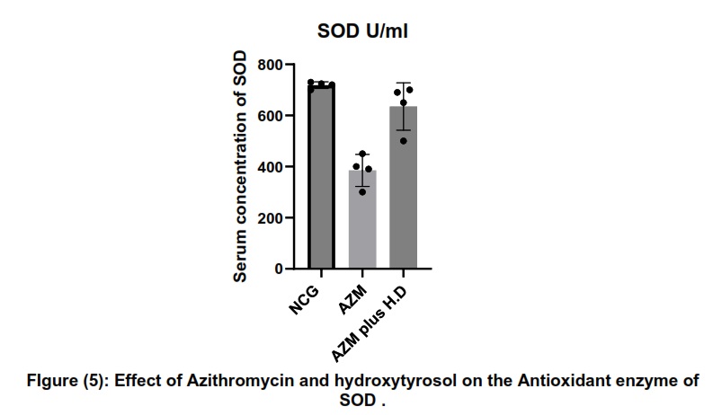
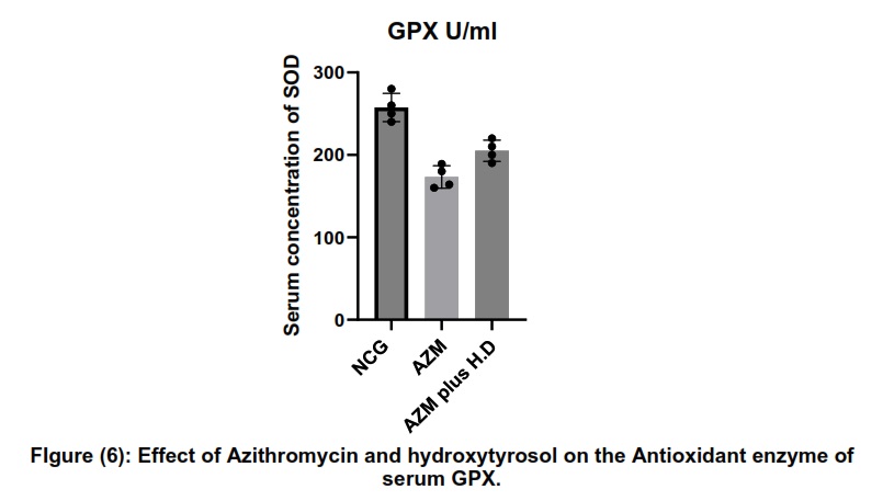
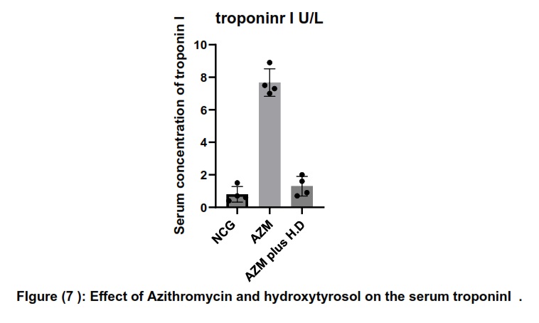
The results of the current study indicated a significant decrease in the antioxidants of the rats exposed to the azithromycin compared with the control, where treatment using the hydroxytyrosol led to an increase in antioxidants, but it remained less compared to the negative control despite the clear improvement in the treatment to recorded mean value (723±24.8) and (164 ±12.4) U/ml for SOD and GPX respectively (figure 5 and 6).On the other hand, there was a significant increase in the troponin I in the rats exposed to the azithromycin, and this indicate the severity of the impact of the heart tissue and the damage that occurred in it, while the treatment with hydroxytyrosol led to a clear decrease and improvement of the heart tissue to recorded mean value (1.47 ±0.3) as compared with azithromycin to recorded mean value (8.25±0.47)(figure7).
Histopathological study
Control group:
The results of histological section for control group were showed normal no pathological lesion (figure 8 A, B, C).
Liver:
T1: The most exanimated section was showed congestion and dilation of sinusoids, with presence of active Kupffer cells (figure 9A).
T2: Histological section was showed mild dilation of sinusoids, with focal mononuclear cells infiltration. (figure9 B).
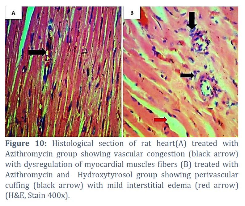
Heart:
T1: The section was showed vascular congestion with dysregulation of myocardial muscles (figure10 A).
T2: The most examined section was showed perivascular cuffing, with mild interstitial edema (figure 10 B).
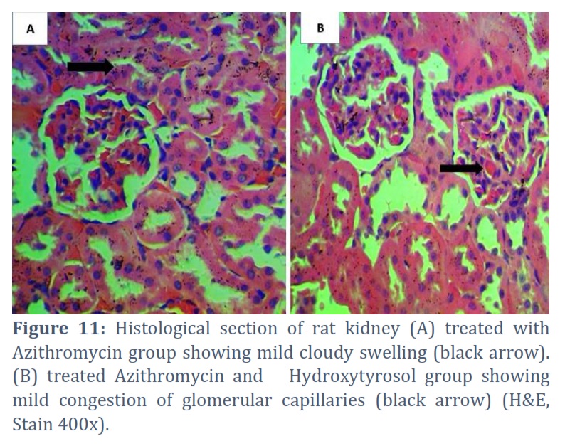
Kidney:
T1: Histopathological result showed mild cloudy swelling (figure 11A).
T2: Most section showed mild congestion of glomerular capillaries (figure 11B).
Figures & Tables
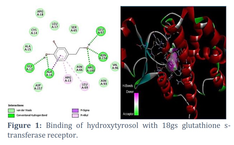
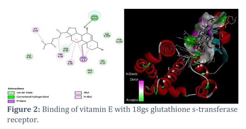
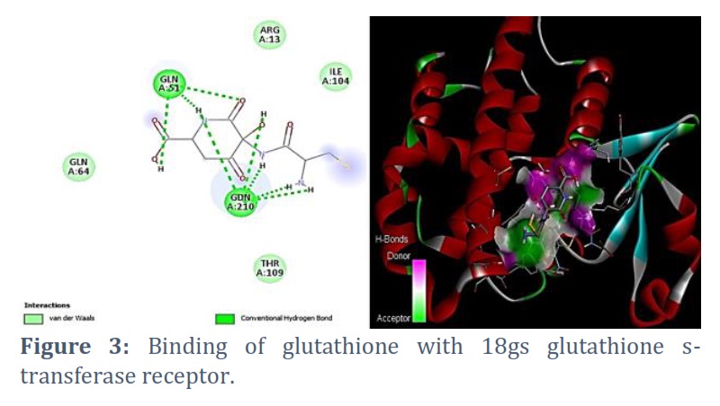
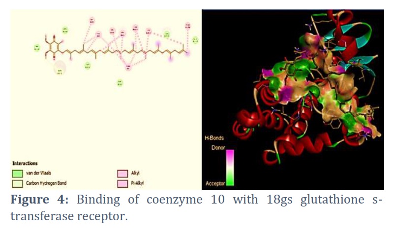
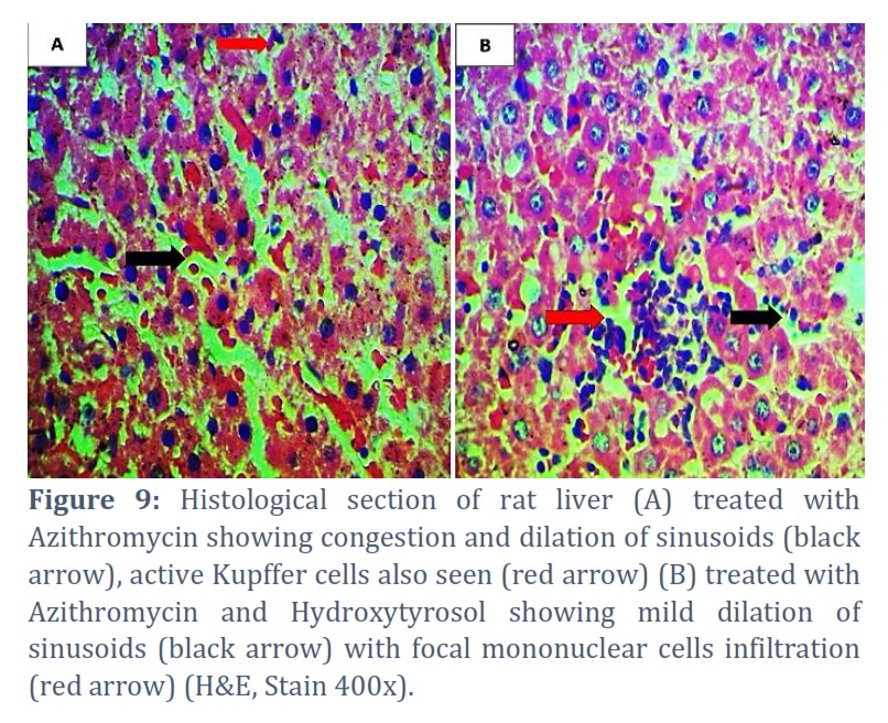
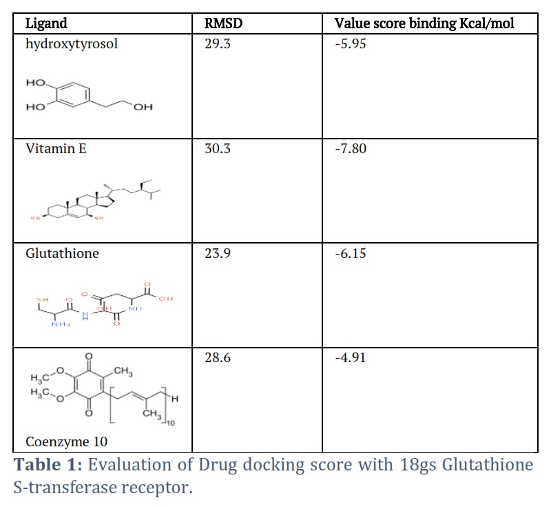
The field of molecular docking has grown over the past several years and recently has started to play a crucial role in drug discovery and development. For predicting protein-ligand complexes, molecular docking is used [12]. The complex shape affects the score function, which predicts the binding affinity of the ligand to the protein. Only the way of evaluating the binding affinity of a ligand using binding energies [13]. Table 1 compares the docking outcomes of 4 molecules. against the target protein 18GS glutathione s-transferase, showing e-values for glutathione of -7.80 and -6.16. In contrast, antioxidant coenzyme Q10 had less binding with 18gs Glutathione S-transferase, with binding measured at -4.9 and RMSD values of 29.3, 30.3, 23.9, and 28.6 Kcal/mol. The enzymes’ ability to generate glutathione-conjugates with compounds that were rich in vitamin E, glutathione, and hydroxytyrosol was demonstrated by docking studies. While hydroxytyrosol treatment recorded mild sinusoidal dilation with focal mononuclear cell infiltration, the present study’s histological section of liver treated with azethromycin showed congestion and dilation of sinusoids, active Kupffer cells. The present study was in agreement with [14] which demonstrated that the combination of hydroxytyrosol and Vit E clear improve hepatic cell via reduce the TGF-induced Human hepatic s Additionally, our data showed that the regime affected the rate of migration and proliferation in TGF-activated HSCs. Outright, these findings imply that the pro-fibrogenic phenotype is present. Hydroxytyrosol exhibits localized mononuclear cell infiltration and moderate sinusoidal enlargement. In HFD mice, oleuropein caused weight reductions in the body, liver, and heart in addition to having anti-inflammatory and antioxidant properties 15]. The former GPx and CAT detoxify hydrogen peroxide, preventing liver and kidney damage even far from the ROS production site, while the former superoxide dismutase enzyme quickly converts superoxide anion to H2O2 and O2. Particularly the GPx is quite effective at low H2O2 concentrations [16, 17]. Recent investigations showed that hydroxytyrosol therapy prevented oxidative stress damage to renal cells and that its metabolites also had protective effects, considerably reducing oxidative damage [18]. The hydroxytyrosol from phenolic-related olive oil acts as a tocopherol regenerator and scavenger of aqueous radicals close to the membrane. In reality, consumption of foods high in antioxidants, such as phenolic compounds, is linked to a lower risk of renal dysfunction, as is well established from clinical research [19]. HT is a prominent phenol found in virgin olive oil. Recent studies by Crupi and Palma [19] revealed that renal cells treated with ochratoxin and protected by HT had increased ROS and LDH levels. HT also prevented the release of LDH into the culture medium, which in turn reduced ROS levels. HT has been found to have a significant protective effect against oxidative cell membrane damage in renal cells. By activating the nuclear factor (erythroid-derived 2)-like 2 (Nrf2) pathways, [20] showed that HT may reduce the oxidative stress brought on by hyperglycemia. They also showed that HT had anti-inflammatory effects by suppressing TNF, IL6, and IL1B. Furthermore, it was found that HT slows the onset and progression of diabetic nephropathy by reducing mesangial cell proliferation, alleviating tubular injury, and enhancing renal dysfunction as measured by markers like tissue oxidative and inflammatory cytokine activities and creatinine in serum and urinary albumin, as well as blood urea nitrogen and diabetic rat models that were left untreated. Group of hydroxytyrosol nanoparticles with normal cardiac muscle fibers and minimal vascular congestion. According to the study, taking azithromycin for 15 days caused a rise in serum levels of lactate dehydrogenase (LDH) and creatine phosphokinase (CPK), which showed that the injured myocardium was releasing these cardiac biomarkers into the bloodstream. Drug dosing also increases plasma levels of MDA and TNF, respectively [21]. The current findings were supported by earlier research that showed an increase in heart damage-related enzymes in rats given azithromycin, including MDA, IL6 and TNF indicators [22]. Recent studies have shown that treating COVID-19 patients with azithromycin, with or without hydroxychloroquine, significantly increases the risk of critical QTc prolongation. The use of azithromycin should be restricted to: Common bacterial infections because of its cardiotoxic effects and lack of evidence of benefit in COVID-19 [23].
Numerous investigations have shown that impairment in hemodynamic and LV contractile function is highly correlated with the myocardial damage caused by isoproterenol in experimental rats [24]. While systolic and diastolic arterial pressure, as well as afterload, significantly improved after HT pre- and co-treatment. Along with a marked decline in serum troponin-T levels and CK-MB, LDH, and AST activity, which are markers of myocardial tissue destruction [25]. Hydroxytyrosol potent binding with 18gs Glutathione S-transferase that to predict potent antioxidant near glutathione and vitamin E and more than Q10 that might be responsible for scavenger free radical and toxic effect of azithromycin.
Conflict of Interest
The authors declare that there is no conflict of interest.
Ghusoon Abdul Kareem Neamah contributed to the histopathological study and writing the manuscript, Ekhlas Abid Hamza Alalwany contributed to oxidative stress study, Fadak Bahaa Rabee, Adnan Mansour Jasim. Ahmed Samir Abukhomra contributed to drag docking, Adnan Mansour Jasim contributed to the design of the study and writing the manuscript.
![]() References
References
- Bertelli M, Kiani AK, Paolacci S, Manara E, Kurti D, Dhuli K, Bushati V, Miertus J, Pangallo D, Baglivo M, Beccari T, Michelini S. Hydroxytyrosol: A natural compound with promising pharmacological activities. Journal of Biotechnology, (2020); 10 (309): 29-33.
- Marcelino G, Hiane PA, Freitas KC, Santana LF, Pott A, Donadon JR, Guimarães RCA. Effects of Olive Oil and Its Minor Components on Cardiovascular Diseases, Inflammation, and Gut Microbiota. Nutrients, (2019); 7; 11(8): 1826.
- Yao Y, Zang Y, Qu J, Tang M, Zhang T. The Toxicity Of Metallic Nanoparticles On Liver: The Subcellular Damages, Mechanisms, And Outcomes. International Journal of Nanomedicine, (2019); 7(14):8787-8804.
- Sultana S, Foti A, Dahl J-U. Bacterial defense systems against the neutrophilic oxidant hypochlorous acid. Infection and immunity, (2020);88(7):e00964-19.
- Jasim AM, Hasan HF, Awady MJ. Preparation of Vorapaxar loaded with Vitamin E TPGS and PVA emulsified PLGA nanoparticles In vitro studies. Research Journal of Pharmacy and Technology, (2019);12(9):4503-10.
- Parnham MJ, Haber VE, Giamarellos-Bourboulis EJ, Perletti G, Verleden GM, Vos R. Azithromycin: mechanisms of action and their relevance for clinical applications. Pharmacology and therapeutics, (2014);143(2):225-45.
- Ray WA, Murray KT, Hall K, Arbogast PG, Stein CM. Azithromycin and the risk of cardiovascular death. New England Journal of Medicine, (2012); 366 (20):1881-90.
- Atli O, Ilgin S, Altuntas H, Burukoglu D. Evaluation of azithromycin induced cardiotoxicity in rats. International journal of clinical and experimental medicine, (2015);8(3):3681.
- Anderson, Handbook of Methods for Oxygen Radical Research" 1985; 317 (R. A. Greenwald, ed.), CRC, Boca Raton, Florida.
- Bancroft, J.D. and Gamble, M.Theory and practice of histology techniques. 2008; p83-134. Churchill Livingstone Elsevier. London.
- Delacre M Leys C, Mora YL, Lakens D. Taking parametric assumptions seriously: Arguments for the use of Welch’s F-test instead of the classical F-test in one-way ANOVA. International Review of Social Psychology, (2019); 32(1):13.
- Kaneria M, Parmar J, Rakholiya K. Molecular docking and drug design of phytoconstituents from Couroupita guianensis–An in-silico perspective. Journal of Pharmacognosy and Phytochemistry, (2019); 8(6): 53-60..
- Maia RT, Nadvorny D. Molecular docking of Anopheles gambiae and Aedes aegypti glutathione S-transferases epsilon 2 (GSTE2) against usnic acid: an evidence of glutathione conjugation. Brazilian Archives of Biology and Technology, (2014);57(5):689-694.
- Panera N, Braghini MR, Crudele A, Smeriglio A, Bianchi M, Condorelli AG, et al. Combination Treatment with Hydroxytyrosol and Vitamin E Improves NAFLD-Related Fibrosis. Nutrients, (2022);14(18):3791.
- Santini SJ, Porcu C, Tarantino G, Amicarelli F, Balsano C. Oleuropein overrides liver damage in steatotic mice. Journal of Functional Foods,(2020); 65:103756;20.
- Ighodaro O, Akinloye O. First line defence antioxidants-superoxide dismutase (SOD), catalase (CAT) and glutathione peroxidase (GPX): Their fundamental role in the entire antioxidant defence grid. Alexandria journal of medicine,(2018);54 (4): 287-293.
- Wang X, Wang Y, Cai Z, Lu X, Li Z, Chen Y, et al. Alterations of IGF-1, complement C3 and superoxide dismutase in patients with moderate-to-severe obstructive sleep apnea hypopnea syndrome. Biomarkers in Medicine, (2018); 12(3):217-28.
- Loru D, Incani A, Deiana M, Corona G, Atzeri A, Melis M. Protective effect of hydroxytyrosol and tyrosol against oxidative stress in kidney cells. Toxicology and industrial health, (2009); 25(4-5):301-10.
- Crupi R, Palma E, Siracusa R, Fusco R, Gugliandolo E, Cordaro M. Protective effect of Hydroxytyrosol against oxidative stress induced by the Ochratoxin in kidney cells: In Vitro and In Vivo study. Frontiers in Veterinary Science, (2020); (7): 136.
- Samir SM, Sheta H, Bakry N. Hydroxytyrosol: A prospective preventive option for diabetic nephropathy in rats. Bulletin of Egyptian Society for Physiological Sciences, (2019);39(1):18-34.
- El-kader A. Evaluation of azithromycin induced cardiotoxicity in male albino rats and the possible protective role of Nigella sativa oil. Egyptian Journal of Histology, (2020); 43(2): 465-76.
- El-Naeem A, Abeer F, Abouelella A, Baset AS. Evaluation of Effect of Azithromycin on The Heart of Adult Male Albino Rats and The Possible Protective role of VIT. C (Histological and Immune-histochemical Study). SVU-International Journal of Medical Sciences, (2022); 5(2): 518-32.
- Farmakis I, Minopoulou I, Giannakoulas G, Boutou A. Cardiotoxicity of azithromycin in COVID-19: an overall proportion meta-analysis. Advances in Respiratory Medicine,(2022);90(3):171-82.
- Kannan MM, Quine SD. Ellagic acid inhibits cardiac arrhythmias, hypertrophy and hyperlipidaemia during myocardial infarction in rats. Metabolism, (2013); 62(1):52-61.
- Evran B, Karpuzoğlu H, Develi S, Kalaz EB, Soluk-Tekkeşin M, Olgaç V. Effects of carnosine on prooxidant–antioxidant status in heart tissue, plasma and erythrocytes of rats with isoproterenol-induced myocardial infarction. Pharmacological Reports, (2014); 66(1):81-6.
This work is licensed under a Creative Commons Attribution-Non Commercial 4.0 International License. To read the copy of this license please visit: https://creativecommons.org/licenses/by-nc/4.0