

Full Length Research Article
The effect of inflammatory cytokines on occurrence of retained placenta in cattle
Nawaf Nooraldeen Dhaher1, Manar Sabah Eesa1, Hayder A. H. Al-Mutar2*, Maythem Abdulealah Ismaeel1
Adv. life sci., vol. 11, no. 2, pp. 414-418, May 2024
*– Corresponding Author: Hayder A. H. Al-Mutar (al_mutar.haydar@covm.uobaghdad.edu.iq)
Authors' Affiliations
2. Department of Surgery and Obstetric, College of Veterinary Medicine, University of Baghdad, Baghdad – Iraq
[Date Received: 15/08/2023; Date Revised: 27/12/2023; Date Available Online: 18/04/2024]
Abstract![]()
Introduction
Methods
Results
Discussion
References
Abstract
Background: In simple terms, retained placenta is a common issue after parturition in cattle that can affect their reproductive ability. In pregnancies with fetal growth restriction (FGR), the placenta has low anti-inflammatory cytokines and high pro-inflammatory cytokines. By looking at variations in cytokine levels in the blood, we can diagnose the condition. The focus of a recent study was to examine the role of some interleukins (Interleukins -10, Interleukins -6, Interleukins -1β), C-reactive protein (CRP) and Tumor Necrosis Factor (TNF)-α, in retained placenta occurrence in cattle.
Methods: The study involved 40 cows, aged 3-8 years in the Salah-Din province. Each animal suffered from retained placenta also eleven healthy cows served as the control group in a period of January to August 2022. The blood serum was analyzed using enzyme immunoassay techniques (ELIZA) to measure the levels of IL-10, IL-6, IL-1β, CRP and TNF-α.
Results: Serum levels of IL-10, IL-6, IL-1β, CRP and TNF-α showed a significant increase in cows experiencing retained fetal membranes compared to healthy cows (2.31±0.11 vs 1.41 ±0.07) (10.48±0.24 vs 5.40±0.19), (13.6±2.1 vs 4±0.9), (0.9±0.02 vs 0.32±0.04) and (60.1 ± 12.79 vs 29.5 ± 16.58) respectively.
Conclusion: from the present study we conclude that IL10, IL6, IL1β and TNFα play an essential component in retained placenta incidence, and the estimation levels of these parameters in serum may be considered good indicator for occurrence in cattle. RT-PCR revealed increased expression of SDHA genes in the maternal compartment of the placenta.
Keywords: Retained placenta; Cytokines; C-reactive protein
Introduction![]()
Retained placenta is described as the failure of cows to expel fetal membranes within approximately 24 hours following giving birth. This condition is a significant post-partum disorder in cattle and can result in reduced reproductive performance [1]. The closure of the cervix shortly after birth may contribute to the retention of fetal membranes [2]. In cows suffering from retained placenta the inflammation and oxidative stress increase [3]. Placenta of fetal growth restriction (FGR) pregnancies have an elevate in pro-inflammatory and decreases of anti-inflammatory cytokines [4]. The concentration difference in cytokines in serum, sweat, saliva and stool give good information for diagnosis many problems [5].
Soluble mediator interleukin 6 is generated quickly in response to tissue damage and infection, triggering both acute phase and immunological responses [6]. Type 2 helper cells (Th2), thymocytes, macrophages, B cells, and monocytes create interleukin-10, a vital pleiotropic immunomodulatory cytokine that is locally supplied from resistant tissues to aid alleviate irritation [7,8]. One of the primary cytokines that induce inflammation is interleukin-1 (IL1). There are two components to it. Because of its pro-inflammatory properties, immune cells are drawn to the area and secondary cytokines are produced, which in turn promote acute phase reactions. Type 2 phospholipase A, cyclooxygenase-2, and inducible IL-1R1 are all induced by nitric oxide synthase, which in turn causes inflammatory reactions (iNOS). The IL-1 precursor cannot attach to the receptor and is hence inactive. A cleavage is required for the compound to become its active form [9, 10].
This study aimed to quantify the contribution of IL10, IL6, IL1β, CRP and TNFα to the incidence of retained fetal membrane in cattle.
Methods![]()
This study, which took place between January and August of 2022, 40 local Holstein cross-breed cows in the Salah-Din region, ranging in age from 3 to 8 years, suffered from retained placenta, while 11 animals had normal placenta expulsion served as the control group. When a cow fails to evacuate her fetal membranes more than 12 hours after giving birth, it is said that she has retained placenta [11]. Every animal underwent a clinical examination to determine whether it was suffering from a metabolic disease or a systemic illness [12,13].
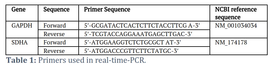
Each experiment animal's jugular vein was used to draw five milliliters of blood, which was then separated into serum using a centrifuge and stored at -20 C until analysis. The following were measured using ELISA kits from United States Biological Company: interleukin-10, IL-6, IL-1β, CRP and TNF-α.two genes used (GAPDH and SDHA) were utilized to assess the integrity of RNA and normalize RT-PCR levels. For GAPDH and SDHA, in specific cow primers (Table 1).
Statistical analysis
The data was presented as mean ± SD. Data evaluated using a one-way ANOVA were deemed significant using the Student's −test if the A value was ≤ 0.01.
Results![]()
Figure 1 shows that in RP cows, the mean values of IL10, IL6 and IL1β were considerably (P<0.01) higher compared to healthy cows (2.31±0.11 versus 1.41±0.07 pg per ml), (13.6±2.1 versus 4±0.9 pg per ml) and (10.48±0.24 versus 5.40±0.19 pg per ml), respectively.
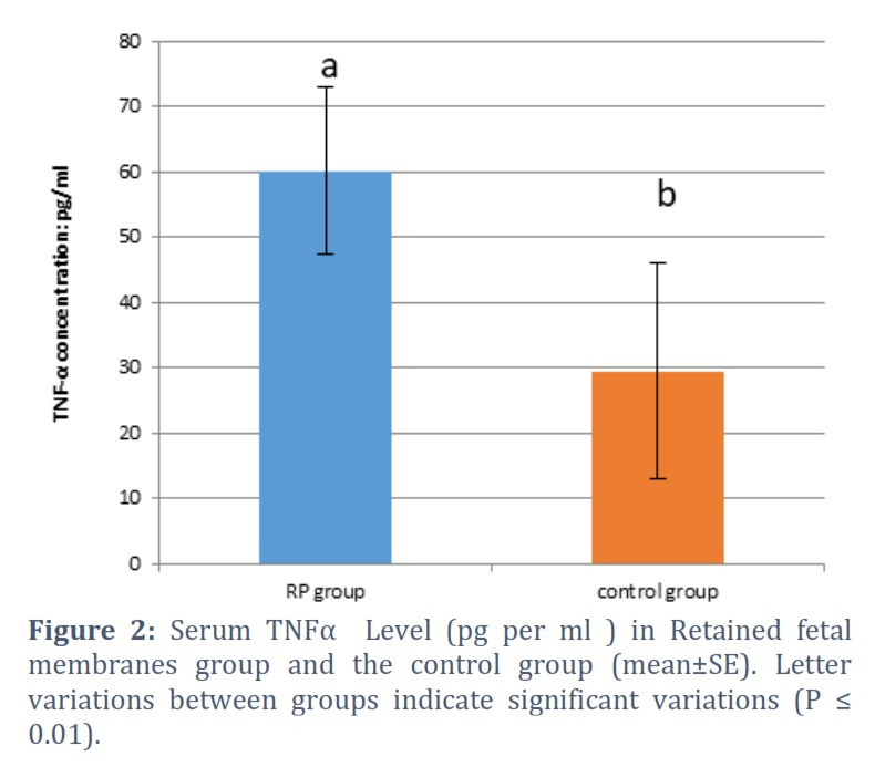
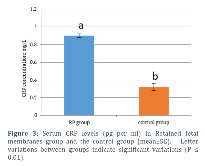
Figures 2 and 3 show that when RP cows were compared to healthy cows, the mean value of CRP was lower (P ≤ 0. 01) in RP cows (0.9±0.02 vs. 0.32±0.04 mg/L, respectively), whereas the mean value of TNF-α was higher (P ≤ 0. 01) in RP cows (60.1 ± 12.79 vs. 29.5 ± 16.58 pg/ml, respectively).
Gene Expression in Retained Placenta in cattle
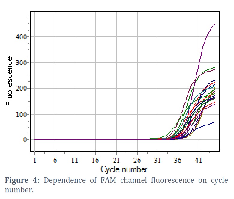
Semi quantitative RT-PCR could detect mRNA expression of SDHA in both paternal and fetal tissue samples , to assess the expression of SDHA polarization markers in mother and fetal cells in the cattle term placenta the level of expression was determined separately in fetal and mother tissues. The results revealed that SDHA expression was considerably higher in both tissues mother than in all other genes (Figure 4).
Figures & Tables
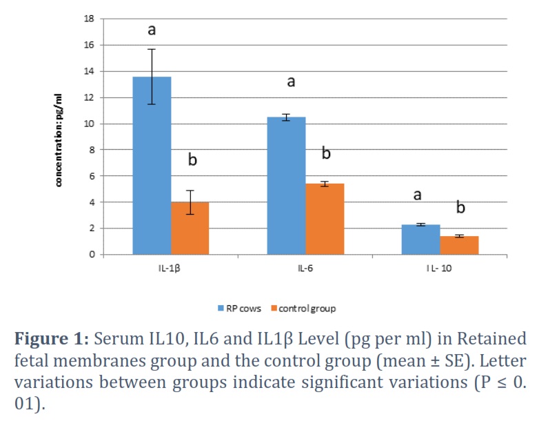
Our investigation found that cows with RP had noticeably higher levels of IL-10, IL-6 and IL-1β than did healthy cows. These differences could be the result of an inflammatory insult that occurred in the mammary gland eight weeks prior to birth. Comparing the results to those of other studies, Trevisi et al. [14] and Sheldon et al. [15] found that elevated levels of inflammatory markers or cytokines in cows during the dry period were linked to inflammatory events or a high incidence of mastitis in the vicinity of parturition. Islam et al. [16] revealed that the IL-10 concentration can be used to identify cows that are prone to reproductive illnesses that may arise after calving, and they proposed that the IL-1 level decline during the two weeks prior to or at parturition can be utilized to do so. According to Ishikawa et al. [17], some retained fetal membranes were linked to higher levels of interleukin 6 prior to calving as opposed to later. C-reactive protein is a pentraxin family plasma protein that is produced by hepatocytes. Its expression is controlled by inflammatory cytokines produced by adipocytes, such as interleukin 6 (IL-6), and it serves primarily as an anti-inflammatory and pro-inflammatory molecule [18, 19]. By attaching phospholipids, phosphocholine, chromatin, fibronectin, and histone, it plays a crucial role in the identification and elimination of infections and damaged cells [20, 21].
In the present study, retained placental cows' CRP levels were considerably lower than those of normal cows. Acute phase proteins, primarily CRP, are more concentrated in retained fetal membranes when mastitis and endometritis are present. This is because lymphocytes and macrophages in retained fetal membranes secrete more IL-6, which causes CRP to bind to the surface of dying cells and make it easier for macrophages to phagocytose these cells, reducing the risk of infection and inflammation in the placental tissue [22, 23]. Activated macrophages, natural killer cells, and T lymphocytes create tumor necrosis factor-alpha (TNF-α), a pleiotropic cytokine that regulates inflammation and is elevated in serum and placental tissue [24]. Several cancers are susceptible to the growth-inhibitory and cytotoxic actions of tumor necrosis factor-alpha. An essential factor in initiating the host’s immune response is the rising levels of tumor necrosis factor-alpha in the serum of cows with retained fetal membranes, which triggers the release of interleukins 1 and 6 [25, 26].
The high level of SDHA expression in fetal and Mother tissues shows that the majority of macrophages are M1 type. In cow, it has been shown that during the second half of pregnancy, the proportion of M2 macrophages increases, contributing to the anti-inflammatory milieu that is essential to prevent fetal rejection [27].
Determining the blood concentrations of interleukins IL-10, IL-6, IL-1β, and TNF-α is a useful method for predicting placental retention in cows since these molecules have a significant role in the development of placental retention. The high expression of SDHA suggests a role for this transcription factor in the overexpression of cytokines.
Acknowledgments
The laboratory facilities were provided by College of Veterinary Medicine, University of Baghdad and Tikrit for which the author is appreciative.
Conflict of Interest
The authors declare that there is no conflict of interest.
Nawaf Nooraldeen and Manar Sabah conceived the project. Hayder Al-Mutar and Maythem Abdulealah supervised the project and provided guidance. All authors wrote, edited, and approved the final manuscript.
![]() References
References
- Raheem KA, Uchechukwu NVS, Odirichukwu E, Onyegbulam OP. Retention in the cow: Report of three cases. Sokoto Journal of Veterinary Sciences, (2016); 14(2): 72-76.
- Baker BC, Heazell AEP, Sibley C. Hypoxia and oxidative stress induce sterile placental inflammation in vitro. Scientific reports, (2021); 11(1): 7281.
- McNaughton AP, Murray RD. Structure and function of the bovine fetomaternal unit in relation to the causes of retained fetal membranes. Vet Record, (2009); 165(21):615–622.
- Sarhat ER, Rmaid ZJ, Jabir TH. Changes of salivary interleukine17, Apelin, Omentin and Vaspin levels in normal subjects and diabetic patients with chronic periodontitis, Annals of Tropical Medicine and Public Health, (2020); 23(1): S404.
- Mohammed IJ, Sarhat ER, Sarhat TR. Assessment of salivary Interleukin (IL)-6, IL-10, Oxidative Stress, Antioxidant Status, pH, and Flow Rate in Dental Caries Experience patients in Tikrit Province. Systematic Reviews in Pharmacy, (2021); 12(1): 55-59.
- Sarhat ER, Mahmood AR. Evaluation of serum concentration Interleukins in Patients with Myocardial Infarction by ELISA Technique. Kirkuk University Journal /Scientific Studies (KUJSS), (2018); 13(1):43-51.
- Ayelign B, Negash M, Andualem H. Association of IL-10 (− 1082 A/G) and IL-6 (− 174 G/C) gene polymorphism with type 2 diabetes mellitus in Ethiopia population. BMC Endocrine Disorders, (2021); 21(70):1-8.
- Souhayla OH, Kreem IA, Sadeq JZ, Ahmed MM. Activity of transaminase enzyme and testosterone hormone in blood of Awassi rams during different season. Asian Pacific Journal of Reproduction, (2017); 6(5): 217-220
- Kim JS, Lee JY, Yang JW, Lee KH. Effenberger M, Szpirt W, Kronbichler A, Shin JI. Immunopathogenesis and treatment of cytokine storm in COVID-19. Theranostics, (2021); 11(1):316-329.
- Emily LB, Vincent JS. Gene-Siew Ngian, Rangi Kandane-Rathnayake,Rachel Mende, Eric F Morand, Tali Lang and James Harris. Analysis of serum interleukin(IL)-1a, IL-1band IL-18 inpatients with systemic sclerosis. Clinical and Translational Immunology, (2019); 8(4):1045.
- Tucho TT, Wahid MA. Economic and reproductive impacts of retained placenta in dairy cows. Journal of Reproduction and Infertility, (2017); 8(1): 18-27.
- Souhayla OH, Hajer KS, Kreem IA. Effect of Dilution, Cooling and Freezing on Physical and Biochemical Properties of Semen for Holstein Bull Born in Iraq. Advances in Animal and Veterinary Sciences, (2016); 4(11) :575-579.
- Al-Anbaghy KI. Shareef KK. Ali AH. Fathalla M. Serum estradiol and progesterone concentrations in cows with and without retained placenta. The Iraqi Journal of Veterinary Medicine, (1989); 13(1): 97–101.
- Trevisi E, Jahan N, Bertoni G, Ferrari A, Minuti A. Pro-inflammatory cytokine profile in dairy cows: consequences for new lactation. Italian Journal of Animal Science, (2015); 14(3):285–292.
- Sheldon IM, Noakes DE, Rycroft AN, Dobson H. Acute phase protein responses to uterine bacterial contamination in cattle after calving. Veterinary Record, (2001); 148(6):172–175.
- Islam R, Kumar H, Nandi S, Mehrotra S. Circulatory level of interleukin-1 in periparturient cows with or without postpartum reproductive diseases. Asian Pacific Journal of Reproduction, (2013); 2(4): 316-320.
- Ishikawa Y, Nakada K, Hagiwara K, Kirisawa R, Iwai H, Moriyoshi M, Sawamukai Y. Changes in interleukin-6 concentration in peripheral blood of pre- and post-partum dairy cattle and its relationship to postpartum reproductive diseases. Journal of Veterinary Medical Science, (2004); 66(11): 1403-1408.
- Kanmani S, Kwon M, Shin MK. Association of C-Reactive Protein with Risk of Developing Type 2 Diabetes Mellitus, and Role of Obesity and Hypertension: A Large Population-Based Korean Cohort Study. Scientific Reports, (2019); 9(4573):1-8.
- Zbaar SA, Sarhat ER, Khalaf SJ. Association of C-Reactive Protein with Risk of Complications of diabetic nephropathy. Egyptian Journal of Chemistry, (2022); 65(8): 483-487.
- Vanderschueren S, Deeren D, Knockaert DC, Bobbaers H, Bossuyt X, Peetermans W. Extremely elevated C-reactive protein. European Journal of Internal Medicine, (2006); 17 (6): 430-4333.
- Julijana S, Jelena R, Katarina B, Milan O, Magbubah E, Sonja Z, Zoran G, Takashi G, Esma RI. Role of C-Reactive Protein in Diabetic Inflammation. Mediators of Inflammation, (2022); 1-15.
- Al-Watar B, Lazim E, Al-Hyani O. Creatine kinase and C reactive protein as an indicator for tissue damage in the retained placenta in cows. Iraqi Journal of Veterinary Sciences, (2021); 35(1): 163-167.
- Amin K, Kauffman CA. Fever of unknown origin: a strategic approach to this diagnostic dilemma. Postgrad Med. (2003); 114(3) :69-75.
- Dos Passos JRR, de Freitas RA, Reppetti J, Medina Y, Dela Justina V, et al. High Levels of Tumor Necrosis Factor-Alpha Reduce Placental Aquaporin 3 Expression and Impair in vitro Trophoblastic Cell Migration. Frontiers in Physiology, (2021); 12:1-10.
- Haimei L, Xinyu L, Limei C, Baowei L, Hang D, et al. Quench-Release-Based Fluorescent Immunosensor for the Rapid Detection of Tumor Necrosis Factor α.ACS Omega., (2021); 6: 31009-31016.
- Jang DI, Lee AH, Shin HY, Song HR, Park JH, et al. The Role of Tumor Necrosis Factor Alpha (TNF-α) in Autoimmune Disease and Current TNF-α Inhibitors in Therapeutics. International Journal of Molecular Sciences, (2021); 22 (2719):1-16.
- Oliveira LJ, McClellan S, Hansen PJ. Differentiation of the endometrial macrophage during pregnancy in the cow. PLoS ONE., (2010); 5(10): 1-12.
This work is licensed under a Creative Commons Attribution-Non Commercial 4.0 International License. To read the copy of this license please visit: https://creativecommons.org/licenses/by-nc/4.0