

Full Length Research Article
Study of the Teratogenic Effects of Antimony on Liver in the Adult Rabbit (Oryctolagus cuniculus)
Ibtisam Khalaf Abd Ali1, Saba Dawod Salman2, Thekra Atta Ibrahim3, Mohammed Nsaif Abbas4*
Adv. life sci., vol. 11, no. 2, pp. 462-469, May 2024
*– Corresponding Author: Mohammed Nsaif Abbas (mohammed.nsaif.abbas@gmail.com)
Authors' Affiliations
2. College of Basic Education, Mustansiriyah University, Baghdad – Iraq
3. Department of Biology, College of Education for Pure Science, University of Diyala – Iraq
4. Materials Engineering Department, College of Engineering, Mustansiriyah University, Baghdad – Iraq
[Date Received: 14/10/2023; Date Revised: 17/03/2024; Date Available Online: 18/04/2024]
Editorial Note on Version of Record
31 May 2025: This article has been corrected. See https://doi.org/10.62940/als.v13i0.4276 for more information.
Abstract![]()
Introduction
Methods
Results
Discussion
References
Abstract
Background: Heavy elements such as antimony greatly affect the environment and living organisms. Antimony is discharged into the environment by mining and industries that use it as pesticides and flame retardants. This activity can lead to environmental pollution, water and soil contamination. Antimony can also accumulate in living organisms and cause negative health effects, such as damage to the respiratory system and skin, and growth abnormalities of animals and plants.
Methods: The primary objective of this investigation was to explore the teratogenic impact of the antimony heavy metal on histological structure of the liver in adult rabbits (Oryctolagus cuniculus). The study included 21 adult white rabbits divided into several groups: the first one is the control group administered physiological saline (0.09% NaCl), the other group administered 20 mg/kg antimony, and the last administered 30 mg/kg antimony over a 30-day period. Following this, postmortem procedures were conducted to extract and fix the liver organ, and tissue sections were prepared.
Result: The results revealed significant histological changes, including distortion and rupture in Glisson’s Capsule, leading to the formation of a sub-capsular space due to its separation from hepatocytes. Additionally, alterations in the radial organization of hepatocytes and pyknosis in the nuclei were observed, characterized by a dark color and reduced size. Karyolysis, where nuclei completely disappeared, and hydropic degeneration in hepatocytes with swollen appearance and dark nuclei due to fluid accumulation were noted. Moreover, an increase in Kupffer cells and blood congestion in the central vein, resulting in dilation compared to the control group, were observed.
Conclusion: Overall, the treatment with antimony at 20 and 30 mg/g doses for 30 days showed profound teratogenic effects on the histological structure of the liver in adult rabbits. These effects are represented by the destruction of various parts of liver, in addition to changes in arrangement, and distortion and rupture of the cells. Furthermore, an increase in Kupffer cells and blood congestion were also recorded.
Keywords: Antimony; Liver; Rabbits; Kupffer cells; Glisson’s capsule; Environmental effects
Introduction![]()
The remediation of environmental pollution plays a pivotal role in safeguarding the well-being and ecological equilibrium of our surroundings, as it precipitates detrimental consequences on ecosystems, wildlife, and human health [1]. Among the diverse categories of environmental contamination, heavy metals occupy a unique and significant role due to their widespread utilization in various industrial and agricultural practices [2]. There is a multitude of environmental pollution remediation techniques available, exhibiting differences in effectiveness, cost, and the requirement for specialized equipment or preparatory procedures [3]. One of the notable and auspicious approaches for addressing a range of environmental contaminants involves heavy metals such as lead, chromium, zinc, iron etc. [4], methyl green stain [5], organic materials [6], inorganic substances of high toxicity like cyanide [7], hardness elements i.e., calcium and manganese [8], resources of eutrophication which are phosphate and nitrate ions [9], humic acid as a type of organic acids [10], and pesticide components [2], from water [11], soil [12], and crude oil [13], is adsorption [14]. Activated carbon stands out as an exceptionally effective material for addressing environmental pollution due to its possession of distinctive attributes that are not present in alternative adsorbents [15]. Notwithstanding, recently, regulatory bodies and researchers have directed greater focus across agricultural and some kinds of industrial residues in exchange for the famous material, i.e., activated carbon [16]. The first choice of these waste was rice husks [17], due to their huge amount remnant. Also, there are watermelon rinds [18], and cellulosic wastes such as banana peels [19], and pomegranate (Punica granatum) peels [20]. Other promising residues, which also gave a good performance were citrus peels like orange [21], and lemon [22]. In addition, domestic wastes such as tea leaves [23], and eggshell [24] were interesting choices for researchers and those concerned in environmental affairs. Furthermore, alga [25], water hyacinth [26], buckthorn leaves [27], and aluminum foil [28] were also used for treatment of contaminated mediums. This can be attributed to their ready accessibility, cost-effectiveness, and minimal toxicity [29]. Furthermore, they do not necessitate intricate manufacturing procedures, in contrast to activated carbon [30]. These waste materials fall within the category of municipal waste and are frequently disposed of at no expense or repurposed using the Zero Residue Level (ZRL) approach [31]. Based on laboratory-scale experimentation, this concept has demonstrated remarkable success in the realm of waste management by effectively transforming hazardous residues into valuable materials, such as additives [32], pesticide [33], ketones [34], or nano-particles [35]. The predicament arises from the limited real-world application of this concept beyond the laboratory setting. Consequently, what were initially non-valuable and environmentally benign adsorption materials in their pristine state undergo a transformation into an environmental concern at their ultimate stage. This transformation occurs as residues and pesticides have the potential to re-enter the environment in novel and potentially more hazardous configurations, rather than serving as a solution to environmental challenges [36]. In spite of the various treatment approaches, established guidelines, and recommended practices, the persistence of heavy metals and their compounds in the environment, owing to their non-biodegradable nature, continues to result in the contamination of natural elements, thereby exerting adverse impacts on both human health and wildlife [18]. Antimony, a heavy metal of particular concern, is extensively utilized in various applications, including alloys, bullets, printing presses, cable sheathing, flame-retardant materials, paints, enamels, as well as in the production of glass and pottery [21]. Nevertheless, the recurring and uncontrolled utilization of this substance gives rise to apprehensions regarding its potential environmental and health repercussions on various organisms, including the human population [22]. Hence, addressing the issue of heavy metal contamination, encompassing antimony specifically, necessitates extensive endeavors and ongoing research aimed at comprehending their mechanisms of impact on both target and non-target organisms within the human body [23]. This pursuit aims to discern effective strategies for mitigating their adverse effects while concurrently attaining equilibrium in alignment with their intended applications [36]. One of the foremost endeavors and investigations in this regard involves histological examinations of organs of living organisms, particularly those susceptible to direct exposure to various pesticide products or their associated residues. Such scientific research is indispensable and pivotal for comprehending the environmental impact of antimony, as well as its effects on living organisms, akin to the scrutiny undertaken for other contaminants like pesticides [37, 38]. The majority of histological investigations concerning the impact of antimony have predominantly centered on murine models, specifically rats [39] and mice [40]. However, a notable void exists in our understanding of the histological ramifications of this heavy metal on rabbits, where the effect known is for testes [41]. The liver is recognized as a pivotal organ responsible for upholding biological equilibrium and essential functions within living organisms. Consequently, any adverse influence on the liver could exert a substantial impact on the well-being of living organisms and the broader ecosystem [42]. The liver is responsible for a multitude of vital functions, encompassing the metabolism of proteins, fats, and carbohydrates, alongside the synthesis of essential plasma proteins like prothrombin factor, fibrinogen factor, and blood coagulation factors. Furthermore, the liver plays a crucial role in blood purification, eliminating bacteria and toxins, while also mitigating the toxic effects of certain pharmaceutical compounds [43]. Numerous chemicals and pharmaceutical compounds are widely recognized for their potential hepatotoxicity, with regulatory directives and guidance issued by the Food and Drug Administration (FDA) cautioning against the hepatotoxic risks associated with these compounds and substances [44]. Currently, pentavalent antimonites, such as sodium stibogluconate (known generically as SSG or Pentostam by GlaxoSmithKline) and meglumine antimonite (marketed as Glucantime™ by Sanofi), enjoy widespread global utilization for the treatment of both Cutaneous leishmaniasis and Visceral leishmaniasis [45]. Considering the aforementioned considerations, the principal aim of the present study is to examine the influence of antimony, a representative heavy metal, on the histological composition of the liver in white rabbits. The study also seeks to precisely elucidate the impact of this heavy metal on the cellular and tissue levels.
Methods![]()
Animals: In this investigation, a total of 21 albino rabbits were obtained from the animal house at the Department of Biology, College of Education for Pure Sciences at the University of Diyala, which also gave the ethical approval of this study. The average weight of animals used ranged between 1250 – 1500 g.
Chemicals: The antimony source used in the current study was antimony trisulfide salt supplied from Sigma-Aldrich company of a gray to black powder. Formalin solution of 10% concentration was prepared by adding 90 ml of distilled water to 10 ml of formaldehyde solution, Eosin stain was prepared by dissolving of one gram of eosin in 99 ml of ethyl alcohol of 70% concentration before adding 0.2 ml glacial acetic acid. Finally, The mixture was then carefully filtered. Harris’s hematoxylin stain was prepared by mixing two solutions, the first one was dissolving 2.5 g of hematoxylin powder in 25 ml of ethyl alcohol. The other solution was prepared by dissolving 50 g of alum in one-half liter of distilled water according to the method described by [44].
Experimental design: The rabbits were subject to random allocation into three distinct groups. The initial group, designated as the control group, consisted of 7 rabbits receiving injections of physiological saline (0.09% NaCl) exclusively. The second group, known as the test group, encompassed 14 rabbits, which were further evenly divided into two sub-groups, each containing 7 rabbits. Within the test group, the rabbits were administered daily doses of antimony at concentrations of 20 and 30 mg/kg of body weight via injection over a span of 30 days.
Methods for estimation of various histological changes: On the final day of the study, the rabbits were anesthetized using chloroform, and the livers were surgically removed. Subsequently, these liver samples were immersed in formalin solution for an overnight fixation period, followed by the preparation of tissue sections using the procedure described by Bancroft and Gamble [46]. The resultant glass sections were then stained using Harris’s Hematoxylin and Eosin (H&E) stain, adhering to the method established by Abd Al-Latif et al., [47]. Finally, the glass sections, treated with Canada balsam, were subjected to testing and imaging using a light microscope equipped with a digital camera.
Results![]()
The observation from the current study recorded significant histological changes in the liver of adult male rabbits exposed to 20 and 30 mg/kg doses of antimony for a period of 30 days.
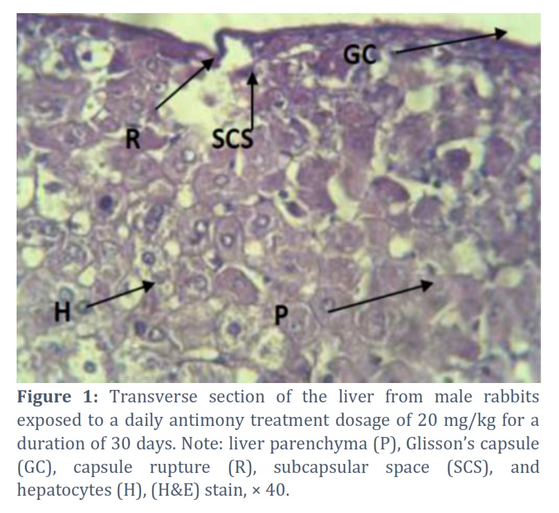
Dose-dependent effect of antimony on Glisson’s capsule and hepatocytes:
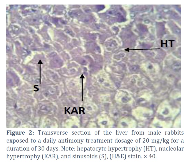
the occurrence of distortion and rupture in the Glisson’s capsule, and its separation from the hepatocytes leading to the appearance of a sub-capsule space and changes in the organization hepatocytes architecture as shown in Figure 1. Whereas, Figure 2 indicates hypertrophy of some hepatocytes, the nucleus of which is Karyomegaly.
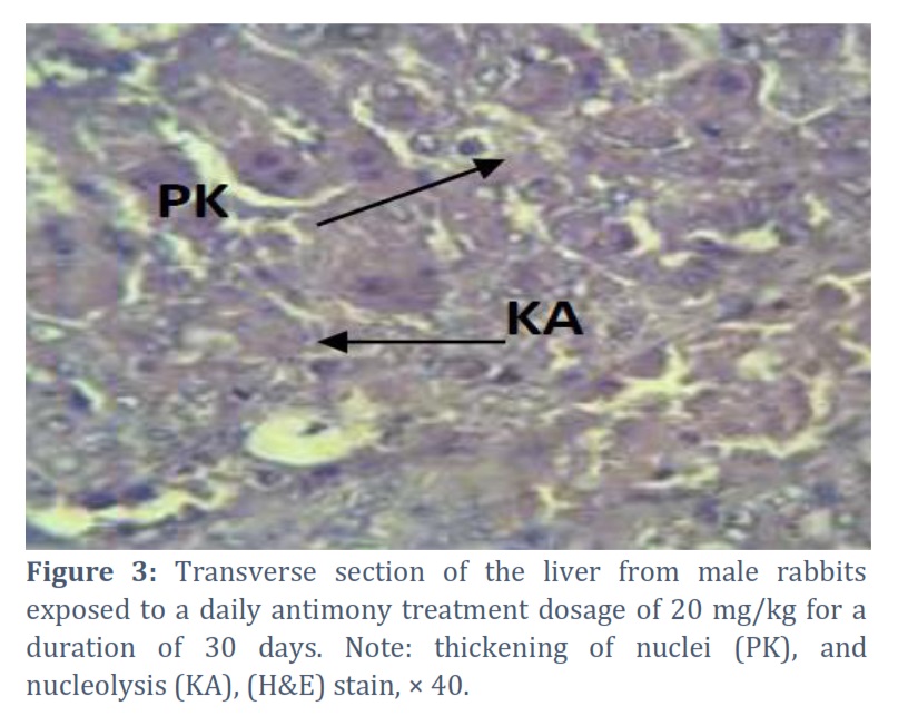
Dose-dependent effect of antimony on Karyolysis and Hydropic degeneration:
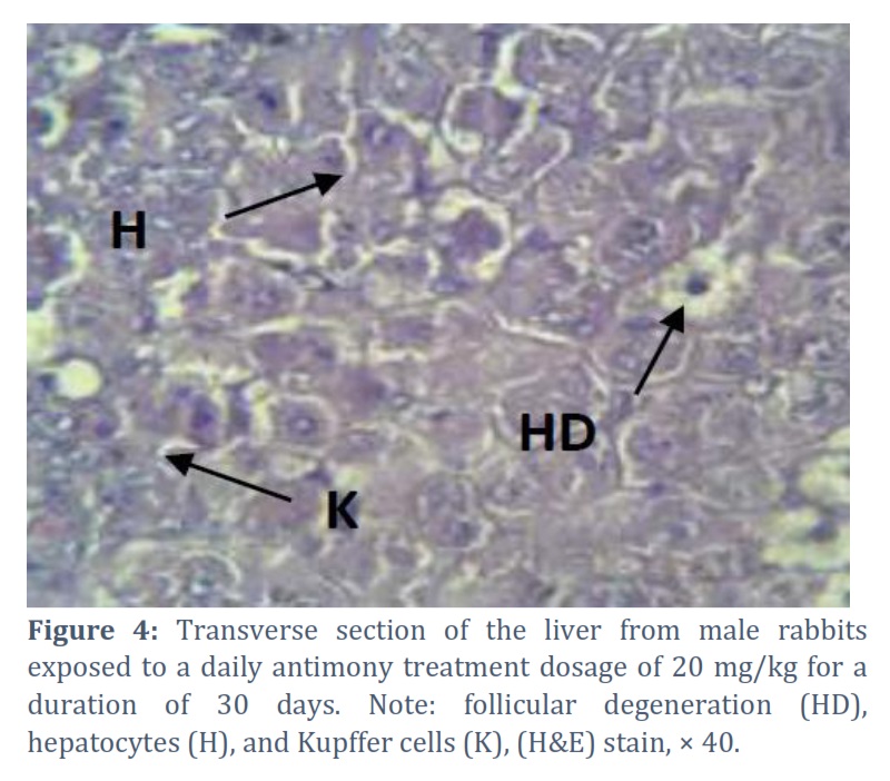
It was also seen that the nuclei of some hepatocytes thickened, where they appeared in a dark color and a small size in the middle of the cells. Karyolysis was also observed in some hepatocytes, where the nuclei completely disappeared, and the cell was seen in a uniform color as in Figure 3. Hydropic degeneration was shown in some liver cells, as it appeared with a swollen appearance with dark-colored nuclei centrally located as a result of fluid accumulation inside them, and an increase in the number of Kupffer cells was also evident as shown in Figure 4.
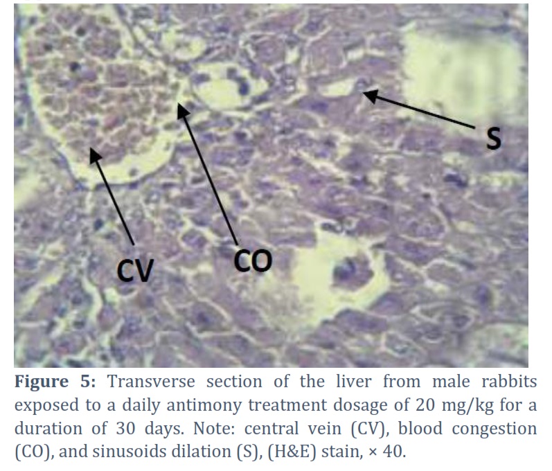
Dose-dependent effect of antimony on central vein and sinusoids
Treatment with antimony led to the occurrence of blood congestion in the central vein and its dilation, as it increased in size compared to the control group, as well as the expansion of the sinusoids located between hepatocytes as in Figure 5.
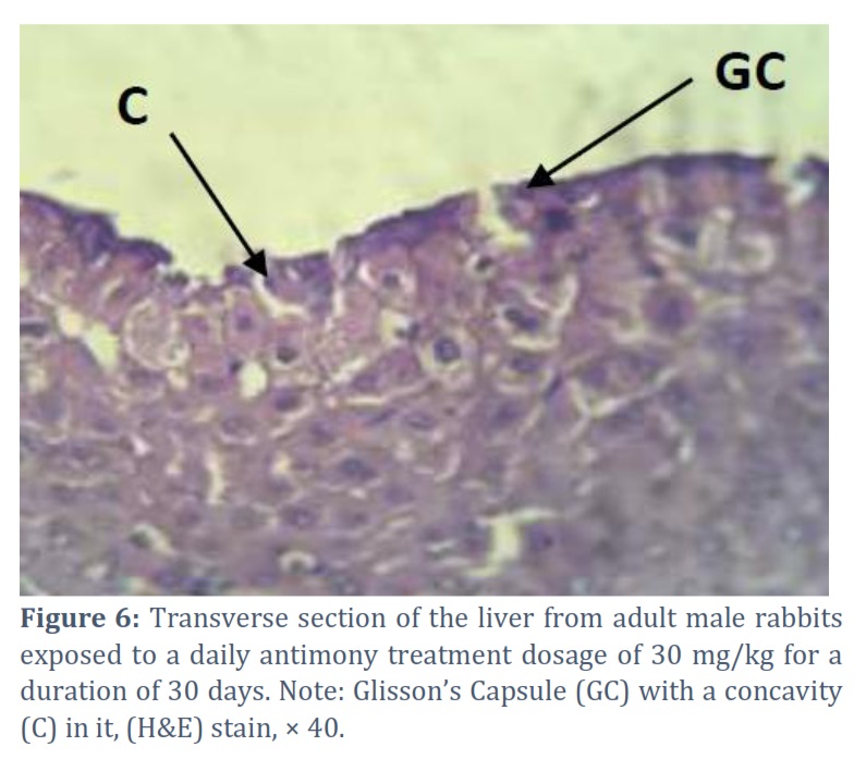
The effect of increasing the dose of antimony on Glisson’s Capsule
Liver of male rabbits of the group treated by a dose of 200 mg/kg of antimony for a period of 30 days, the pathological alterations intensified in the capsule of Glisson’s Capsule, where the natural convex disappeared and the appearance of a concave in it as in Figure 6.
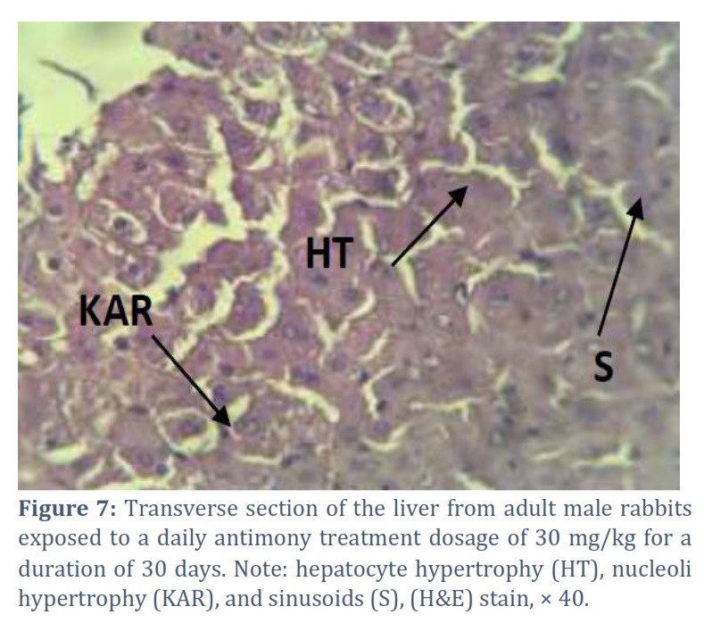
The effect of increasing the dose of antimony on Hypertrophy some hepatocytes
Hypertrophy was also observed in some hepatocytes, where an increase in the size of the cells appeared, as well as karyomegaly hypertrophy of the nuclei of some hepatocytes as in Figure 7.
The effect of increasing the dose of antimony on thickening of the nuclei of hepatocytes and Karyolysis:
An increase in the thickening of the nuclei of hepatocytes was observed, as it appeared darker and smaller in size. Karyolysis of some hepatocytes was also evident as shown in Figure 8.
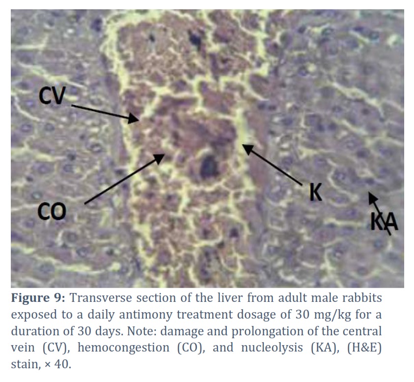
The effect of increasing the dose of antimony on damaged the central vein:
Also, treatment with antimony damaged the central vein, as it changed its normal circular shape to an irregular elongated oval shape, and congestion was observed in it as in Figure 9.
Figures & Tables

The results of this study showed damage to the Glisson’s capsule, as the sub capsular space appeared, and the normal convex shape of the capsule almost disappeared. This result is in agreement with the study of Tan et al., [48] who indicated that malignant tumors, cirrhosis, and infection lead to scattering of the capsular circumference and the loss of its normal convex shape. Lee et al., [49] showed that the effect on the capsular and subcapsular space may be due to pathological conditions such as congestion and inflammation. The changes that occur in the dynamic movement of blood in the liver, such as the obstruction of the portal vein due to its congestion, are one of the reasons that are observed in cases not related to malignant liver tumors. The damage to the capsular shape and the appearance of the subcapsular space may be due to the negative effect of pyridine on this outcome. Hydropic degeneration was also seen in hepatocytes when treated with antimony, as this degeneration led to hypertrophy of hepatocytes. This is consistent with what was stated by Damjanov [50], who confirmed that hydropic degeneration ion occurs in active organs such as the liver as it is the central organ for metabolic processes, and degeneration appears when a liver injury occurs, as it turns into necrosis if it is not treated. On the other hand, the results indicated the occurrence of ballooning degeneration in hepatocytes, as this degeneration is a form of cellular death. This is similar to what Lock et al., [51] found in their study on rat livers exposed to Hexachlorobutadiene (HCBD). Where they confirmed the occurrence of degeneration as a result of the entry of fluids in a large amount into the cell to increase potassium and sodium ions, and this increase can cause damage to the cell membrane. While Malhi et al., [52] indicated that hepatocellular damage and death have the ability to stimulate immune responses, the damage and stress in the hepatocyte results in the stimulation of special cells associated with autoimmunity such as natural killer cells (NK) and Kupffer cells. Changes have been reported on the nuclei of some hepatocytes in the liver of adult rabbits, and this result is consistent with what was stated Jarrar and Taib [53]. They showed through their study of the chronic toxic effect of lead on the mouse liver, that it causes an increase in cellular and nuclear activity in the process of detoxification, and a change in the nuclei of hepatocytes occurs. Whereas Wang et al., [54] confirmed this case also in other organs such as the brain, skeletal muscles and the heart. The polyploid cells become pyramidal in shape and have low proliferative capacity, and the more polyploidy is observed, it is evidence of significant tissue damage. Moreover, this study recorded the presence of pyknosis in some nuclei of hepatocytes. This result is consistent with the results of Kumar et al., [42] where they mentioned that the thickening of the nuclei usually occurs when the cell, after a cellular injury as a result of a toxic substance, undergoes necrosis, which is represented by swollen degeneration, after which the cell undergoes chromatin condensation and thickening. In addition, Karyolysis of some hepatocytes was observed in this result, as the cell appeared in a uniform color due to the complete disappearance of the nucleus. These outputs agree with what was mentioned by Pandey et al., [55] about condensing substances in dead cells, as they confirmed that chromatin degeneration of dead and necrotic cells and destructive fragmentation caused nucleolysis as a result of chemical toxicity. The cause may be the degeneration and enlargement of hepatocytes, the thickening and decomposition of the nuclei treated with antimony, and its clear tissue damage to the hepatocytes that affected the normal shape of the liver. The appearance of widening in the sinuses was observed, and this is consistent with what was stated by Brancatelli et al., [56] who explained that the lack of venous blood flow and the blockage of the central vein led to an increase or congestion in venous pressure, which leads to the occurrence of widening of the sinuses. Congestion in the central veins was also seen, as it was clearly evident that blood accumulated in these vessels, and this is consistent with what they indicated Brancatelli et al., [56], as they showed that the lack of blood flow leads to congestion in the blood vessels and liver board. In a related context, Hassan and Sahi [57] explained in their study on the histopathological changes in the liver and kidneys of mice when exposed to zinc sulfate toxicity, the occurrence of congestion in the veins as a result of decreased metabolism due to hypothyroidism or due to lack of blood flow. The observed congestion may have occurred in the central veins in the current result as a result of inflammation resulting from antimony toxicity.
Conflict of Interest
The authors declare that there is no conflict of interest.
I.K. Abd Ali and T.A. Ibrahim designed the research, while T.A. Ibrahim and S.D. Salman conducted the experiments and carried out laboratory procedures. M.N. Abbas contributed to chemicals preparation and performed the technical writing of the manuscript.
![]() References
References
- Khaleel LR, Al-Hermizy SM, Abbas MN. Statistical Indicators for Evaluating the Effect of Heavy Metals on Samaraa Drug Industry Water Exposed to the Sun and Freezing. Tropical Journal of Natural Product Research, (2022); 6(12): 1969-1974.
- Rajaa N, Kadhim FJ, Abbas MN, Banyhussan QS. The improvement of concrete strength through the addition of sustainable materials (agro-waste loaded with copper ions). 3rd International Conference for Civil Engineering Science (ICCES 2023), IOP Conf. Series: Earth and Environmental Science, (2023); 1232, 012038, 9 Pages.
- Abbas MN, Ali ST, Abbas RS. Rice Husks as a Biosorbent Agent for Pb+2 Ions from Contaminated Aqueous Solutions: A Review. Biochemical and Cellular Archives, (2020); 20(1): 1813-1820
- Hashem NS, Ali GAA, Jameel HT, Khurshid AN, Abbas MN. Heavy Metals Evaluation by Atomic Spectroscopy, for Different Parts of Water Hyacinth (Eichhornia crassipes) Plants Banks of Tigris River. Biochemical and Cellular Archives, (2021); 21(2): 3813-3819.
- Alalwan HA, Mohammed MM, Sultan AJ, Abbas MN, Ibrahim TA, Aljaafari HAS, Alminshid AA. Adsorption of methyl green stain from aqueous solutions using non-conventional adsorbent media: Isothermal kinetic and thermodynamic studies. Bioresource Technology Reports, (2021); 14, Article number: 100680.
- Abbas MN, Alalwan HA. Catalytic Oxidative and Adsorptive Desulfurization of Heavy Naphtha Fraction. Korean Journal of Chemical Engineering, (2019); 12(2): 283-288.
- Alalwan HA, Abbas MN, Alminshid AH. Uptake of Cyanide Compounds from Aqueous Solutions by Lemon Peel with Utilising the Residue Absorbents as Rodenticide. Indian Chemical Engineer, (2020); 62(1): 40-51.
- Ibrahim SA, Hasan MB, Al-Tameemi IM, Ibrahim TA, Abbas MN. Optimization of adsorption unit parameter of hardness remediation from wastewater using low-cost media. Innovative Infrastructure Solutions, (2021); 6(4) Article number: 200.
- Abbas MN. Phosphorus removal from wastewater using rice husk and subsequent utilization of the waste residue. Desalination and Water Treatment, (2015); 55(4): 970-977.
- Abbas MN, Abbas FS. Application of Rice Husk to Remove Humic Acid from Aqueous Solutions and Profiting from Waste Leftover. WSEAS Transactions on Biology and Biomedicine, (2014); 11:62-69.
- Abbas MN, Abbas FS. Iraqi Rice Husk Potency to Eliminate Toxic Metals from Aqueous Solutions and Utilization from Process Residues. Advances in Environmental Biology, (2013); 7(2): 308-319.
- Abbas MN, Al-Madhhachi AT, Esmael SA. Quantifying soil erodibility parameters due to wastewater chemicals. International Journal of Hydrology Science and Technology, (2019); 9(5): 550-568.
- Ali GAA, Ibrahim SA, Abbas MN. Catalytic Adsorptive of Nickel Metal from Iraqi Crude Oil using non-Conventional Catalysts. Innovative Infrastructure Solutions, (2021); 6(7): 1-9.
- Abbas MN, Abbas FS. The Predisposition of Iraqi Rice Husk to Remove Heavy Metals from Aqueous Solutions and Capitalized from Waste Residue. Research Journal of Applied Sciences, Engineering and Technology, (2013); 6(22): 4237-4246.
- Maddodi SA, Alalwan HA, Alminshid AH, Abbas MN. Isotherm and computational fluid dynamics analysis of nickel ion adsorption from aqueous solution using activated carbon. South African Journal of Chemical Engineering, (2020); 32: 5-12.
- Abbas MN, Abbas FS. The Feasibility of Rice Husk to Remove Minerals from Water by Adsorption and Avail from Wastes. Research Journal of Applied Sciences, WSEAS Transactions on Environment and Development, (2013); 9(4): 301-313.
- Alalwan HA, Abbas MN, Abudi ZN, Alminshid AH. Adsorption of thallium ion (Tl+3) from aqueous solutions by rice husk in a fixed-bed column: Experiment and prediction of breakthrough curves. Environmental Technology and Innovation, (2018); 12: 1-13.
- Abbas MN, Nussrat TH. Statistical Analysis of Experimental Data for Adsorption Process of Cadmium by Watermelon Rinds in Continuous Packed Bed Column. International Journal of Innovation, Creativity and Change, (2020); 13(3): 124-138.
- Abdullah WR, Alhamadani YAJ, Abass IK, Abbas MN. Study of chemical and physical parameters affected on purification of water from inorganic contaminants. Periodicals of Engineering and Natural Sciences, (2023); 11(2): 166-175.
- Hamdi GM, Abbas MN, Ali SAK. Bioethanol Production from Agricultural Waste: A Review. Journal of Engineering and Sustainable Development, (2024); 28(2): 233–252.
- Hasan MB, Al-Tameemi IM, Abbas MN. Orange Peels as a Sustainable Material for Treating Water Polluted with Antimony. Journal of Ecological Engineering, (2021); 22(2): 25-35.
- Al-Hermizy SMM, Al-Ali SIS, Abdulwahab IA, Abbas MN. Elimination of Zinc Ions (Zn+2) from Synthetic Wastewater Using Lemon Peels. Asian Journal of Water, Environment and Pollution, (2022); 19(5): 79-85.
- Al-Ali SIS., Abudi ZN, Abbas MN. Modelling and Simulation for the use of Natural Waste to Purified Contaminated Heavy Metals. Journal of the Nigerian Society of Physical Sciences, (2023); 5(1): Article No.: 1143.
- Ali SAK, Almhana NM, Hussein AA, Abbas MN. Purification of Aqueous Solutions from Toxic Metals using Laboratory Batch Mode Adsorption Unit Antimony (V) Ions as a Case Study. Journal of Green Engineering (JGE), (2020); 10(11): 10662-10680.
- Abbas MN, Al-Hermizy SMM, Abudi ZN, Ibrahim TA. Phenol Biosorption from Polluted Aqueous Solutions by Ulva lactuca Alga using Batch Mode Unit. Journal of Ecological Engineering, (2019); 20(6): 225–235.
- Ali GAA, Abbas MN. Atomic Spectroscopy Technique Employed to Detect the Heavy Metals from Iraqi Waterbodies Using Natural Bio-Filter (Eichhornia crassipes) Thera Dejla as a Case Study. Systematic Reviews in Pharmacy, (2020); 11(9): 264-271.
- Alhamd SJ, Abbas MN, Manteghian M, Ibrahim TA, and Jarmondi KDS. Treatment of Oil Refinery Wastewater Polluted by Heavy Metal Ions via Adsorption Technique using Non-Valuable Media: Cadmium Ions and Buckthorn Leaves as a Study Case. Karbala International Journal of Modern Science, (2024); 10(1): 1-18.
- Ghulam NA, Abbas MN, Sachit DE. Preparation of synthetic alumina from aluminium foil waste and investigation of its performance in the removal of RG-19 dye from its aqueous solution. Indian Chemical Engineer, (2020); 62(3): 301-313.
- Abbas MN, Ibrahim SA. Catalytic and thermal desulfurization of light naphtha fraction. Journal of King Saud University – Engineering Sciences, (2020); 32(4): 229-235.
- Abbas MN, Al-Tameemi IM, Hasan MB, Al-Madhhachi AT. Chemical Removal of Cobalt and Lithium in Contaminated Soils using Promoted White Eggshells with Different Catalysts; South African Journal of Chemical Engineering, (2021); 35: 23-32.
- Abbas FS, Abdulkareem WS, Abbas MN. Strength Development of Plain Concrete Slabs by the Sustainability Potential of Lead-Loaded Rice Husk (LLRH). Journal of Applied Engineering Science, (2022); 20(1): 160-167.
- Abdulkareem WS, Aljumaily HSM, Mushatat HA, Abbas MN. Management of Agro-Waste by Using as an Additive to Concrete and Its Role in Reducing Cost Production: Impact of Compressive Strength as a Case Study. International Journal on “Technical and Physical Problems of Engineering” (IJTPE), (2023); 54, 15(1): 62-67.
- Abd ali IK, Ibrahim TA, Farhan AD, Abbas MN, Study of the effect of pesticide 2,4-D on the histological structure of the lungs in the albino mice (Mus musculus). Journal of Pharmaceutical Science and Research, (2018); 10(6): 1418-1421.
- Abbas MN, Ibrahim SA, Abbas ZN, Ibrahim TA. Eggshells as a Sustainable Source for Acetone Production. Journal of King Saud University – Engineering Sciences, (2021); 34(6): 381-387.
- Alminshid AH, Abbas MN, Alalwan HA, Sultan AJ, Kadhome MA. Aldol condensation reaction of acetone on MgO nanoparticles surface: An in-situ drift investigation. Molecular Catalysis, 501, Article No.: 111333.
- Alwan EK, Hammoudi AM, Abd IK, Abd Alaa MO, Abbas MN. Synthesis of Cobalt Iron Oxide Doped by Chromium Using Sol-Gel Method and Application to Remove Malachite Green Dye. NeuroQuantology, (2021); 19(8): 32-41.
- Ibrahim TA, Mahdi HS, Abbas RS, Abbas MN, Study the Effect of Ribavirin Drug on the histological structure of the testes in Albino mice (Mus musculus). Journal of Global Pharma Technology, (2020); 12(02 Suppl.): 142-146.
- Ibrahim TA, Mohammed AM, Abd ali IK, Abbas MN, Hussien SA. Teratogenic Effect of Carbamazepine Drug on the Histological Structure of Testes in the Albino Mouse (Mus musculus). Indian Journal of Forensic Medicine & Toxicology, (2020); 14(4): 1829-1834.
- Rashedy AH, Solimany AA, Ismail AK, Wahdan MH, Saban KA. Histopathological and functional effects of antimony on the renal cortex of growing albino rat. International journal of clinical and experimental pathology, (2013); 6(8): 1467–1480.
- Coelho DR, De-Oliveira A, Parente T, Leal BS, das Chagas LF, Oliveira TN, Saint’Pierre TD, Paumgartten F. In vivo and in vitro effects of pentavalent antimony on mouse liver cytochrome P450s. Human & experimental toxicology, (2017); 36(1): 33–41.
- Abbas RS, Ali IKA, Abdulwahab IA, Ibrahim TA. Evaluation the effect of alcoholic extract of ruta graveolens plant on the histological structure of lungs in the rabbit. International Journal of Drug Delivery Technology, (2020); 10(4): 612-616.
- Kumar A, Sasmal D, Sharma N, Mechanism of deltamethrin induced thymic and splenic toxicity in mice and its protection by piperine and curcumin: in vivo study. Drug and chemical toxicology, (2018); 41(1), 33–41.
- Ali ST, Qadir HT, Moufak SK, Al-Badri MAM, Abbas MN. A Statistical Study to Determine the Factors of Vitamin D Deficiency in Men the City of Baghdad as a Model. Indian Journal of Forensic Medicine & Toxicology, (2020); 14(1): 691-696.
- Reuben A, Koch D, Lee W. Drug-induced acute liver failure: Results of a U.S. multicenter, prospective study. Hepatology, (2010); 52(6):2065-2076.
- Radha MJ, Nazzal MF, Ibrahim TA. Evaluated Serum Levels for Ige, Cytokines Il-6 And Il-10 in Patients With Eczema in City of Baqubah. Biochemical and Cellular Archives, (2019); 19(2): 3469–3471.
- Bancroft JD, Gamble M. Theory and Practices of Histological Technique. (2008). 6th edition. Churchill Livingstone, Elsevier, Philadelphia.
- Abd Al-Latif FS, Ibrahim TA, Abbas MN. Revealing Potential Histological Changes of Deltamethrin Exposure on Testicular Tissue in Albino Rabbits (Oryctolagus cuniculus). Advancements in Life Sciences, 10(4): 619-626.
- Tan G, Miranda R, Sutherland T. Cases of hepatic capsular retraction: a pictorial essay. Insights into Imaging, (2016); 7(6): 831-840.
- Lee J, Kim S, Kwack S, Kim C, Moon T, Lee S, Cho M, Kang D, Kim G. Hepatic Capsular and Subcapsular Pathologic Conditions: Demonstration with CT and MR Imaging. RadioGraphics, (2008); 28(5):1307-1323.
- Damjanov I. Pathology Secrets. (2009). 3rd ed. Philadelphia: Mosby, Inc., an affiliate of Elsevier Inc: pp v+528.
- Lock E, Ishmael J, Pratt I. Hydropic change in rat liver induced by hexachloro-1: 3-butadiene. Journal of Applied Toxicology, (1982); 2(6): 315-320.
- Malhi H, Gores G, Lemasters J. Apoptosis and necrosis in the liver: A tale of two death? Hepatology, (2006); 43(1): 31-44.
- Jarrar B, Taib N. Histological and histochemical alterations in the liver induced by lead chronic toxicity. Saudi Journal of Biological Sciences, (2012); 19(2); 203-210.
- Wang M, Chen F, Lau J, Hu Y. Hepatocyte polyploidization and its association with pathophysiological processes. Cell Death and Disease, (2017); 8(5): e2805.
- Pandey G, Srivastava DN, Madhuri S. A standard hepatotoxic model produced by paracetamol in rat. Toxicology International, (2008); 15(1): 69-70.
- Brancatelli G, Furlan A, Calandra A, Dioguardi Burgio M. Hepatic sinusoidal dilatation. Abdominal radiology (New York), (2018); 43(8): 2011–2022.
- Hassan IJ, Sahi IS. Histopathological Changes in the Liver and Kidney of Albino Mice on Exposure to Zinc Toxicity, Indian Journal of Public Health Research & Development, (2019); 10(7): 596-600.
This work is licensed under a Creative Commons Attribution-Non Commercial 4.0 International License. To read the copy of this license please visit: https://creativecommons.org/licenses/by-nc/4.0