

Full Length Research Article
Simultaneous detection of phylogroups and ESBL genes in E. coli using Multiplex PCR
Bushra B. Patoli1*, Atif A. Patoli1, Iram Nabi Begum2, Zarafshan Majeed1
Adv. life sci., vol. 11, no. 2, pp. 470-476, May 2024
*– Corresponding Author: Bushra B. Patoli (bushrapatoli@gmail.com)
Authors' Affiliations
2. Centro de salud Las Albarizas (Distrito Costa del Seol) Marbella. Province Malaga – Spain
[Date Received: 17/10/2023; Date Revised: 07/02/2024; Date Available Online: 18/04/2024]
Abstract![]()
Introduction
Methods
Results
Discussion
References
Abstract
Background: Escherichia coli (E. coli) strains harbor various allelic versions of beta lactamase genes and their identification using conventional phenotypic tests is a tedious and time consuming task. In the present study, multiplex PCR is performed for the simulatanoues detection of E. coli phylogroups and extended-spectrum β-lactamase- (ESBL) genes.
Methods: A total of 128 E. coli isolates from urine samples were screened for antibiotic resistance and expression of ESBL activity using phenotypic and genotypic methods. Uniplex and multiplex PCRs were used to detect E. coli phylogroup detrminants and blaCTX-M-15, blaOXA-1 and TEM1 genes. Chi Square test of independence was employed for evaluating significant levels at P value < 0.05.
Results: Phylogroup B2 was detected as the predominant group (36%) followed by group D (30%), A (25%) and B1 (9%). The highest resistance was seen against nalidixic acid (100%) and the lowest against amoxicillin-clavulanic acid (55%). Significant P values were observed for resistance against cefotaxime and cefepime in the phylogroup B2 while resistance against cefoxitin, sulfamethoxazole and fosfomycin was significantly associated with group D. Combination disc diffusion test (CDDT) showed ESBL activity in 42% E. coli isolates. A significant association of blaCTX-M-15 gene was observed for phylogroup B2 (P = 0.007). Moreover, a combination genotype of blaCTX-M-15 and TEM1 was also found statistically prevalent in phylogroup B2 (P = 0.006).
Conclusion: The study highlights the alarming rise in antibiotic resistance and delineates B2 a predominant phylogoup with a high prevalence of blaCTX-M-15 and TEM1 genes in urinary E. coli isolates.
Keywords: UTI; E. Coli; Phylogenetic groups; ESBL genes; Multiplex PCR
Introduction![]()
Co-existence of Escherichia coli (E. coli) as a commensal constitutes an integral part of the natural microflora in many mammalian gastrointestinal tracts [1]. The metabolic diversity in E. coli allows the organism to maintain its homeostasis in diverse environments. Genetic accretions, pathological adaptation to the host immunity and ability to translocate at multiple niches facilitates the transit from a commensal to the pathological state [2]. Heterogeneity in E. coli pathotypes makes it a most common intestinal and extra-intestinal pathogen [3]. Extra-intestinal pathogenic E. coli (ExPEC) are commonly associated with bacteremia, neonatal meningitis and urinary tract infections (UTIs) [4]. ExPEC affect large population with UTI throughout the globe [5]. It is very important to detect prevalent E. coli pathotype associated with UTIs and its epidemiology in UTI infections for monitoring and management of infection at the regional and global scale. Based on the detection of three genetic markers, i.e. chuA, yjaA, and TspE4.C2, Clermont et al., provided a rapid and convenient tool to categorize E. coli into four distinct phylogroups (i.e. A, B1, B2 and D) [6]. Phylogroups B2 and D are the most frequent ExPEC isolates from UTI with virulent determinants whereas E. coli belong to the phylogenetic groups A and B1 are usually witnessed as avirulent species [1,6,7]. However, resistance to multiple antibiotics is widely reported for both the pathogenic [7] and commensal E. coli [8]. The resistance against β-lactam antibiotics is mainly manifested through the production of β- lactamase enzymes. These enzymes are classified into different groups based on their phenotypic/functional characteristics (i.e. Group 1-3) and their amino acid sequences (i.e. A-D classes) [9]. Group 2 (classes A and D) includes extended spectrum of β- lactamase (ESBL) enzymes with the tendency to hydrolyze Penicillin, cephalosporins and monobactams. Since last few decades several variants have been observed for harboring ESBL genes like blaCTX-M, blaOXA, blaSHV and blaAmpC and a huge diversity has been reported across the world including Pakistan. Co-existence of one or combinations of these genes was also reported worldwide indicating prevalence of different functional groups of β-lactamase enzymes is an emerging resistant trait in E. coli pathotypes [10]. In the present study we tried to explore the association of different phylogroups with antimicrobial resistance and the related ESBL genes such as; TEM1, blaOXA-1, blaCTX-M-15. We propose here that in comparison to traditional phenotypic methods, multiplex PCR could be a reliable method for the direct detection of E. coli pathotype and the associated ESBL genotype. Previous studies showed multiplex PCR an ideal choice for the detection of multiple β-lactamase genes or their variants in single run [11,12]. In the present study, we optimized the protocol of multiplex PCR for simultaneous detection of E. coli phylogroup (A, B1 B2 & D) and the most prevalent blaCTX-M-15, blaOXA-1 genes in a single reaction. We propose this multiplex PCR assay would help in probing the molecular epidemiology, diagnosis and management of E. coli infections in the community.
Methods![]()
Isolation and Identification of E. coli
Urine specimens with bacterial count of more than 105 Colony Forming Units (CFU) per ml were considered positive for bacterial UTIs. The isolation of E. coli from urine samples was performed in collaboration with Diagnostic research laboratory at Civil hospital Hyderabad, during the period of February, 2022 to July, 2022. A total of 475 urine samples were processed to isolate E. coli either on MacConkey’s agar medium/CLED agar medium (Oxoid, UK). E. coli were recovered from 128 samples and their identity was confirmed with biochemical phenotype of lactose and indole poitive but citrtae negative. All of the 128 isolates were stock cultured in 50 % glycerol for the antimicrobial susceptibilty check and molecular characterization in the Molecular Microbiology and Genetics Research laboratory at the Institute of Microbiology, University of Sindh, Jamshoro.
Antibiotic Susceptibility test and phenotypic detections of ESBL
Antibiotic susceptibility test was performed according to the CLSI guidelines on Muller Hinton using Kirby Baur-disc diffusion method [13]. Antibiotics (n=18) used from varying groups include, Beta-lactamase inhibitors [piperacillin tazobactam (TZP), amoxicillin-clavulanic acid (AMC)]; Cephalosporins [ceftazidime (CAZ), cefotaxime (CTX), ceftriaxone (CRO), cefpodoxime (CPD), cefepime (FEP), cefoxitin (FOX)]; Fluoroquinolones [ciprofloxacin (CIP), nalidixic acid (NA)]; Aminoglycosides [amikacin (AK)]; chloramphenicol (CM); trimethoprim sulfonamides (SXT); imipenem (IMP) and fosfomycin (FOS) (Oxoid, UK). ESBL phenotype were confirmed by standard double disc synergy test (DDST) and combination disc diffusion tests (CDDT) using ceftazidime (30 µg), cefotaxime (30 µg), ceftriaxone (30 µg), cefpodoxime (10 µg) antibiotic discs with 27 mm apart in synergy with AMC (30 µg) at center. Additional discs of cefepime (30 µg) was also placed. All the results were interpreted according to the CLSI guidelines and a published data for Enterobacteriaceae [14]. Expansion in the inhibition zone towards β-lactam inhibitor (AMC) for the otherwise decreased susceptibility to ceftazidime, cefotaxime, ceftriaxone, cefpodoxime and cefepime was considered a positive synergy test for ESBL production.
Detection of phylogroup markers and β-lactamase genes
Genomic DNA was extracted from E. coli isolates using boiling method as described before (15). Each isolate was subject to triplex PCR using protocol by Clermont et al, to detect different phylogenetic groups of the E. coli strains [6]. For the detection of ESBL genes, initially single PCR reactions were set with one set of primers and for multiplex PCR five different pairs of primers were used.
Single PCR reactions were set with the specific primer at their respective annealing temperature to yield the PCR products of TEM1 (297 bp); blaCTX-M-15 (586 bp) and blaOXA-1 (814bp) separately. For multiplex PCR five pairs of primers including blaCTX-M-15, blaOXA-1, ChuA, yjA and TspE4.C2 were added for simultaneous detection of phylogroup and ESBL genes. Due to the close proximity in base pair size of TEM1 to chuA, a separate PCR reaction was performed to detect TEM-1 β-lactamase. Primers for the genes are presented in Supplementary Table 1. PCR reactions contained 4 μl of GoTaq PCR master mix (Promega), 0.5 μl of each forward and reverse primers, 2 μl bacterial DNA extract and water required to adjust the total 50 μl volume. Thermocycler was programmed as follows: initial denaturation at 95 °C for five minutes followed by 25 consecutive cycles at 95 °C for 30 seconds 55 °C for 30 seconds and 72 °C for 1 minute with a final 2 minutes extension at 72 °C. The amplified products were run on 2% agarose gel electrophoresis in Tris–borate–EDTA (TBE) buffer and visualized after ethidium bromide (EtBr) staining in gel doc system (BioBase).
Statistical analysis
The statistical analysis was performed using SPSS (IBM) version 21 and online statistical calculator [Select Statistical Services UK (www.select-statistics.co.uk)]. Chi square test for independence was used to assess differences by assuming cut-off P value of < 0.05 as significant.
Results![]()
Phylogenetic determination of E. coli isolates using triplex PCR
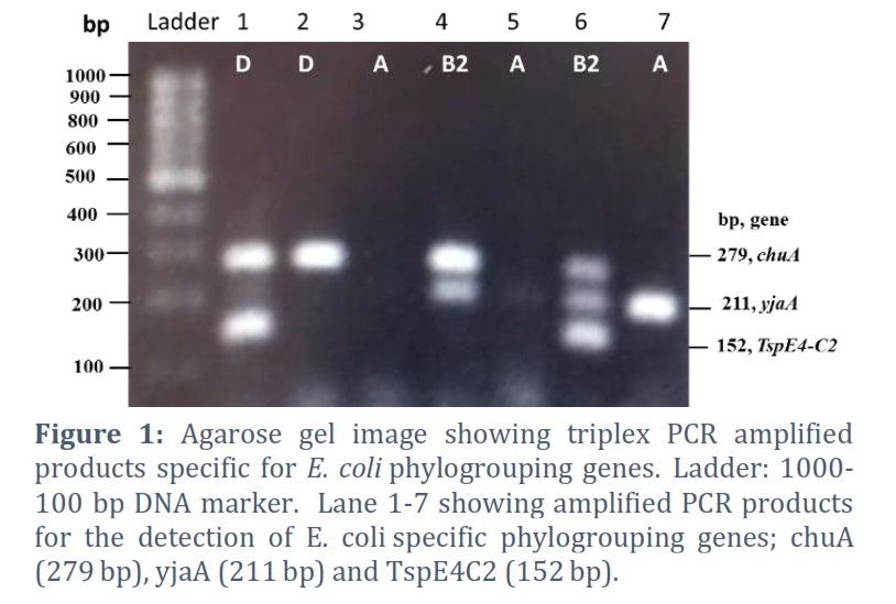
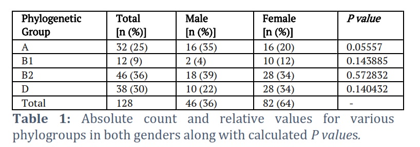
A total of 128 E. coli isolates were categorized into the four different phylogenetic groups (A-D) according to the original Clermont’s phylogenetic scheme [6]. A representative gel image displaying the amplification of phylogenetic specefic genes is shown in Figure 1. Triplex PCR targeting chuA, yjaA and TspE4.C2 DNA fragment indicated phylogroup B2 as the predominant group (n = 46, 36%), followed by group D (n = 38, 30%), A (n =3 2, 25%) and B1 (n = 12, 9%) (Table 1). From the 128 E. coli isolates, 36% (n = 46) were isolated from male and 64% (n = 82) were from female group. Isolates belonging to phylogroup A and B2 were more prevalent in male group of patients (35 and 39% respectively) while the isolates belonging to phylogroup B1 and D were more prevalent in female group of patients (12 and 34% respectively). The P value (< 0.05) determined for each phylogroups were non-significant indicating that their prevalence is independent of gender (Table 1).
The Phylogenetic groups were assigned according to the following genotypes: group A (chuA−/ yjaA−/ TspE4. C2−or yjaA +), group B1 (chuA−/TspE4.C2+), group B2 (chuA+/yjaA+ or chuA+/yjaA+/TspE4.C2+), group D (chuA+/yjaA−or chuA+ /TspE4.C2+), as per the original Clermont’s scheme [6].
Prevalence of antibiotic resistance pattern and ESBL phenotype in different phylogroups
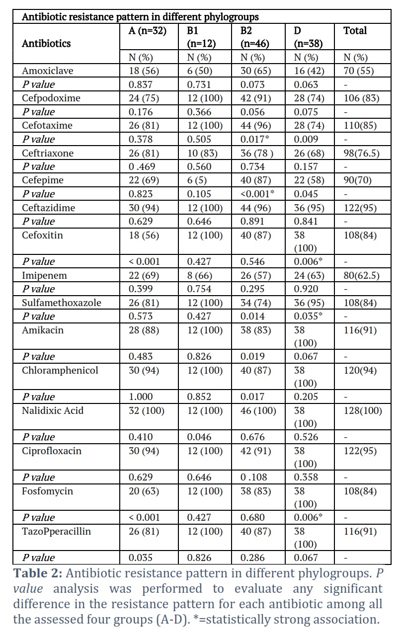
Except co-amoxiclave, more than 70% resistance was witnessed against each antibiotic used in this study (Table 2). All the isolates were resistant to nalidixic Acid (100%) followed by ciprofloxacin and ceftazidime (95%). The lowest percentage of resistance was seen against co-amoxiclave 55% followed by imipenem (62.5%). Isolates of the phylogroup B2 expressed statistically significant dependence for cefotaxime (p = 0.017) and cefipime (p = 0.001) resistance. Similarly, the isolates belonging to Phylogroup D showed a statistically significant dependence for cefoxitin (p = 0.006), sulfomethoxazol (p = 0.035) and fosfomycin (p = 0.006) resistance. A significant independence was seen for cefoxitin (p = 0.001), fosfomycin (p = 0.001) and tazobactam (p = 0.035) in phylogroup A, sulfamethaxasole (p = 0.014), amikacin (p = 0.019), chloramphenicol (p = 0.017) in phylogroup B2, and cefotaxime (p = 0.009) and cefepime (p = 0.045) resistance in phylogroup D isolates (Table 2).
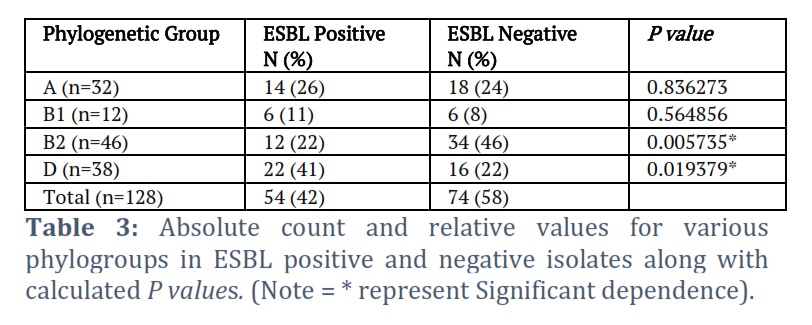
Combination disc diffusion test (CDDT) showed extended spectrum of β- lactamase (ESBL) activity in 54 E. coli isolates (42%) whereas 74 (58%) were detected ESBL negative (Figure S1). A chi-square test calculation and P value analysis was performed to evaluate any significant difference between ESBL phenotype and the phylogroups (A-D). Table 3 shows a significant association of ESBL positive with phylogroup D (p = 0.019) and non-ESBL phenotype with phylogroup B2 (p = 0.005).
Multiplex PCR for detection of ESBL genes and phylogroups
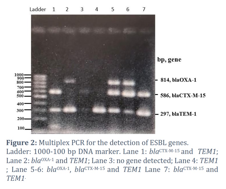
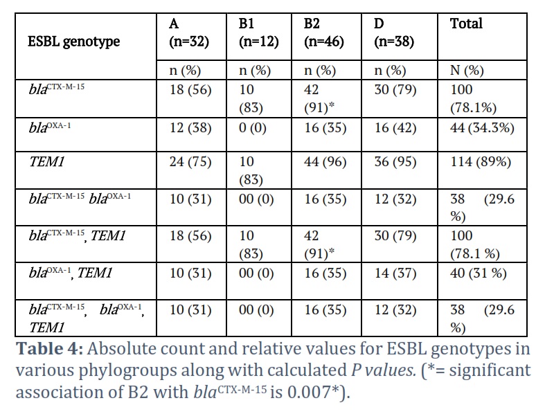
Different combinations of ESBL genes were visualized after the multiplex PCR amplification of TEM1, blaCTX-M-15 and blaOXA-1 genes (Figure 2). ESBL genes were identified in 90.6% (114/128) E. coli strains. TEM1 was observed as the most prevalent genotype (89%) followed by blaCTX-M-15 (78.1%) and blaOXA-1 (34.3%) (Table 4).
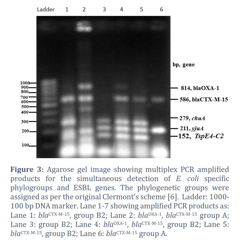
In Figure 3, multiplex PCRs show simultaneous detection of blaCTX-M-15 and blaOXA-1 ESBL genes and E. coli phylogroups using three identification markers (chuA , yjaA and TspE4.C2). A significant association was seen for the presence of blaCTX-M-15 gene in the isolates belonging to phylogroup B2 (p = 0.007), while none of the other phylogroups showed significant association for the presence of blaCTX-M-15 , blaOXA-1 or TEM1 genes (Table 4). A combination of four different genotype patterns is analyzed in the individual phylogroups. Mixed genotypes of blaCTX-M-15, blaOXA-1 and TEM1 were detected among 100/128 (78.1%) E. coli isolates. A duo set of blaCTX-M-15 and TEM1 was seen as the most predominant gene combination in the all assessed phylogroups (A-D). β-lactamase genes in triplet combination were largely observed among the members of group B2 (n = 16, 35%), however, none of the group B1 members was found to display the triplet or dual presence of genes except blaCTX-M-15 and TEM1 pattern (n = 12). A combination of blaCTX-M-15 and TEM1 was observed statistically prevalent ESBL genotype [n=42 (91%), P value = 0.006] in B2 phylogroup (Table 4).
Figures & Tables
Distribution of E. coli phylogroups and the epidemiology varies in different regions across the globe. For example, in Iran more than 50% of E. coli isolates belong to phylogroup D [16] whereas in Ethiopia and China prevalence of phylogroup B2 is observed as the highest followed by group D [17,18] which is similar to our findings in the current study. Interestingly, specific phylogroups have been shown missing in specific studied population, like B2 in Nigeria and South korea and D in Benin, Africa [19].
One of the limitations in the present study is that we used original triplex PCR method which classified E. coli into four phylogroups (A, B1, B2 and D). Clermont’s initial phylogroup scheme is advanced to quadruple
cost . This validation study showed that 80–85% of the phylogroup assignments using the Clermont’s original method are correct. For strains with group B2 genotype, the accuracy of assigning phylogroup is found 95% valid [22].
In the present study, B2 was detected as the highest phylogroup 36% (n=46), followed by group D 30% (n=38), in agreement with the findings that UTI is mainly associated with group B2 and D [6,23]. However, it is notably different from the other study conducted in Pakistan where phylogroup B2 is the predominant group followed by groups A and B1 [23].
In the present study, extreme antibiotic resistance was noticed against different groups of antibiotics. Almost all the E. coli isolates were resistant to nalidixic acid and ciprofloxacin, once considered an ideal choice for the treatment of uncomplicated UTI and pyelonephritis [24]. More or less 70-95 % E. coli strains were resistant to second, third and fourth generation cephalosporin; protein and folate synthesis inhibitors which is alarmingly higher than previously reported in Pakistan [25, 26]. Although the genetic localization of these resistance determinants was not the focus of this study, however restricted use of these antibiotics can limit the dissemination and curtail the cost of E. coli associated UTI management. Carbapenem resistance is swiftly emerging in the clinical isolates of E. coli [27]. In the present study, 62.5% E. coli isolates were found resistant to imipenem which is similar to the findings in Iran where carbapenemase production was detected in 63.2% E. coli strains [28]. Intriguingly, in the current findings, lowest resistance was observed for amoxiclave (54%) and it is also comparatively lower than the other studies conducted in Pakistan [29, 30].
To investigate any significant associations of β-lactamase genes, the individual and a combination set of ESBL genotypes were analyzed in each phylogroup. Statistically significant association was seen for the presence of blaCTX-M-15 – TEM1 genes in group B2 while none of the other phylogroups showed significant association to any one of the assesed ESBL genes. The blaCTX-M-15 fall into functional group A according to the Bush and Jacoby classification [09] and are characterized by increased hydrolysis of oxyimino-β-lactams (i.e. cefotaxime, ceftazidime, ceftriaxone, cefepime, aztreonam) which is also in agreement with our phenotypic observations.
The current study propose multiplex PCR can be used as a rapid diagnostic method to delineate the molecular epidemiology of ESBL genes and E. coli phylogroups. Moreover, a frightening increase in antibiotic resistance also suggests to explore the alternate therapeutic agents with maximum efficacy for the managemnet of bacterial infections in the community.
Acknowledgement
We show our gratitude to Diagnostic and Research Labortary (Civil Hospital, Hyderabad) for providing a platform for this work. We are also grateful to University of Sindh to facilitate us to execute this research study.
Ethical Statement/ Study Approval
The study was approved from the Institutional Review Board at its 141 meeting held on 11-12-2021, vide resolution No. 12 (25), University of Sindh, Jamshoro. A verbal consent was taken from each patient before inclusion to this study. Each Patient’s name was given a unique identification number to include the information about their gender and age.
Conflict of Interest
The authors declare that there is no conflict of interest.
1. Dr. Bushra B. Patoli designed and supervised the reserach work and wrote a manusript.
2. Dr. Atif A. Patoli did the statistical analysis and helped in performing PCR experiments.
3. Dr. Iram Nabi Begum helped in interpretation and writing of the results and reviewed the manuscript.
4. Zarafshan Majid processed E. coli isoaltes and performed ESBL work.
![]() References
References
- Escobar-Páramo P, Grenet K, Le Menac’h A, Rode L, Salgado E, Amorin C, et al. Large-scale population structure of human commensal Escherichia coli isolates. Applied and Environmental Microbiology, (2004); 70(9):5698–700.
- Kaper JB, Nataro JP, Mobley HLT. Pathogenic Escherichia coli. Nature Reviews Microbiololgy, (2004); 2(2):123–40.
- Lindstedt BA, Finton MD, Porcellato D, Brandal LT. High frequency of hybrid Escherichia coli strains with combined Intestinal Pathogenic Escherichia coli ( IPEC ) and Extraintestinal Pathogenic Escherichia coli ( ExPEC ) virulence factors isolated from human faecal samples. BMC Infectious Diseases, (2018); 18(1):1–12.
- Proença JT, Barral DC, Gordo I. Commensal-to-pathogen transition: One-single transposon insertion results in two pathoadaptive traits in Escherichia coli-macrophage interaction. Scientific Reports, (2017); 7(1):1–12.
- Poolman JT, Wacker M. Extraintestinal pathogenic Escherichia coli, a common human pathogen: challenges for vaccine development and progress in the field. The Journal of Infectious Diseases, (2016); 213(1):6–13.
- Clermont O, Bonacorsi S, Bingen E. Rapid and simple determination of the Escherichia coli phylogenetic group. Applied and Environmental Microbiology, (2000); 66(10):4555–8.
- Iranpour D, Hassanpour M, Ansari H, Tajbakhsh S, Khamisipour G, Najafi A. Phylogenetic groups of escherichia coli strains from patients with urinary tract infection in Iran based on the new Clermont phylotyping method. BioMed Research International, (2015); (2015): 5–12.
- Sacher-Pirklbauer A, Klein-Jöbstl D, Sofka D, Blanc-Potard AB, Hilbert F. Phylogenetic groups and antimicrobial resistance genes in escherichia coli from different meat species. Antibiotics, (2021); 10(12): 1543
- Bush K, Jacoby GA. Updated functional classification of β-lactamases. Antimicrobial Agents and Chemotherapy, (2010); 54(3):969–76
- Liaqat Z, Khan I, Azam S, Anwar Y, Althubaiti EH, Maroof L. Isolation and molecular characterization of extended spectrum beta lactamase producing Escherichia coli from chicken meat in Pakistan. PLoS ONE, (2022) ;17(6):1–15.
- Ellington MJ, Kistler J, Livermore DM, Woodford N. Multiplex PCR for rapid detection of genes encoding acquired metallo-β-lactamases. Journal of Antimicrobial Chemotherapy, (2007); 59(2):321–2.
- Dallenne C, da Costa A, Decré D, Favier C, Arlet G. Development of a set of multiplex PCR assays for the detection of genes encoding important β-lactamases in Enterobacteriaceae. Journal of Antimicrobial Chemotherapy, (2010); 65(3):490–5.
- Walker RD. Standards for antimicrobial susceptibility testing. American journal of veterinary research, (1999); 60: 1034.
- Drieux L, Brossier F, Sougakoff W, Jarlier V. Phenotypic detection of extended-spectrum β-lactamase production in Enterobacteriaceae: Review and bench guide. Clinical Microbiology and Infection, 2008; 14(1):90–103.
- Changkaew K, Intarapuk A, Utrarachkij F, Nakajima C, Suthienkul O, Suzuki Y. Antimicrobial resistance, extended-spectrum b-Lactamase productivity, and class 1 integrons in Escherichia coli from healthy swine. Journal of Food Protection, (2015); 78(8):1443–50.
- Halaji M, Fayyazi A, Rajabnia M, Zare D. Phylogenetic Group Distribution of Uropathogenic Escherichia coli and Related Antimicrobial Resistance Pattern : A Meta-Analysis and Systematic Review. Frontiers in Cellular and Infection Microbiology, (2022); 12:1–14.
- Dadi BR, Abebe T, Zhang L, Mihret A, Abebe W, Amogne W. Distribution of virulence genes and phylogenetics of uropathogenic Escherichia coli among urinary tract infection patients in Addis Ababa, Ethiopia. BMC Infectious Diseases, (2020); 20(1):108.
- Li B, Sun JY, Han LZ, Huang XH, Fu Q, Ni YX. Phylogenetic groups and pathogenicity island markers in fecal escherichia coli isolates from asymptomatic humans in china. Applied and Environmental Microbiology, (2010); 76(19):6698–700.
- Munkhdelger Y, Gunregjav N, Dorjpurev A, Juniichiro N, Sarantuya J. Detection of virulence genes, phylogenetic group and antibiotic resistance of uropathogenic Escherichia coli in Mongolia. The Journal of Infection in Developing Countries, (2017);11(1):51–7.
- Clermont, O., Christenson, J.K., Denamur, E. and Gordon, D.M. A new E. coli phylo-typing method. Environmental Microbiology Reports, (2013); 5: 5865.
- Khairy RM, Mohamed ES, Abdel Ghany HM, Abdelrahim SS. Phylogenic classification and virulence genes profiles of uropathogenic E. coli and diarrhegenic E. coli strains isolated from community acquired infections. PLoS ONE, (2019); 14(9): 0222441.
- Gordon, D.M., Clermont, O., Tolley, H. and Denamur, E. Assigning Escherichia coli strains to phylogenetic groups: multi-locus sequence typing versus the PCR triplex method. Environmental Microbiology, (2008); 10: 2484-2496.
- Bashir S, Haque A, Sarwar Y, Ali A, Anwar MI. Virulence profile of different phylogenetic groups of locally isolated community acquired uropathogenic E. coli from Faisalabad region of Pakistan. Annals of Clinical Microbiology and Antimicrobials, (2012); 11:1–6.
- Blango MG, Mulvey MA. Persistence of uropathogenic Escherichia coli in the face of multiple antibiotics. Antimicrobial Agents and Chemotherapy, (2010); 54(5):1855–63.
- Bashir S, Haque A, Sarwar Y. Prevalence of Integrons and Antibiotic Resistance among Uropathogenic Escherichia Coli from Faisalabad Region of Pakistan. Archives of Clinal Microbiology, (2015); 6(4):1–7.
- Rehman N, Azam S, Ali A, khan I, Asghar M, Ali M, et al. Molecular epidemiology of antibiotic-resistant genes and potent inhibitors against TEM, CTX-M-14, CTX-M-15, and SHV-1 proteins of Escherichia coli in district Peshawar, Pakistan. Saudi Journal of Biological Sciences, (2021); 28(11):6568–81.
- Li F, Ye K, Li X, Ye L, Guo L, Wang L, et al. Genetic characterization of Carbapenem-Resistant Escherichia coli from China, 2015–2017. BMC Microbiology, (2021); 21(1):1–7.
- Nasrollahian S, Halaji M, Hosseini A, Teimourian M, Armaki MT, Rajabnia M, et al. Genetic Diversity, Carbapenem Resistance Genes, and Biofilm Formation in UPEC Isolated from Patients with Catheter-Associated Urinary Tract Infection in North of Iran. International Journal of Clinical Practice, (2022); 2022: 62.
- Ali I, Rafaque Z, Ahmed I, Tariq F, Graham SE, Salzman E, et al. Phylogeny, sequence-typing and virulence profile of uropathogenic Escherichia coli (UPEC) strains from Pakistan. BMC Infectious Diseases, (201); 19(1):1–9.
- Khan FZ, Nawaz T, Mirani ZA, Khan S, Raza Y, Kazmi SU. Study of class integrons in multidrug-resistant uropathogenic Escherichia coli isolated from different hospitals in Karachi. Iranian Journal of Basic Medical Sciences, (2018); 21(10):1079–82.
This work is licensed under a Creative Commons Attribution-Non Commercial 4.0 International License. To read the copy of this license please visit: https://creativecommons.org/licenses/by-nc/4.0