

Full Length Research Article
Correlation of SNR Value on DPOAE Examination with HSP70 Levels in Blood and HSP70 Expression in Cochlea of Noise Model Rattus norvegicus as an Indicator of Inner Ear Damage
Florensia Elita Pratiwi1, Tengku Siti Hajar Haryuna1*, Indri Adriztina1, Khalisanni Khalid2
Adv. life sci., vol. 11, no. 2, pp. 322-328, May 2024
*– Corresponding Author: Tengku Siti Hajar Haryuna (tengkusitihajarharyuna@usu.ac.id)
Authors' Affiliations
2. Malaysian Agricultural Research and Development Institute (MARDI), MARDI Headquarters, Persiaran MARDI-UPM, 43400 Serdang, Selangor – Malaysia
[Date Received: 17/01/2023; Date Revised: 29/09/2023; Date Available Online: 18/04/2024]
Abstract![]()
Introduction
Methods
Results
Discussion
References
Abstract
Background: Cellular stress caused by noise-induced hearing loss can be observed through the damage of cochlear hair cells, mechanically and metabolically. SNR value on the DPOAE examination assesses the sensory function of cochlear outer hair cells. HSP70 as marker of cellular stress can be identified from its levels in blood and its expression in the cochlea.
Methods: Three groups of rats were used for the noise intervention: Group 1 was the control group, Group 2 had a noise exposure of 100 dB, and Group 3 received a noise exposure of 110 dB. DPOAE examination was then conducted, blood samples from rats’ tail were collected after a noise treatment, followed by the measurement of HSP70 levels by ELISA readings. Rats were terminated afterwards and immunohistochemical examination of the cochlear organ of Corti was performed to calculate the expression of HSP70. The data obtained were analyzed using SPSS.
Results: A decrease occurred in the values of SNR and an increase occurred in HSP70 levels in blood along with its expression in the cochlea of noise model Rattus norvegicus, and there is a correlation between the three.
Conclusion: The decrease in SNR values, increase in HSP70 levels in blood, and increase in HSP70 expression in the cochlear organ of Corti of Rattus norvegicus, proves that noise-induced hearing loss triggers the production of HSP70 as a cytoprotective agent in preventing inner ear damage.
Keywords: DPOAE; Rattus norvegicus; NIHL; Heat Shock Protein 70
Introduction![]()
Noise induced hearing loss (NIHL) is the most common form of sensorineural hearing loss in adults, which is highly complex consisting of interaction between environmental and genetic factors [1, 2]. The World Health Organization (WHO) estimated that more than 466 million of the world population (6,12%) experienced hearing loss in 2018 [3], of which 22% were related to noise-induced hearing loss in worksites [4]. The prevalence of NIHL is occurred more frequently in men than women and the risk of its occurrence increases with age [2].
Pathogenesis of noise-induced hearing loss is a cellular mechanism [5] where mechanical destruction occurs which causes structural and functional loss of cochlear hair cells effectively as well as impaired metabolic activity [1], in which noise exposure can cause inner ear damage either temporary or permanent changes in hearing threshold [2]. This event also causes molecular disturbance, which are changes occurring in hair cells, auditory nerves, and other structures in the cochlea. Excessive surge of Ca2+ in cells can trigger ROS accumulation and stimulates the activation of cell death and cell damage signaling pathways [5]. This causes the cochlea to protect itself with a cytoprotective response through the expression of Heat Shock Protein (HSP70) chaperon molecule from the heat shock transcription factor (Hsf1) gene, a subgroup of 70 kDa molecular weight HSP family [6]. HSP was first discovered by Ritossa in 1962 in the salivary glands of Drosophila melanogaster larvae to maintain eukaryotic and prokaryotic homeostasis [6] at the cellular level when a stress signal is found, such as excessive noise [7-10]. Whereas the extracellular HSP70 molecules activate the production of proinflammatory cytokines as a stress signal on the innate and acquired immune system [11], in order to avoid cell damage [8]. There are studies which proved that the HSP70 haplotype mutation can affect a person’s susceptibility to NIHL [1] while still considering individual factors such as smoking habits and high blood pressure [12].
By analyzing otoacoustic emission (OAE), inner ear damage can be determined. As a sign of the cochlear outer hair cells' sensory activity, the OAE interaction is an acoustic emission generated by the electro-motile active vibrations of the cells. [13]. This study uses DPOAE (Distortion Product Otoacoustic Emission) which is a response that occurs when the cochlea is stimulated simultaneously by two pure tones with a frequency ratio of 1.25 [14]. The results of the signal to noise ratio (SNR) are used as an indicator to assess inner ear damage in hearing loss incidence [15].
Prolonged exposure to noise rises the risk of NIHL, as stated by prior study that 89.2 dBA noise exposure for 8 hours a day caused NIHL in workers [16]. The Occupational Safety and Health Administration (OSHA) mentioned the importance of using hearing protection equipment for workers exposed to noise that exceeds the 90 dBA limit for 8 hours or more daily [17].
NIHL can actually be prevented, as there can be a transient threshold shift (TTS) change. On the other hand, if it has decreased due to an increase in the permanent threshold of hearing (PTS), then there has been an active stimulation of intracellular stress pathways, resulting in irreversible and treatment-unresponsive apoptosis and necrosis of the outer hair cell’s structure [5].
Derived from the statement of problems explained above, the researchers are intended to discover the correlation of the signal to noise ratio (SNR) values with HSP70 levels in the blood and HSP70 expression in the cochlear organ of Corti of Rattus norvegicus due to noise exposure as an indicator of inner ear damage.
Methods![]()
A veterinarian certified as healthy, 27 male rats of the pure strain Rattus norvegicus, weighing between 200 and 300 grams, participated in the investigation, which involved a randomized posttest-only control group experimental setup in a laboratory. The environment was conditioned with the cage temperature maintained at 200C-260C, air humidity of 30-70%, an ensured light source, along with adequate food and drink according to the 2011 Guide for the Care and Use of Laboratory Animals. Breeding and treatment were carried out at the Animal House in Faculty of Mathematics and Natural Sciences, Universitas Sumatera Utara (FMIPA USU). This research has received approval from the Research Ethics Committee of Universitas Sumatera Utara in 2020 with the number 230/KEP/USU/2020.
Three groups, each with nine rats, were created out of the experimental group. Groups 2 and 3 of rats were exposed to noise levels of 100 dB and 110 dB, respectively. Group 1 served as a control group that did not experience any noise. Noise exposure was performed by placing the rat in a box with a size of (64.5 x 45 x 40) cm made of foam-coated cork. A hole was constructed at the bottom of the box to measure the noise level, and a speaker was fixed to the box cover's roof. The noise level was measured at eight sites where the rat cage will be placed using a sound level meter (Sound Level Meter TENMARS TM-102), with a noise differential not exceeding 1 dB. The noise was recorded at a frequency of 1–10,000 Hz, and the intensity (dB) was adjusted using an amplifier in accordance with the volume knob and sound level meter. The noise was delivered for the duration of two hours every day for two days. DPOAE examination (Elios Elito Otodia Echodia Ltd., London, UK) was done three times, which were before noise exposure (Day 0), two hours after noise exposure on the second day (Day 2), and two days after noise exposure on the second day finished (Day 4). Before the examination, the rats were given intraperitoneal injections of xylazine (10 mg/kgBW) and ketamine (90 mg/kgBW) to induce anesthesia [18].
Blood samples from the rats’ tail were collected before and after the noise exposure. Blood was let frozen for 30 minutes before centrifuged at 1000 g for 5 minutes at 40C to remove residual debris. The supernatant was placed in a clean tube and then stored at -200C. Reagents, working standards, and samples were put into the wells provided, followed by adding a washing buffer, HSP70 abcam antibody (ab133061) and HRP conjugate anti-IgG antibody (horseradish peroxidase), TMB substrate solution (tetramethylbenzidine), as well as the stopping solution (Wampole) into each well in sequence in order to be read immediately. The bound enzyme depends on the amount of serum HSP70. Thus, when given the substrate, the enzyme will process the substrate and a color change will occur in the liquid in the well with color gradations according to the concentration of serum HSP70 contained. Based on the ELISA reader, readings on the wells containing 0-7 standards, with a wavelength of 450 nm, there will be quantitative absorbance results obtained with standard dilutions.
Temporal bone necropsy was carried out and the rats were euthanized following DPOAE evaluation and blood sample. After being fixed with a 10% formalin buffer solution, the temporal bone was exposed to EDTA for four weeks in order to promote decalcification. Following paraffin block preparation, each tissue sample will be sliced into thick 4 μm sections and placed on slides for hematoxylin-eosin and immunohistochemical staining. This will allow for the assessment of HSP70 expression in the Corti cochlear organ using primary and secondary Anti-HSP70 antibodies [3A3] (ab5439) and biotinylated secondary antibodies. Two independent examiners (a researcher and an anatomical pathologist) used the double-blind approach to calculate HSP70 expression using an Olympus XC 10 microscope with 10x magnification. The cytoplasmic brown color’s area (P) and intensity (I) scores were used to determine the expression of HSP70. There were four categories used to categorize the distribution of brown color in the external appearance of the cells: distribution score 0 = no cells stained brown, 1 = <10% of cells stained brown, 2 = 11-50% of cells stained brown, 3 = 51-80% of cells stained brown, and 4 = >81% of cells stained brown. Immuno-reactive scores range from 0 to 12, and intensity scores are as follows: 1-3, or 0 = unblemished, 1 = weak intensity, 2 = moderate intensity, and 3 = strong intensity. The immune-reactive score is calculated by multiplying P and I [19].
Data were analyzed using ANOVA or Kruskal Wallis test and correlation test to see the correlation between SNR and HSP70 levels in the blood and HSP70 expression in the cochlear organ of Corti. Statistical test is considered significant if p < 0,05. Data analysis was carried out using SPSS 25.0 program.
Results![]()
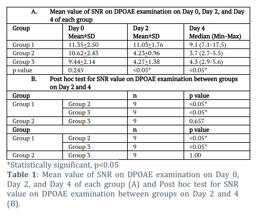
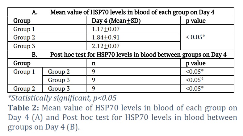
The study found that the SNR value was higher in the control group (group 1) compared to the treatment groups (groups 2 and 3). This was consistent with the levels of HSP70 in the blood and HSP70 expression in the organ of Corti, which were lower in the control group than in the treatment groups. In Table 1, a significant difference in the mean value of SNR on Day 2 and Day 4 of each group is shown (p < 0.05). It can be seen that there is a significant difference in SNR values on Day 2 and Day 4 between Group 1 and Group 2 as well as Group 1 and Group 3 (p < 0.05), and no significant difference between Group 2 and Group 3 (p > 0.05). Table 2 shows significant differences in HSP70 levels in blood between Group 1, 2 and 3 on Day 4 (p < 0.05). It is shown that there are significant differences in HSP70 levels in blood in all groups on Day 4 (p < 0.05).
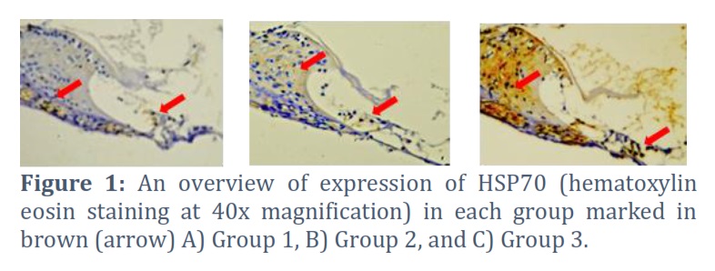
Assessment of HSP70 expression in cochlear outer hair cells of Rattus norvegicus rats began by hematoxylin eosin staining followed by immunohistochemical staining, resulting in an increase of HSP70 expression in the group of rats exposed to noise, both in the group with 100 dB noise exposure (Group 2) and 110 dB (Group 3). In Fig. 1, the brown color of the cytoplasm demonstrates the expression of HSP70, indicating that Group 3 has a more intense brown color than Groups 2 and 1 (control). Higher noise intensity was accompanied by an increase in the average value of HSP70 expression.
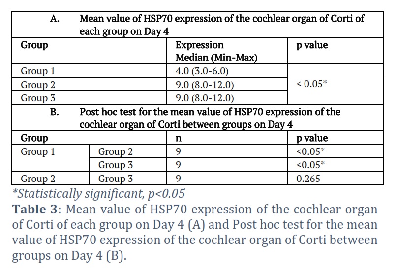
It is shown in Table 3 that there is a significant difference in the mean value of HSP70 expression in all groups (p < 0.05). The relevancy of the significant difference in HSP70 expression in all groups can be assessed from the table below. It can be seen that there is a significant difference in the mean value of HSP70 expression between Group 1 with Group 2 and 3 (p < 0.05), and no significant difference between Group 2 and Group 3 (p > 0.05).
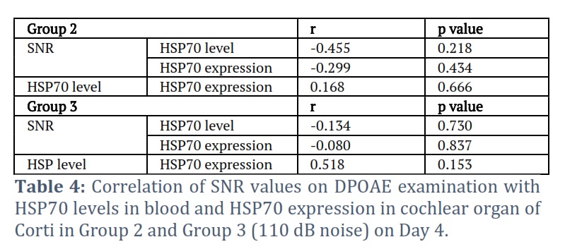
It can be identified from Table 4 that in both groups (Group 2 and Group 3) on Day 4, the correlation of SNR with HSP70 levels and HSP70 expression has a negative r value coefficient, indicating a negative direction of correlation. Whereas the value of r on the correlation of HSP70 levels with HSP70 expression is positive, it can be stated that the direction of the correlation is positive. The p value of the correlation of SNR with HSP70 levels, correlation of SNR with HSP70 expression, and correlation of expression with levels of HSP70 in both groups on Day 4 shows that the correlation is insignificant.
Figures & Tables
In this study, noise levels of 100 dB and 110 dB were applied to Rattus norvegicus rats for two hours on two consecutive days. In addition, this study also examined HSP70 levels in blood and HSP70 expression in the cochlear organ of Corti of Rattus norvegicus. This study is expected to identify indications of impaired hearing function, as shown by the correlation between decreased SNR values, increase of HSP70 levels in blood, and increased expression of HSP70 in the cochlear organ of Corti due to noise exposure.
This study found that at 100 dB and 110 dB noise exposure, the SNR value on the fourth day did not return to normal, in which PTS had occurred. It is in line with a study done by Kim et al., which stated that 100 dB noise exposure given on male Balb/C rats for 3 hours triggers the production of ROS and inflammatory proteins in the cochlear, caused a permanent shift in the hearing threshold when examined 10 days after noise stimulation was given. Meanwhile, exposure to 110 dB noise for 4 hours can induce glutamate excitotoxicity which causes swelling of the cochlear nerve terminals. It is an acute response to noise, which begins with loss of the presynaptic ribbons and postsynaptic terminals of the inner hair cells [20, 21]. In their research, Mannstrom et al., explained that when exposed to 110 dB noise intensity for 90 minutes, female Sprague-Dawley albino rats already experienced PTS on the first exposure, while 104 dB noise exposure within 90 minutes was the highest intensity and longest exposure time which can be repeated up to 6 times without causing PTS and rats became resistant to high-intensity noise exposure [21]. Amanipour et al., developed PTS of 20 dB 4 weeks after adult rats were exposed to 110 dB noise for 45 minutes, subsequently an improvement happened 1 year later where PTS was found to be less than 10 dB [22].
Another consequence of noise exposure was an increase in calcium in the outer hair cells after acoustic overstimulation [2] which caused increased expression of HSP70 [6], and apoptosis as well as activation of cell death pathways [5, 6]. It is in accordance with this study where noise exposure given to experimental animals, both at an intensity of 100 dB and 110 dB, lead to the increased production of HSP70 both its levels in blood and its expression in cochlear organ of Corti, as evidence of its role as a chaperon protein that works in overcoming cell damage. Comparing the group without noise exposure, higher noise intensity was associated with higher levels of HSP70 in the blood and higher expression of HSP70 in the cochlear organ of Corti.
A study by Reastuty et al., found that the cochlear organ of Corti had lower levels of Superoxide Dismutase (SOD) expression, and the Distortion Product Otoacoustic Emission (DPOAE) test showed lower Signal to Noise Ratio (SNR) values. Nevertheless, when Corti's rat cochlear organ was subjected to noise, there was a rise in the expression of malondialdehyde (MDA). The study also discovered that the SNR value and SOD expression had a positive association (r=0.733, p=0.025), and the SNR value and MDA expression had a negative correlation (r=-0.678, p= 0.045). Consequently, it can be concluded that alterations in the function of outer hair cells in rats exposed to noise are correlated with the oxidative and antioxidant status of the auditory organ of Corti [23]. Another study found a correlation between the Signal to Noise Ratio (SNR) value (r= 0.792, p= 0.011) and the expression of nuclear factor erythroid 2-related factor 2 (NRF2) in the auditory organ of rats subjected to 110 dB noise. This demonstrates that endogenous antioxidants are still powerless to aid in the restoration of cochlea damage in rats exposed to noise [24].
Research by Turkyilmaz, 2011 in Karafakioglu, 2019, done in broiler chickens (Ross 308) that were exposed to acute noise of 80, 100, and 120 dB for 15 minutes resulted in increased cholesterol levels, while 120 dB noise increased blood sugar levels and reduced the number of leukocytes. Meanwhile, daily noise exposure of 75 dB for 1 month in male Wistar albino, significantly increased the number of red blood cells [25]. Demirel et al., also reported that rats subjected to noise at 100 dB for four hours per day for twenty days were able to elevate stress indices in their blood, including GSH-Px (glutathione peroxidase), NO (nitric oxide), and MDA (malondialdehyde) [26]. This indicates that noise exposure causes metabolic disturbances which leads to the increase of HSP70 levels in blood as an effort to prevent systemic damage.
Pawlak-Osinska et al., found an increase in circulating immune complexes and free HSP70 protein in the blood of patients with sudden sensorineural hearing loss [27]. Additionally, this study found an association between elevated blood levels of HSP70 and elevated HSP70 expression in the Corti cochlear organ of Rattus norvegicus. This finding highlights the role HSP70 plays in preserving homeostasis and promoting cell repair in tissues subjected to noise stress. This is in accordance with the study of Gunther et al., which stated the occurrence of a significant increase in HSP70 levels in the peripheral blood circulation of patients and an increase in HSP70 expression in patients with squamous cell and adeno NSCLC (non-small cell lung carcinoma), inflammatory diseases such as COPD (chronic obstructive pulmonary disease) and pneumonia [28].
Intracellular HSP70 protein functions in preventing apoptosis and inflammation in lung, liver, and kidney tissues, where its concentration increases when exposed to bacterial endotoxins in the event of sepsis and is then carried in exosomes from the intracellular out through the cell membranes, leading to an increase of its levels in the extracellular [29] to modulate the immune system [8]. According to this study, there is a positive correlation between the levels of HSP70 in blood and the expression of HSP70 in the cochlear organ of Corti.
In this study, a negative correlation was found between SNR value with HSP70 expression in the cochlear organ of Corti and HSP70 levels in blood of Rattus norvegicus exposed to 100 dB and 110 dB noise. Droge, 2002 in Kurabi et al., stated that noise causes damage to cochlear hair cells where ROS free radicals are formed with its chemical reactions associated with DNA, proteins, cytosolic molecules, cell surface receptors, and membrane lipids that affect intracellular processes. ROS formation occurred in the cochlea after exposure to damaging noise [5]. HSP70 inhibits cochlear hair cell damage by initiating protein folding, refolding misfolded peptides, degrading irreversible proteins [7], as well as modulating the inflammatory response, hence it plays a very important role in the cell stress response [29]. The mechanism above was proven from this study where the noise-exposed group was characterized by hair cell damage and discovery of an increase in HSP70 and a decrease in SNR value. Thus, it can be stated that if a decrease in the SNR value occurs, the value of HSP70 expression and levels will increase. When noise occurs, the SNR value tends to decrease [15]. In addition, under the same conditions, intracellular HSP70 expression increases along with an increase of its levels in blood [11]. The occurrence of noise-induced hearing loss as measured by the SNR value [15] is also influenced by individual genetic factors on noise stress resistance [30]. No homeostasis occurred on exposure to noise of 100 dB and 110 dB, where the cochlear organ of Corti was damaged, which was characterized by SNR value that did not return to normal and increased HSP70 levels in blood and HSP70 expression in the cochlear organ of Corti.
Oftentimes, hearing loss due to noise is not realized by the sufferer despite the occurrence of molecular changes in hair cells which is characterized by an increase in HSP70 [2]. HSP in this case is highly important as a marker of stress, such as noise [10], in helping to restore damaged proteins, stabilize protein structures, and play a role in immunological activity [11]. NIHL is usually found to have occurred in a state of severe disturbance [25]. It can be stated that the high rate of disability caused by noise over the years can cause permanent hearing loss if not prevented or treated properly [15]. This will reduce the quality of life for patients who are still productive [25]. Therefore, this study suggests the need for DPOAE examination in individuals who are at risk of experiencing noise-induced hearing loss as an early prevention of the occurrence of NIHL.
Acknowledgement
Researcher would like to thank Lembaga Penelitian Universitas Sumatera Utara (LP USU) who has supported the funding of this research in accordance with the TALENTA Research Agreement Letter of Universitas Sumatera Utara for Fiscal Year 2020 No. 4142/UN5.1.R/PPM/2020, April 27, 2020.
Conflict of Interest
The authors declare that there is no conflict of interest.
Tengku Siti Hajar Haryuna, corresponding author is contributed to the conception and design of the study, responsible for performing all experiments.
Indri Adriztina participated in the material preparation and data collection.
Florensia Elita Pratiwi, participated in data analysis, wrote the first draft of the manuscript
Khalisanni Khalid, critically reviewed the manuscript.
All authors contributed to the writing and editing of the manuscript. All authors have read and approved the final manuscript.
![]() References
References
- Chang NC, Ho CK, Lin HY, Yu ML, Chien CY et al. Association of polymorphisms of heat shock protein 70 with susceptibility to noise-induced hearing loss in the Taiwanese population. Audiology and Neurotology, (2011); 16(3):168-74.
- Le TN, Straatman LV, Lea J, Westerberg B. Current insights in noise-induced hearing loss: a literature review of the underlying mechanism, pathophysiology, asymmetry, and management options. Journal of Otolaryngology-Head & Neck Surgery, (2017); 46(41); 1-15.
- World Health Organization. Addressing the rising prevalence of hearing loss. Accessed from: http://www.who.int/pbd/deafness/estimates/en/, (2018)
- Mahboubi H, Zardous S, Oliaei S, Pan D, Bazargan M, Djalilian HR. Noise-induced hearing threshold shift among US adults and implications for noise-induced hearing loss: National Health and Nutrition Examination Surveys. European Archives of Oto-Rhino_Laryngology. (2013); 270(2): 461-7.
- Kurabi A, Keithley EM, Housley GD, Ryan AF, Wong ACY. Cellular mechanisms of noise-induced hearing loss. Hearing Research, (2017); 349: 129-37.
- Ghazaei C. Role and mechanism of the HSP70 molecular chaperone machine in bacterial pathogens. Journal of Medical Microbiology, (2017); 66(3): 259-65
- Ikwegbue PC, Masamba P, Oyinloye BE, Kappo AP. Roles of Heat Shock Proteins in Apoptosis, Oxidative Stress, Human Inflammatory Diseases, and Cancer. Pharmaceuticals, (2017); 11(2): 1-18
- De Maio A. Extracellular HSP70: export and function. Current Protein and Peptide Science, (2014); 15(3): 225-31
- Masser AE, Kang W, Roy J, Kaimal JM, Quintana-Cordero J, et al. Cytoplasmic protein misfolding titrates HSP70 to activate nuclear Hsf1. eLife Research Article, (2019); 1-27
- Hassan AM, Hosny AN, Solimon FE. Occupational Hearing Loss Due to Noise Exposure and its Relation with Heat Shock Protein 70 and its Antibody. Medical Journal Cairo University, (2010); 78(1): 511-16
- Tukaj S. Heat Shock Protein 70 as a Double Agent Acting Inside and Outside the Cell: Insights into Autoimmunity. International Journal of Molecular Sciences, (2020); 21(15): 1-13
- Ahsan, M and Waheed, F. Contribution of common Single Nucleotide Polymorphisms in Noise-induced hearing loss. Pak-Euro Journal of Medical and Life Sciences, (2020); 3(3): 125-35.
- Zimatore G, Stanzial D, Orlando MP. Otoacoustic Emissions. Research and Applications Intech, (2013); 204-23
- Najarkola MSA, Khavanin A, Mirzaei R, Salehnia M, Muhammanejad A, et al. ‘Noise-induced outer hair cells’ dysfunction and cochlear damage in rabbits’. Iranian Red Crescent Medical Journal, (2012); 14(10): 647–656.
- Wu YH, Stangl E, Chipara O, Hasan SS, Welhaven A, Oleson J. Characteristics of Real-World Signal to Noise Ratios and Speech Listening Situations of Older Adults With Mild to Moderate Hearing Loss. Ear and hearing, (2018); 39(2): 293-304
- Jabbari K, Nassiri P, Monazzam Esmaeelpour MR, Azam K, Faridan M, Heidari L. The Relationship between Occupational Noise Exposure and Noise Induced Hearing Loss (NIHL) in Small-Scale Industries: A Case Study in the City of Damavand, Iran. Biotechnology and Health Sciences, (2016); 3(4): 49-56.
- Le Prell CG, Miller JM. The role of oxidative stress in hearing loss. Oxidative Stress and Antioxidant Protection: The Science of Free Radical Biology and Disease, (2016); 8: 115-31. John Wiley & Sons, Inc
- Etten V. Recommended Methods of Anesthesia, Analgesia, and Euthanasia for Laboratory Animal Species. Albert Einstein College of Medicine, Institute for Animal Studies, (2014); 460(718): 839-7100.
- Fedchenko N, Reifenrath J. Different approaches for interpretation and reporting of immunohistochemistry analysis results in the bone tissue- a review. Diagnostic Pathology 9, (2014); (221): 1-12
- Kim MS, Kwak S, Baek H, Li Z, Choe SK, Song K. Protective Effect of Yang Mi Ryung Extract on Noise-Induced Hearing Loss in Mice. Evidence-Based Complementary and Alternative Medicine, (2017); 2017: 1- 11.
- Mannstrom P, Kirkegaard M, Ulfendahl M. Repeated Moderate Noise Exposure in the Rat-an Early Adulthood Noise Exposure Model. Journal of the Association for Research in Otolaryngology, (2015); 16(6): 763-72.
- Amanipour RM, Zhu X, Duvey G, Celarine S, Walton JP, Frisina RD. Noise-Induced Hearing Loss in Mice: Effects of High and Low Levels of Noise Trauma in CBA Mice, (2018); 1210-13
- Reastuty R, Haryuna TSH. Correlation of SOD and MDA Expression in the Organ of Corti and Changes in the Function of Outer Hair Cells Measured by DPOAE Examination in Noise -Exposed Rat Cochlea. Reports of Biochemistry & Molecular Biology, (2021); 10(1): 41-49
- Amellya D, Haryuna TSH, Riawan W. Correlation of signal to noise ratio (SNR) value on distortion product otoacoustic emission (DPOAE) and expression of nuclear factor erythroid 2-related factor 2 (NRF2) in cochlear organ of Corti in rat exposed to noise. Medicinski Glasnik, (2021); 18(1): 102-106.
- Karafakioglu YS. Effect of lipoic acid on noise induced oxidative stress in rats. Saudi Journal Biology Science, (2018); 26(5): 989-94.
- Demirel R, Mollaoğlu H, Yeşilyurt H, Üçok K, Ayçiçek A, et al. Noise Induces Oxidative Stress in Rat. European Journal General Medicine, (2009); 6(1): 20-4.
- Pawlak-Osinska K, Golda R, Osinski S, Kazmierczak H, Krumrych, W. Circulating Immune Complexes and Heat Shock Protein 70 in the Sera of Patients with Sudden Sensorineural Hearing Loss. The Journal of International Advanced Otology, (2018); 14(3): 426-31.
- Gunther S, Ostheimer C, Stangl S, Specht HM, Mozes P, et al. Correlation of HSP70 serum levels with gross tumor volume and composition of lymphocyte subpopulations in patients with squamous cell and adeno non-small cell lung cancer. Frontiers in Immunology, (2015); 6: 1-12
- Sulzbacher MM, Ludwig MS, Heck TG. Oxidative stress and decreased tissue HSP70 are involved in the genesis of sepsis: HSP70 as a therapeutic target. Revista Brasileira de Terapia Intensiva, (2020); 32(4): 585-91
- Li Y, Yu S, Gu G, Chen G, Zheng Y et al. Polymorphisms of heat shock protein 70 genes (HSPA1A, HSPA1B and HSPA1L) and susceptibility of noise-induced hearing loss in a Chinese population: A case-control study. PLoS One, (2017); 12(2): 1-12.
This work is licensed under a Creative Commons Attribution-Non Commercial 4.0 International License. To read the copy of this license please visit: https://creativecommons.org/licenses/by-nc/4.0