

Full Length Research Article
miRNA-181a and miRNA-128 expression levels as a predictive marker for response in treating Acute Lymphoblastic Leukemia
Zainab Kadhim Abdul-Hussein, Ibrahim A. Altamemi*
Adv. life sci., vol. 12, no. 3, pp. 534-539, August 2025
*- Corresponding Author: Ibrahim A. Altamemi (Email: ibrahim.altamemi@qu.edu.iq)
Authors' Affiliations
[Date Received: 13/12/2024; Date Revised: 28/02/2025; Available Online: 31/10/2025]
Abstract![]()
Introduction
Methods
Results
Discussion
References
Abstract
Background: The genesis of Acute Lymphoblastic Leukemia (ALL) has been established to involve the workings of certain microRNAs (miRNAs), such as miR-181a and miR-128. As such, science research has been exploring their potential as biological markers for various therapeutic uses, such as disease detection, relapse likelihood prediction, and monitoring of patient treatment response. Moreover, identifying additional miRNA biomarkers can also enable scientists to develop new treatment approaches in their entirety.
Methods: This study examined 100 participants, including 70 ALL patients—i.e., 40 in remission, 20 who relapsed, and 10 who were resistant—and 30 healthy controls. Researchers employed RT-qPCR to identify and quantify expressions of miR-181a and miR-128 in blood samples and statistically compared expressions in all these groups.
Results: The expression analysis revealed significant differences between the study groups for both miRNA-181a and miRNA-128 (p < 0.001). The highest peak level of expression was noted in the relapse group in comparison to the response, resistant, and control participants. Furthermore, in relapse, response, and resistant groups, there was significantly elevated expression (p < 0.05), and control levels were lowest of all (p < 0.05).
Conclusion: Quantitation of expression levels of miR-181a and miR-128 holds great promise in predicting treatment response in ALL patients. More importantly, miR-181a shows promise as a specific and sensitive biomarker in predicting resistance and relapse in adult ALL patients.
Keywords: Pseudomonas aeruginosa, Efflux pump inhibitors, MexXY-OprM efflux system, Conessin
Introduction![]()
Acute Lymphoblastic Leukemia (ALL) originates as an uncontrollable disorder of cells as lymphoid progenitors (blasts) multiply uncontrollably to override normal maturation of blood cells. It becomes impossible to have therapeutic control over malignant B or T lymphoblasts due to their uncontrollable division, whereby immature abnormal lymphocytes and their progenitors substitute for elements in the bone marrow, affecting other lymphoid organs to establish an independent disease course [1-3]. Multiagent chemotherapy has improved treatment outcomes in ALL patients such that an estimated 90% of adult patients can achieve complete remission (CR). Early treatment response predictors play an important role in terms of guiding therapy, as relapse and drug resistance remain significant clinical problems [4,5]. MicroRNAs (miRNAs), small modulatory RNA molecules, have pivotal functions in regulating expressions of genes during normal hematopoiesis and leukemogenesis of hematopoietic cells. Transcript profiles of both miR-181 and miR-128 have been validated as potential diagnostic as well as prognostic ALL biomarkers. These miRNAs are expressed at high levels in early B-cell phases during lymphocyte maturation and diminish in subsequent maturation phases. Notably, maximum miR-181 transcription occurred during double-positive T-cells, during which it represses essential targets including BCL-2, CD89, EGR1, and the T-cell receptor [6,7]. The two paralogs of miR-128, i.e., miR-128-1 and miR-128-2, have been part of establishing ALL pathological features. Key cell functions, including cell growth and division, programmed cell death, and cell commitment to fate, have been regulated strictly through miRNAs. Study outcomes revealed ALL patients could be differentiated clearly from normal healthy control individuals due to significantly different expressions of miR-128, as different expressions of this miRNA have been related to disease aggressiveness and treatment responses [8,9]. Additional evidence also revealed that miRNAs are good indicators to detect minimum residual disease to predict relapse and assist therapeutic choices based on this prediction. Studies also indicated functions of miR-181 and miR-128 in ALL drug sensitivity and resistance mechanisms through apoptotic-related and drug efflux genes regulations [10,11]. Additionally, these miRNAs have also been related to signal transduction cascades, including PI3K/AKT and TGF-β, showing their potential as biomarkers and targets for therapy. Knowledge of these specific molecular pathways is essential to have superior therapeutic options [12-14].
This study explores potential therapeutic response predictive biomarker applications of miR-181a and miR-128 expression profiles in adult ALL patients, owing to several roles assumed by these latter in treatment effectiveness and disease initiation. The results of this study can refine patient categorization procedures in use and offer potential new therapeutic strategy developments [15,16].
Methods![]()
2021 to October 2022 at Hematology Center, Baghdad Teaching Hospital. The research included 70 Iraqi ALL patients and 30 healthy control participants, all in an age category of 14–72 years. Confirmation of diagnosis in patients was carried out based on different methods, i.e., medical history screening, physical checkup, peripheral blood smear, aspiration analysis of bone marrow, flow cytometry, and cytogenetic studies. All these diagnoses were strictly carried out in accordance with WHO 2016 classification guidelines. The 30 healthy control participants, in an equivalent category (14–72 years), were also included as a control set. Ethical approval was taken from Iraqi Ministry of Health Ethics Committee, and informed written consent was also taken from all participants before enrollment.
The study population was classified into three classes, and the first class included 40 ALL patients who achieved complete remission (CR) and continued under maintenance therapy. The second group included 30 patients, comprising 10 resistant cases and 20 relapsed cases. The third group consisted of 30 healthy volunteers who served as controls.
Blood Sample collection and preservation, RNA preparation, and cDNA synthesis
Quantitative analysis of miRNA-128 and miRNA-181a expression was performed using RT-qPCR, normalized against the U6 housekeeping gene. The analysis was conducted on whole blood samples (1 mL blood combined with 1 mL TRIzol reagent) collected from both patients and control subjects using Real-Time PCR methodology.
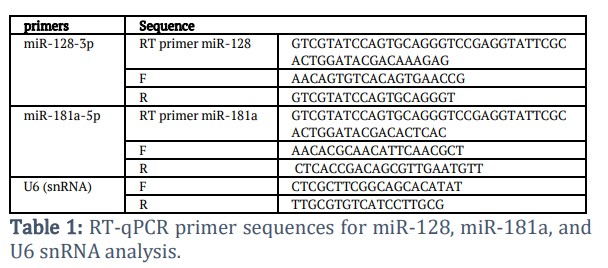
Special primers exist for measuring microRNA expression quantities. Table 1 contains the sequences that demonstrate the use of RT primers during reverse transcription steps alongside forward and reverse PCR primers required for amplifying miR-181a-5p, miR-128-3p, and U6 (snRNA). U6 (snRNA) operated as a built-in control reference for normalization purposes. This table presents abbreviations and sequences for Reverse Transcription and Forward and Reverse primers used in Polymerase Chain Reactions on Homo sapiens small nuclear RNA and Homo sapiens sequences
Total RNA was extracted from whole blood samples using the TRIzol kit according to the manufacturer's guidance. The RNA quality and concentration were measured by a spectrophotometer, which measured absorbance at 260/280 nm. After extraction, cDNA synthesis for miRNA-128 and miRNA-181a was conducted using the cDNA master mix. The qPCR master mix was prepared by using the GoTaq® qPCR Master Mix Promega kit, which was dependent on SYBR Green dye detection of gene amplification in the Real-Time PCR system.
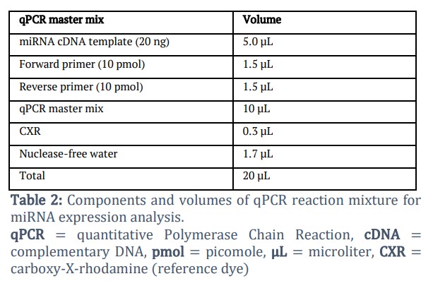
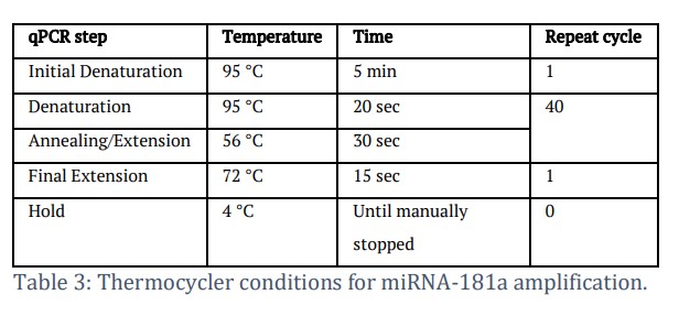
Table 2 displays the composition of the qPCR master mix, which was utilized for miRNA expression quantification. The master mix composition for qPCR reactions included 20 µL of solutions containing specific concentrations of template cDNA, together with primers and other necessary constituents. The thermocycler protocol mentioned in Table 3 was used after loading the qPCR plate.
Results![]()
Comparison of the levels of miRNA-128 and miRNA-181a according to treatment response
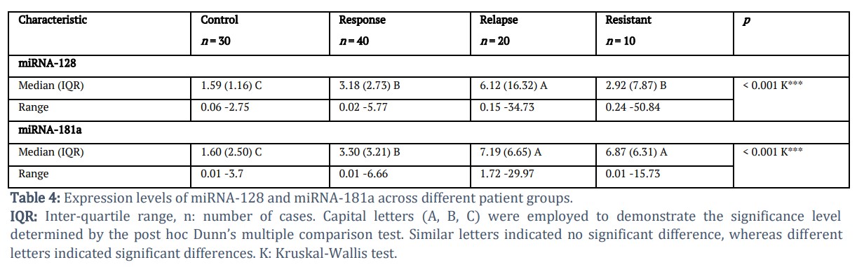
Laboratory analyses showed whole blood miRNA-128 concentrations had statistically significant group discrepancies (p < 0.001), presenting systematic distribution patterns (Table 4). The relapse group possessed elevated miRNA-128 levels, with the response and resistant groups showing the second and third highest levels, while the control group demonstrated the lowest levels. The variance between the response group and resistant group was insignificant (p > 0.05), but the difference was significantly lower than that of the relapse group (p < 0.05) and considerably elevated than the control group (p < 0.05).
The research data pointed to meaningful variations in miRNA-181a expression across groups (p < 0.001) (Table 4). The relapse group had the highest miRNA-181a levels, followed by the resistant and response groups, while the levels of the control group remained lower. The difference between the response group and resistant group was significant (p < 0.05), and the response group was significantly lower in value than that of the relapse group (p < 0.05) and significantly higher than that of the control group (p < 0.05).
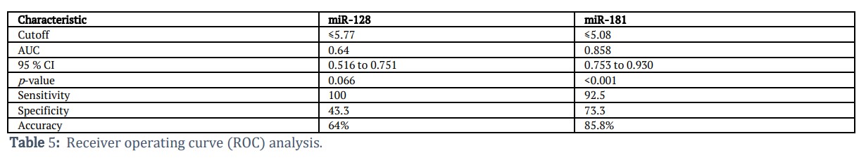
The results of Table 5 showed that the AUC value for miR-128 indicates poor to fair diagnostic ability, while for miR-181, the diagnostic ability is excellent. The cutoff value indicates the threshold above which samples were classified as positive for each microRNA. The 95% confidence interval (CI) value shows that results may be unreliable for miR-128, showing weak diagnostic performance, while results are reliable for miR-181, showing strong diagnostic performance of the microRNA. The p-value for miR-128 is not statistically significant, but it is highly significant for miR-181, showing that it is a real diagnostic marker. The accuracy showed that miR-128 showed modest diagnostic performance, while miR-181 showed high diagnostic performance. The high sensitivity and low specificity, along with broad AUC and wide CI value for miR-128, suggest that it is not a durable standalone biomarker. On the contrary, good sensitivity and specificity values, along with excellent AUC and narrow CI values, indicate that miR-181 is a strong and reliable diagnostic biomarker for ALL.
Figures & Tables
The present study showed that miRNA-128 expression profiles in the relapsed and resistant patients were significantly upregulated in these groups compared with the healthy control group, which is consistent with earlier studies. This differentially expressed pattern implies miR-128 may be used as a biomarker of ALL diagnosis confirmation, treatment response follow-up, and early relapse prediction [13,17,18]. A study by Yamada et al., [19] explained that oncogenic miRNA overexpression through altered methylation profiles was demonstrated in the case of miR-128. Specifically, augmented expression was caused by demethylation of the miR-128 promoter region. In vitro studies discovered that FADD gene expression was hindered by miR-128, which has a vital role in FAS-mediated apoptosis. At the experimental level, the cell survival was encouraged by the overexpression of miR-128 [19]. Previous studies found an association between miR-128 and the hypomethylation of CpG, which was found to be significant in leukemia pathogenesis [14,20]. Also, miR-128 could be significant in the regulation of PI3K-AKTmTOR signaling pathway through decreasing expression of PTEN [21]. On the contrary, previous studies showed a relationship between the miRNA-128 expression in malignant lymphopoiesis, which may be related to drug resistance mechanisms. Glucocorticoids (GCs) monotherapy induces apoptosis in lymphoid lineage cells and is therefore used in the therapy of acute lymphoblastic leukemia (ALL) and related malignancies. The 24-hour therapy resulted in significant downregulation of miR-128-2 levels in children with ALL. The important part of glucocorticoids monotherapy is that adjusting miR-128-2 states of expression could possibly further improve disease management and outcomes for patients. Overexpression of these miRNAs may play a role in ALL development, and they may be helpful as therapeutic targets. Several key pathways show that they might be a way to treat: miRNAs targeting the MYC genes, CDX2 transcription factors, and the XIST long non-coding RNA could be particularly important for B-ALL development. Furthermore, NOTCH1 is an important target because of the gain-of-function mutations leading to T-ALL, while NOTCH1 and NOTCH2 are involved in B-cell neoplasia. [22,23]. Also, results of the current study are well aligned with El Desoky et al., [7], and with Wallaert et al., [24], which reported overexpression of the closely related paralog miR-181b. Although miR-181a has been reported as a tumor suppressor in pediatric ALL, similar regulatory roles may apply in adult cases, consistent with the present study’s findings. It is implicated in ALL pathogenesis and used as a biomarker for diagnostics and relapse detection, whereas it is used for forecasting the fate response in induction therapy. Because GCs could induce apoptosis in lymphoid lineages, they are used as a curative measure for precancerous diseases, chronic lymphocytic leukemia, acute lymphocytic leukemia (ALL), and other cancerous diseases. The therapy for these blood cancers is almost entirely dependent upon GCs because they can induce apoptosis (programmed cell death) in lymphoid cells. The therapeutic validation of glucocorticoids in ALL and associated conditions mainly comes down to their ability to kill cells uniquely in the lineage of lymphoid cells, which makes them essential in the treatment protocol [25]. High levels of miR-128 and miR-181 were shown to be diagnostic biomarkers for ALL. These unique microRNA expression profiles enable more accurate patient classification and risk stratification that may be beneficial to improve therapeutic outcomes in ALL treatment.
miR-181a functions as a tumor suppressor in pediatric ALL through its interaction with its target gene, Smad7. This gene (Smad7), which is overexpressed in these circumstances, is a negative feedback regulator of TGF-β1 signaling and can be involved as an intermediary of other signaling pathways to TGF-β1. This implies that the overexpression of Smad7 may provide therapeutic usefulness in ALL treatment [26].
Circulating microRNAs have been reported as potential biomarkers for residual leukemia monitoring. Blood-based measurement of miR-128-3p confers predictive value for day 15 flow cytometry MRD outcome, seven days earlier than flow. Although these microRNA markers show lower sensitivity as compared to day 15 bone marrow flow cytometry MRD analysis, significant downregulation of both miR-128-3p and miR-181b-5p was noted during the first week of treatment [27]. Recent research offers compelling evidence that miRNAs have an important role in controlling the treatment responsiveness of ALL patients through the regulation of numerous transport proteins, particularly focusing on ATP-binding cassette (ABC) transporters [16,28]. Researchers have shown that exosomes can be transfected with miR-181a inhibitors, and this leads to the inhibition of exo-miR-181a activity. This inhibition indirectly reduced B-ALL cell proliferation and offered a potential approach for the development of targeted and efficient B-ALL treatment strategies [29]. Clinical studies investigating transport miRNA therapy are currently in progress and are anticipated to yield further valuable data [30].
It is concluded that the expression level of miR-181a and miR-128 fold change has a great value in predicting patient response to chemotherapy, and miR-181a was considered a specific and sensitive biomarker for resistant and relapse group adult patients with ALL.
Acknowledgement
The authors would like to thank the staff at Baghdad Teaching Hospital/Hematology Center for their assistance in sample collection and patient recruitment. The authors also appreciated the cooperation of all patients and healthy volunteers who participated in this study.
Author Contributions
Zainab Kadhim Abdul-Hussein: Conceptualization, methodology, investigation, data collection, formal analysis, and writing of original draft.
Ibrahim A. Al-Tamemi: Supervision, validation, methodology review, writing, review, editing, and project administration.
The authors have read and approved the final manuscript and accept responsibility for ensuring the accuracy and integrity of all aspects of the research. The authors also commit to investigating and resolving any questions that may arise regarding any part of the work.
The authors declare no conflict of interest.![]()
References
- Terwilliger T, Abdul-Hay M. Acute lymphoblastic leukemia: a comprehensive review and 2017 update. Blood cancer journal, (2017); 7(6): e577-e577.
- Becerra E.B., García-Alcocer G. (2021) MicroRNAs and Their Role in Acute Lymphoblastic Leukemia. Acute Leukemias. London, United Kingdom: IntechOpen. pp. 109.
- Lazuwardi RA, Andarsini MR, Hernaningsih Y. Clinical and laboratory effects of exchange transfusion in pediatric acute lymphoblastic leukemia with hyperleukocytosis. Paediatrica Indonesiana, (2023); 63(6): 464-471.
- Al-Hashimi MM. Incidence of childhood leukemia in Iraq, 2000-2019. Asian Pacific Journal of Cancer Prevention, (2021); 22(11): 3663-3670.
- Mjali A, Matti BF, Kareem Y, Hasan DA, Alharganee A, et al. Hyper-CVAD protocol versus UKALL protocol and the minimal residual disease status in adult acute lymphoblastic leukemia patients. International Journal of Medical Research & Health Sciences, (2020); 9(10): 1-7.
- Abou Dalle I, Jabbour E, Short NJ. Evaluation and management of measurable residual disease in acute lymphoblastic leukemia. Therapeutic advances in hematology, (2020); 11(2020): 1-13.
- El Desoky A, Badrawy H, Abd El Razik DI, Riad KF, Abdelhamid ON, et al. Predictive value of miRNA-181a in pediatric acute lymphoblastic leukemia. Journal of Cancer Therapy, (2020); 11(11): 673-682.
- Luan C, Yang Z, Chen B. The functional role of microRNA in acute lymphoblastic leukemia: relevance for diagnosis, differential diagnosis, prognosis, and therapy. OncoTargets and therapy, (2015); 8(2015): 2903-2914.
- Neilson JR, Zheng GX, Burge CB, Sharp PA. Dynamic regulation of miRNA expression in ordered stages of cellular development. Genes & development, (2007); 21(5): 578-589.
- Dawidowska M, Jaksik R, Drobna M, Szarzyńska-Zawadzka B, Kosmalska M, et al. Comprehensive investigation of miRNome identifies novel candidate miRNA-mRNA interactions implicated in T-cell acute lymphoblastic leukemia. Neoplasia, (2019); 21(3): 294-310.
- Duyu M, Durmaz B, Gunduz C, Vergin C, Yilmaz Karapinar D, et al. Prospective evaluation of whole genome microRNA expression profiling in childhood acute lymphoblastic leukemia. BioMed research international, (2014); 2014(1): 967585.
- Rzepiel A, Kutszegi N, Gézsi A, Sági JC, Egyed B, et al. Circulating microRNAs as minimal residual disease biomarkers in childhood acute lymphoblastic leukemia. Journal of translational medicine, (2019); 17(2019): 1-16.
- Shafik RE, Abd El Wahab N, Senoun SA, Ebeid E, El Taweel MA. Expression of micro-RNA 128 and let-7b in pediatric acute lymphoblastic leukemia cases. Asian Pacific journal of cancer prevention, (2018); 19(8): 2263-2267.
- Akpinar F, Polat A, Balci YI, Akça H, Şenol H, et al. Microrna expression profiles and changes with treatment on childhood leukemias. UHOD-Uluslararasi Hematoloji-Onkoloji Dergisi, (2020); 30(2): 72-80.
- Grobbelaar C, Ford AM. The role of MicroRNA in paediatric acute lymphoblastic leukaemia: challenges for diagnosis and therapy. Journal of Oncology, (2019); 2019(1): 8941471.
- Nemes K, Csóka M, Nagy N, Márk Á, Váradi Z, et al. Expression of certain leukemia/lymphoma related microRNAs and its correlation with prognosis in childhood acute lymphoblastic leukemia. Pathology & Oncology Research, (2015); 21(2015): 597-604.
- Vardhana S, Younes A. The immune microenvironment in Hodgkin lymphoma: T cells, B cells, and immune checkpoints. Haematologica, (2016); 101(7): 794-802.
- Han BW, Feng DD, Li ZG, Luo XQ, Zhang H, et al. A set of miRNAs that involve in the pathways of drug resistance and leukemic stem-cell differentiation is associated with the risk of relapse and glucocorticoid response in childhood ALL. Human molecular genetics, (2011); 20(24): 4903-4915.
- Yamada N, Noguchi S, Kumazaki M, Shinohara H, Miki K, et al. Epigenetic regulation of microRNA-128a expression contributes to the apoptosis-resistance of human T-cell leukaemia jurkat cells by modulating expression of fas-associated protein with death domain (FADD). Biochimica et Biophysica Acta (BBA)-Molecular Cell Research, (2014); 1843(3): 590-602.
- Mi S, Lu J, Sun M, Li Z, Zhang H, et al. MicroRNA expression signatures accurately discriminate acute lymphoblastic leukemia from acute myeloid leukemia. Proceedings of the National Academy of Sciences, (2007); 104(50): 19971-19976.
- Palumbo T, Faucz FR, Azevedo M, Xekouki P, Iliopoulos D, et al. Functional screen analysis reveals miR-26b and miR-128 as central regulators of pituitary somatomammotrophic tumor growth through activation of the PTEN–AKT pathway. Oncogene, (2013); 32(13): 1651-1659.
- Martinez N, Almaraz C, Vaqué J, Varela I, Derdak S, et al. Whole-exome sequencing in splenic marginal zone lymphoma reveals mutations in genes involved in marginal zone differentiation. Leukemia, (2014); 28(6): 1334-1340.
- Rainer J, Ploner C, Jesacher S, Ploner A, Eduardoff M, et al. Glucocorticoid-regulated microRNAs and mirtrons in acute lymphoblastic leukemia. Leukemia, (2009); 23(4): 746-752.
- Wallaert A, Van Loocke W, Hernandez L, Taghon T, Speleman F, et al. Comprehensive miRNA expression profiling in human T-cell acute lymphoblastic leukemia by small RNA-sequencing. Scientific reports, (2017); 7(1): 7901.
- Gutierrez-Camino A, Lopez-Lopez E, Martin-Guerrero I, Piñan MA, Garcia-Miguel P, et al. Noncoding RNA–related polymorphisms in pediatric acute lymphoblastic leukemia susceptibility. Pediatric research, (2014); 75(6): 767-773.
- Nabhan M, Louka ML, Khairy E, Tash F, Ali-Labib R, et al. MicroRNA-181a and its target Smad 7 as potential biomarkers for tracking child acute lymphoblastic leukemia. Gene, (2017); 628(2017): 253-258.
- Anelli L, Zagaria A, Specchia G, Musto P, Albano F. Dysregulation of miRNA in leukemia: exploiting miRNA expression profiles as biomarkers. International Journal of Molecular Sciences, (2021); 22(13): 7156.
- Kibria G, Hatakeyama H, Harashima H. Cancer multidrug resistance: mechanisms involved and strategies for circumvention using a drug delivery system. Archives of pharmacal research, (2014); 37(2014): 4-15.
- Haque S, Vaiselbuh SR. Silencing of exosomal miR-181a reverses pediatric acute lymphocytic leukemia cell proliferation. Pharmaceuticals, (2020); 13(9): 241.
- Yin JAL, O'Brien MA, Hills RK, Daly SB, Wheatley K, et al. Minimal residual disease monitoring by quantitative RT-PCR in core binding factor AML allows risk stratification and predicts relapse: results of the United Kingdom MRC AML-15 trial. Blood, The Journal of the American Society of Hematology, (2012); 120(14): 2826-2835.
This work is licensed under a Creative Commons Attribution-Non Commercial 4.0 International License. To read the copy of this license please visit: https://creativecommons.org/licenses/by-nc/4.0

