

Full Length Research Article
Anticancer Activity of Aqueous and Chloroform Extract of Spirulina platensis against SK-GT-4 human cancer cells
Rasha. N. Aljabery1, Mohammed A. Auda1*, Hussain Yousif Al-Rekabi2
Adv. life sci., vol. 12, no. 3, pp. 569-576, August 2025
*- Corresponding Author: Mohammed A. Auda (Email: moh.ajah@sci.utq.edu.iq)
Authors' Affiliations
2. Technical Institute Al-Nassiriah, Southern Technical university – Iraq
[Date Received: 14/12/2024; Date Revised: 24/02/2025; Available Online: 31/10/2025]
Abstract![]()
Introduction
Methods
Results
Discussion
References
Abstract
Background: Algal derived natural products proved very encouraging in cancer therapy, especially in overwhelming the proliferation and metastasis. Against global health issue that cancer represents, and the side-effects that in so many cases are a feature of established treatments, increasing attention falls on alternative treatments. Deploying bioactive agents from the natural world like these aids efforts to look beyond the traditional drug-based approach.
Methods: The cytotoxicity of Spirulina platensis extracts was assessed by MTT assay. Target cells were incubated with different extract concentrations for 72 hours under standard conditions (37 °C, 5 % CO₂). The bioactive components present in these extracts were assessed using GC-MS analysis.
Results: Chloroform extract and aqueous extract obtained from Spirulina platensis had cytotoxic activity against SK-GT-4; .cytotoxicity of the extracts increased in a concentration-dependent manner. The esophageal cancer SK-GT-4 cell line was more sensitive to extracts than in normal HBL100 cell line. An experiment that made use of gas chromatography-mass spectrometry (GC-MS) discovered that an extract of the plant in its aqueous form had a total of 35 bioactive chemicals, whilst an extract of the plant in its chloroform form included a total of 84 bioactive phytochemical components.
Conclusion: The findings of this study make it abundantly evident that Spirulina platensis, despite the fact that it is a natural product, has the potential to be further developed as an anticancer medicine.
Keywords: Anticancer, SK-GT-4 cell line, Gas chromatography-mass spectrometry, Spirulina platensis
Introduction![]()
A correlation was found in several research works between the anticancer properties of algae and their high antioxidant activity. Research revealed that the extracts of the brown algae Dictyota dichotoma displayed considerable levels of cytotoxic activity and anticancer activity [1].
This was the case despite the fact that the extracts had a strong antioxidant power. This conduct is certainly due to hydrophobic anticancer substances, since this seems to be the most likely explanation. It is important to note that some bacteria that live in symbiosis with algae, in addition to medicinal algae, can yield compounds that have the potential to be cytotoxic and anticancer in nature. This is something that should be taken into consideration. One of the kinds of algae known as Spirulina platensis is among those that showed a substantial influence on the treatment and inhibition of cancer. Arthrospira platensis, better known to mankind under the name Spirulina platensis, represents the group of cyanobacteria—blue-green algae that are made up of multicellular organisms. Cyanobacteria are sometimes referred to by their common name, blue-green algae. Because of its broad nutrient profile consisting of 50–70% protein (on a dry-weight basis), unsaturated fatty acids, liposoluble vitamins, minerals, photosynthetic pigments, storage polysaccharides, β-carotene, astaxanthin, phycocyanobilin, Spirulina platensis is widely recognized for its nutritional value [2,3]. The vast majority of these compounds have significant therapeutic potential that might be used for the treatment of inflammatory disorders, cancers, high blood pressure, obesity, high cholesterol, and cardiovascular diseases (CVDs) [4]. It has been shown that certain of these chemicals lower the likelihood of developing cancer [5].
In addition to providing protection from the formation of cancer cells, Spirulina platensis also behaves as an agent that provides protection from chemotherapy, as shown by the findings of five separate studies. Several particular medications, whether natural or synthetic, have the capacity to block or even reverse the evolution of carcinogenic pathways prior to the formation of cancer [6]. These drugs have the ability to do this regardless of whether the drug originated in nature or a laboratory. Hosseini et al. (2013) mentioned in their study that the high protein and polyunsaturated fatty acid content of the Spirulina platensis algae, in particular the high linolenic acid concentration, imparts several medicinal actions on the product. These therapeutic properties are vital in the treatment of several conditions, like anticancer, antiviral, and antibacterial action, as well as antioxidant, immunomodulatory, and anti-heavy metal activity. It is speculated that the large amount of linolenic acid present in the plant is what is responsible for these pharmacological effects [7].
El-Beltagi et al. (2020) mentioned in their study that Spirulina platensis extract has the potential to kill cancer cell lines, and the results showed that it has cytotoxic properties. The CACO, HCT-116, and HepG2 cancer cell lines were investigated. These three distinct cancer cell lines had varied IC₅₀ values, which were 21.8, 14.3, and 11.3 µg/ml, respectively. The presence of antioxidant plant pigments (chlorophyll, phycocyanin, and carotenoids) and polysaccharides was shown to be responsible for this effect. It has been suggested that an extract made from Spirulina platensis might be included in the production of anticancer medications. The pigment known as phycocyanin may sometimes be discovered in blue-green algae [8].
Cancer treatments, which are often much more hazardous than the illness itself, may also have unintended side effects. When using natural medicines for the treatment or prevention of illness, reducing the likelihood of experiencing negative responses becomes achievable. According to a study, the influence that Spirulina platensis has on the immune system, its ability to repair DNA, and its antioxidant attributes may provide it with anti-cancer properties. According to the amount of work that has already been done, there is an obvious need for more investigation. An alternate method of treating cancer will have its major research emphasis directed towards the production of novel, wholly natural anticancer compounds that are obtained from algae.
The primary focus of the study is whether or not Spirulina platensis can slow or stop the cellular growth of cancer.
Methods![]()
Spirulina platensis
Alibaba Company was contacted for Spirulina platensis purchase in powder form. For further usage, a refrigerator was employed to store the powdered sample.
Preparation of extracts
To extract ethanolic extract of Spirulina platensis, the Soxhlet method was used, in which 20 g of Spirulina platensis powder was placed in the thimble, and 250 mL of 70% ethanol was poured into the round-bottom flask of the Soxhlet apparatus. The extraction was performed at 50°C for 3-5 hours, then dried at 40°C. The ethanolic extract served as the primary extract and was subsequently fractionated by solvent–solvent partitioning into chloroform and aqueous phases for comparative evaluation. The alcoholic extract was initially mixed with 25 ml of distilled water and similar concentration of chloroform in a separating funnel to achieve phase partitioning into organic and aqueous layers. This process was repeated after the remaining layer of liquid was added with 30 ml chloroform, with constant agitation on a magnetic stirrer for 48 hours. This final mixture was returned to a separating funnel, and the chloroform (organic) phase was separated and concentrated to obtain the chloroform extract. The remaining aqueous phase was further treated with equal volume of ethyl acetate and stirred for 48 more hours. This mixture was subjected to separation with the aid of a funnel, and clear aqueous and ethyl acetate layers were recovered. The aqueous layer was finally dried to obtain the aqueous extract.
Gas Chromatography-Mass spectrometry analysis (GC-MS)
The aqueous and chloroform extracts of Spirulina platensis were analyzed for chemical composition with gas chromatography (GC) in an Agilent 7890 B system that was integrated with an Agilent 5977 A mass selective detector (MSD). This analytical instrument, operated using Mass Hunter GC/MS Acquisition software (USA), was established at Basra Oil Company’s Nahran Omar Laboratories. The apparatus was equipped with a 5 DB-MS capillary column of fused silica, with a length of 30 meters, film thickness of 0.25 μm, and 0.32 mm internal diameter. Carrier gas used was helium with 99.9% purity. Compound resolution was in compliance with the following GC-MS thermal gradient: oven temperature was maintained constant at 40°C for 5 minutes, following which it was ramped up to 150°C and subsequently to 250°C at a rate of 4°C per minute. Compounds identified from chromatograms were calculated in proportion to their respective relative peak areas and expressed in percentages [9]. When identical compounds were detected at multiple retention times, each peak was retained as a separate entry to reflect the raw GC-MS output. The presence of repeated compound names therefore represents multiple detection peaks of the same compound at different retention times. To ensure data accuracy, identical compounds identified at different peaks (e.g., oleic acid, neophytadiene, dodecanoic acid) were verified by name and molecular weight. Each compound detected at multiple retention times was retained as a separate entry to reflect the raw GC–MS output. Repeated compound names represent distinct elution peaks of the same compound rather than consolidated totals. Samples (1 µL) were injected in split mode (10:1) using electron ionization (EI, 70 eV). The mass range scanned was 50–600 m/z.
Cytotoxic test
SK-GT-4 human esophageal carcinoma cells and HBL-100 normal human breast epithelial cells were provided by the College of Education for Pure Sciences, Basra University, tissue culture lab. Cells were cultured in DMEM supplemented with 10 % FBS and 1 % penicillin–streptomycin, maintained at 37 °C in a humidified 5 % CO₂ incubator. In order to screen for cell proliferation and viability, we used the in vitro, reliable, and sensitive MTT assay. This in vitro assay is routinely employed to identify alterations in cell health, like inhibition of cell proliferation, apoptotic cell death, or necrotic cell death, based on the assessment of the metabolic activity of cells [10]. MTT is used in these assays. In an incubator set to 37 degrees Celsius with 5% carbon dioxide, 96-well plates were seeded with 1.0 x 10⁴ cells per well, and the culture was allowed to continue for 24 hours. The cells were treated with extracts made from chloroform and aqueous medium at a number of different concentrations, including 15.5, 31, 62.5, and 125 μg/ml. Each concentration of extracts was analyzed in four duplicate sets in order to make the results exact and reproducible. Treated cells remained in standardized conditions of 37°C and 5% CO₂ for 72 hours. After the stipulated time had elapsed, the supernatant was removed, and 100 μl of MTT solution was added. The mixture was then placed in an incubator set to 37°C for a period of 4 hours. After that, 100 µL of dimethyl sulfoxide (DMSO) was added to the mixture and stirred for ten minutes at room temperature. At a wavelength of 620 nm, an ELISA reader was used to obtain the absorbance reading. The following equation was utilized to assess the percentage of viable cells at each concentration:
viability % = the absorbance of treated / the absorbance of control × 100.
The half-maximal inhibitory concentration (IC50) for each extract was determined through GraphPad Prism software [11].
Statistical analysis
Mean and standard deviation (SD) are used to describe the findings. To find differences between the treated and control cell lines, one-way ANOVA was used. The values of P < 0.05 were considered statistically significant for the data analysed. Post-hoc analysis was performed using Tukey’s multiple comparison test in GraphPad Prism and significance was set at P < 0.05.
Results![]()
GC-MS Analysis
GC-MS analysis conducted for both chloroform and aqueous extracts of Spirulina platensis indicated that the chloroform extract contained 84 bioactive compounds, while the aqueous extract contained 35 compounds, as shown in Figures 1 and 2. nitial GC-MS output revealed repeated detections of certain compounds (e.g., oleic acid, neophytadiene, dodecanoic acid) at multiple retention times. To maintain transparency in data presentation, compounds detected at multiple retention times were retained as separate entries. These repeated names reflect distinct GC-MS peaks of the same compound rather than mathematically consolidated values. The major constituents in the chloroform extract were hexadecanoic acid, ethyl ester (14.77%), neophytadiene (9.90%), oleic acid (6.87%), (E)-9-octadecenoic acid, ethyl ester (4.56%), and phytol (3.10%). Detailed identification and area percentage of each compound are listed in Table 1, and representative chemical structures are shown in Table 2. In contrast, the aqueous extract had significant amounts of 9-octadecenoic acid, methyl ester (23.17%), hexadecanoic acid, methyl ester (18.18%), dodecanoic acid, methyl ester (11.68%), and methyl tetradecanoate (8.22%).
Cytotoxicity Assay and Cell Viability
The in vitro cytotoxicity of Spirulina platensis extracts was assayed comparatively against the SK-GT-4 and the HBL-100 cells.
Chloroform Extract
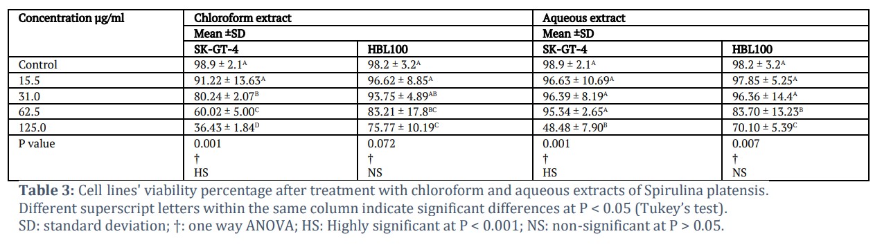
The cell viability of SK-GT-4 significantly decreased with growing quantity of extract (from 91.22% at 15.5 µg/ml to 36.43% at 125 µg/ml) as presented in Table 3. In contrast, normal HBL100 cells showed less reduction in viability (from 96.62% to 75.77%) over the same concentration range, as shown in Table 3. The IC₅₀ value for SK-GT-4 cells treated with chloroform extract was 53.83 µg/ml.
Aqueous Extract
In the SK-GT-4 cancer cells, viability remained largely unchanged at concentrations up to 62.5 µg/ml but dropped to 48.48% at 125 µg/ml (Table 3). The HBL100 cell line demonstrated minimal effects at lower doses, with a decrease to 70.10% viability only at the highest concentration (125 µg/ml), as shown in Table 3. The IC₅₀ for the aqueous extract was 72.84 µg/ml.
Figures & Tables
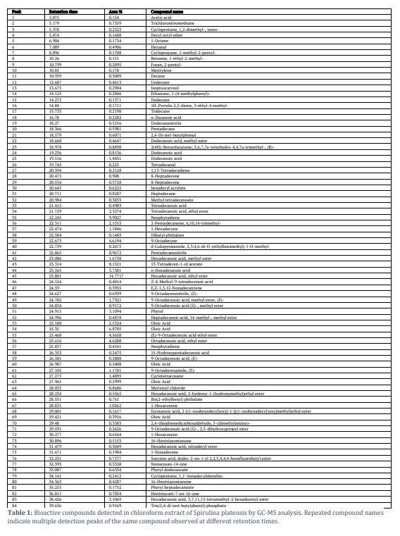
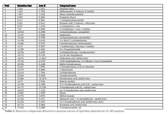
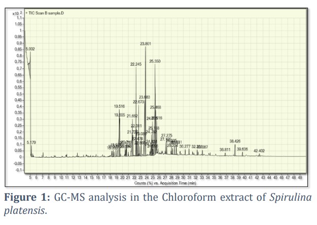
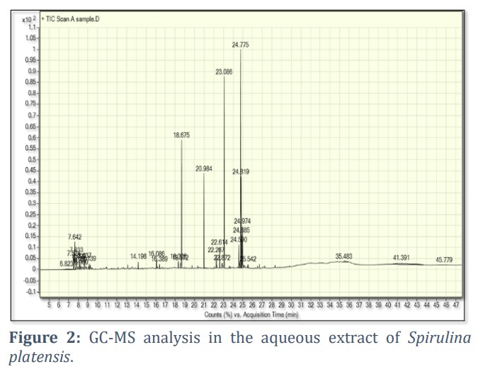
The occurrence of several bioactive compounds was established by the GC-MS analysis in both chloroform and aqueous extracts of Spirulina platensis, which were compared against the NIST library and standard references for compound identification [12]. The chloroform extract showed a broader chemical diversity (84 compounds) than the aqueous extract (35 compounds), as detailed in Figures 1 and 2 and Tables 1 & 2. Major constituents in the chloroform extract included hexadecanoic acid, ethyl ester (14.77%), neophytadiene (9.90%), oleic acid (6.87%), (E)-9-octadecenoic acid, ethyl ester (4.56%), and phytol (3.10%). The aqueous extract contained dominant compounds such as 9-octadecenoic acid methyl ester (23.17%) and hexadecanoic acid methyl ester (18.18%). These results align with the findings of El Din SM (2019) [13].
Cytotoxicity results indicated a concentration-dependent reduction in viability of SK-GT-4 cancer cells treated with the chloroform extract, with significant effects observed at concentrations ≥ 31.0 µg/ml (Table 3). In contrast, HBL100 normal cells showed minimal cytotoxicity under the same treatment (Table 3). The calculated IC50 for SK-GT-4 was 53.83 µg/ml. These outcomes are consistent with those reported by MZ MS (2012), who demonstrated selective cytotoxicity of chloroform microalgal extracts on cancer cell lines [14]. The observed anticancer effects may be attributed to compounds such as 9-octadecenoic acid, oleic acid, ethyl ester of hexadecanoic acid, and n-hexadecanoic acid [15-23]. The anticancer potential of fatty acids, including hexadecanoic acid, has been reported in various seaweed species and is associated with induction of apoptosis through activation of caspase-3, caspase-8, and Bax proteins [24,25].
Furthermore, studies demonstrated the cytotoxic effect of Scenedesmus sp. and S. obliquus extracts against HepG2, MCF7, and CT116 cancer cell lines [26,27]. The presence of fatty acid esters enhances the biological activity of algal extracts [28,29]. Aqueous extract treatment also influenced cell viability, albeit to a lesser extent. Only the highest concentration (125 µg/ml) significantly reduced viability in SK-GT-4 cells (Table 3), with an IC50 value of 72.84 µg/ml. HBL100 cells showed a more marked viability reduction at concentrations ≥ 62.5 µg/ml (Table 3). These results agree with Abo El-Sayed B (2022), who observed modest anticancer activity in aqueous extracts [30], and with MZ MS (2012), who validated the anticancer potential of microalgae [14]. Bioactive constituents in the aqueous extract include methenamine, N-hydroxy-N-methyl, methyl tetradecanoate, phytol, behenic alcohol, and 9-octadecenoic acid (Z)-methyl ester [31]. These compounds likely contribute to the extract’s cytotoxicity. As indicated by the spectrum of alcohols, phenols, esters, and fatty acids identified, these components may be responsible for modulating anticancer effects [32]. This suggests that crude extracts of Spirulina platensis could serve as valuable resources for anticancer drug development.
Acknowledgement
The University of Thi-Qar has funded this effort so that it may be included in their various research and higher study projects.
Author Contributions
Mohammed A. Auda conceptualized the review, developed the framework, and provided oversight for the manuscript. Rasha N. Aljabery conducted the primary literature search, data analysis, synthesis, and background sections, while Hussain Yousif Al-Rekabi focused on the discussion and future perspectives. The revision, correction, and final version approval of the manuscript were conducted by all authors equally.
The authors declare no conflict of interest in the publication of this manuscript.![]()
References
- El-Shaibany A, Al-Habori M, Al-Maqtari T, Al-Mahbashi H. The Yemeni Brown Algae Dictyota Dichotoma Exhibit High In Vitro Anticancer Activity Independent of Its Antioxidant Capability. BioMed research international, (2020); 2020(1): 1-9.
- Alvarenga RR, Rodrigues PB, Cantarelli VD, Zangeronimo MG, Silva Júnior JW, et al. Energy values and chemical composition of spirulina (Spirulina platensis) evaluated with broilers. Revista Brasileira de Zootecnia, (2011); 40(5): 992-996.
- Juneja A, Ceballos RM, Murthy GS. Effects of environmental factors and nutrient availability on the biochemical composition of algae for biofuels production: a review. Energies, (2013); 6(9): 4607-4638.
- Hamed I. The evolution and versatility of microalgal biotechnology: A review. Comprehensive reviews in food science and food safety, (2016); 15(6): 1104-1123.
- 5.Grawish ME. Effects of Spirulina platensis extract on Syrian hamster cheek pouch mucosa painted with 7, 12-dimethylbenz [a] anthracene. Oral Oncology, (2008); 44(10): 956-962.
- 6.O’Shaughnessy JA, Kelloff GJ, Gordon GB, Dannenberg AJ, Hong WK, et al. Treatment and prevention of intraepithelial neoplasia: an important target for accelerated new agent development: recommendations of the American association for cancer research Task force on the Treatment and Prevention of intraepithelial neoplasia. Clinical Cancer Research, (2002); 8(2): 314-346.
- Marzieh Hosseini S, Shahbazizadeh S, Khosravi-Darani K, Reza Mozafari M. Spirulina platensis: Food and function. Current Nutrition & Food Science, (2013); 9(3): 189-193.
- El-Beltagi HS, Dhawi F, Ashoush IS, Ramadan K. Antioxidant, anti-cancer and ameliorative activities of Spirulina platensis and pomegranate juice against hepatic damage induced by CCl4. Notulae Botanicae Horti Agrobotanici Cluj-Napoca, (2020); 48(4): 1941-1956.
- Abd Alzahra EM. Detecting of Chemical compounds of aqueous and alcoholic extracts of damasConocarpus lancifolus Engl. leaves using GC-MS technique. Journal of Education for Pure Science, (2022); 12(1): 29-39.
- Bahuguna A, Khan I, Bajpai VK, Kang SC. MTT assay to evaluate the cytotoxic potential of a drug. Bangladesh Journal of Pharmacology, (2017); 12(2): 115-118.
- Chen Z, Bertin R, Froldi G. EC50 estimation of antioxidant activity in DPPH assay using several statistical programs. Food chemistry, (2013); 138(1): 414-420.
- Mlozi SH, Mmongoyo JA, Chacha M. GC-MS analysis of bioactive phytochemicals from methanolic leaf and root extracts of Tephrosia vogelii. Scientific African, (2022); 16(2022): e01255.
- El Din SM, Hussein MH, Hamouda RA, Shehawy MA, Abd El Maksoud AI. Bioactive potentiality of some secondary metabolites extracted from microalga Spirulina platensis. Journal of Chemical and Pharmaceutical Research, (2019); 11(10): 22-35.
- MZ MS, MS MH, MD S, HY A. Screening of anticancer activities of crude extracts of unicellular green algae (Chlorella vulgaris) and filamentous blue green algae (Spirulina platensis) on selected cancer cell lines. Journal of Academia, (2012); 2(2012): 38-42.
- Cherchi G, Deidda D, Gioannis BD, Marongiu B, Pompei R, et al. Extraction of Santolina insularis essential oil by supercritical carbon dioxide: influence of some process parameters and biological activity. Flavour and Fragrance Journal, (2001); 16(1): 35-43.
- Aly AA, Maraei RW, Ali HG. Fatty acids profile and chemical composition of Egyptian Moringa oleifera seed oils. Journal of the American Oil Chemists' Society, (2016); 93(3): 397-404.
- Srivastava R, Mukerjee A, Verma A. GC-MS analysis of Phytocomponents in, pet ether fraction of wrightia tinctoria seed. Pharmacognosy Journal, (2015); 7(4): 249-253.
- Jeeshna MV, Paulsamy S. Phytochemistry and bioinformatics approach for the evaluation of medicinal properties of the herb, Exacum bicolor Roxb. International Research Journal of Pharmacy, (2011); 2(2011): 163-168.
- Patil A, Jadhav V. GC-MS analysis of bioactive components from methanol leaf extract of Toddalia asiatica (L.). International Journal of Pharmaceutical Sciences Review and Research, (2014); 29(1): 18-20.
- Flefel EM, El-Sayed WA, Mohamed AM, El-Sofany WI, Awad HM. Synthesis and anticancer activity of new 1-thia-4-azaspiro [4.5] decane, their derived thiazolopyrimidine and 1, 3, 4-thiadiazole thioglycosides. Molecules, (2017); 22(1): 170.
- Adeyemi JO, Onwudiwe DC, Ekennia AC, Anokwuru CP, Nundkumar N, et al. Singh M, Hosten EC. Synthesis, characterization and biological activities of organotin (IV) diallyldithiocarbamate complexes. Inorganica Chimica Acta, (2019); 485(2019): 64-72.
- Radhakrishnan S, Reddivari L, Sclafani R, Das UN, Vanamala J. Resveratrol potentiates grape seed extract induced human colon cancer cell apoptosis. Front Biosci (Elite Ed), (2011); 3(4):1509-1512.
- Sangpairoj K, Settacomkul R, Siangcham T, Meemon K, Niamnont N, et al. Hexadecanoic acid-enriched extract of Halymenia durvillei induces apoptotic and autophagic death of human triple-negative breast cancer cells by upregulating ER stress. Asian Pacific journal of tropical biomedicine, (2022); 12(3): 132-140.
- Cardoso C, Ripol A, Afonso C, Freire M, Varela J, et al. Fatty acid profiles of the main lipid classes of green seaweeds from fish pond aquaculture. Food Science & Nutrition, (2017); 5(6): 1186-1194.
- Sharifi S, Mostafavi PG, Tarasi R, Moradi AM, Givianrad MH, et al. Purified compounds from marine organism sea pen induce apoptosis in human breast cancer cell MDA-MB-231 and cervical cancer cell HeLa. European journal of pharmacology, (2020); 877(2020): 173075.
- Custódio L, Soares F, Pereira H, Barreira L, Vizetto-Duarte C, et al. Fatty acid composition and biological activities of Isochrysis galbana T-ISO, Tetraselmis sp., and Scenedesmus sp.: Possible application in the pharmaceutical and functional food industries. Journal of Applied Phycology, (2014); 26(1): 151-161.
- Abd El Baky HH, El-Baroty GS, Ibrahem EA. Antiproliferation and antioxidant properties of lipid extracts of the microalgae Scenedesmus obliquus grown under stress conditions. Der Pharma Chemica, (2014); 6(5): 24-34.
- Hamad GM, Abd El-Baky N, Sharaf MM, Amara AA. Volatile compounds, fatty acids constituents, and antimicrobial activity of cultured Spirulina (Arthrospira fusiformis) isolated from Lake Mariout in Egypt. The Scientific World Journal, 2023; 2023(1): 9919814.
- Morsi HH, El-Sabbagh SM, Mehesen AA, Mohamed AD, Al-Harbi M, et al. Antibacterial activity of bioactive compounds extracted from the Egyptian untapped green alga Rhizoclonium hieroglyphicum. Water, (2023); 15(11): 2030.
- El-Sayed B, El-Feky AE, Mounier A, Reda M. C-Phycocyanin, anticancer activity and nutritional value of mass-produced Spirulina platensis. Egyptian Journal of Chemistry, (2022); 65(11): 611-625.
- Gong JE, Kim JE, Park SH, Lee SJ, Choi YJ, et al. Anti‑tumor effects of an aqueous extract of Ecklonia cava in BALB/cKorl syngeneic mice using colon carcinoma CT26 cells. Oncology reports, (2023); 49(6): 128.
- Mofeed J, Deyab MA, Abd El-Halim EH. Anticancer activity of some filamentous cyanobacterial isolates against Hep-G2 and MCF-7 cancer cell lines. International Journal of Life Sciences, (2018); 8(1):10-17.
This work is licensed under a Creative Commons Attribution-Non Commercial 4.0 International License. To read the copy of this license please visit: https://creativecommons.org/licenses/by-nc/4.0

