

Full Length Research Article
Analysis of mRNA Expression for Injury-Age Estimation
Nusrat Saba1, Saadia Noreen1, Mubarak Ali Anjum2, Saqib Ali3, Muhammad Jawad1*, Allah Rakha1
Adv. life sci., vol. 7, no. 1, pp. 16-19, November 2019
*- Corresponding Author: Muhammad Jawad (Email: m.jawad@uhs.edu.pk)
Authors' Affiliations
2. Institute of Agricultural Sciences, Punjab University, Lahore, Pakistan
3. Centre of Agricultural Biochemistry and Biotechnology, University of Agriculture, Faisalabad, Pakistan
Abstract![]()
Introduction
Methods
Results
Discussion
References
Abstract
Background: Determination of wound age is pivotal in forensic medical sciences, criminal and civil cases for the construction of crime scene and answering the questions like, time of infliction, manner of wound infliction, how long the person survives after infliction of wound and characterizing antemortem or postmortem wounds. The findings differ considerably among individuals due to the biological variations. Previous developed method in the injury-age determination is clinical, microscopic, enzymatic reaction at the wound margins, histological, and immunohistochemistry with the pitfalls associated with it.
Methods: This study is conducted on blunt injuries, particularly lacerated wound (type of wound inflicted by blunt weapon) to analyze the different expression pattern in injury-age up to 72hrs in total 21 individuals randomly grouped in different time intervals. To determine the time of injury, transcript abundance of mRNA of Fibronectin (FBN), IL1β, VEGFA, and GM-CSF was analyzed by real time polymerase chain reaction. 18S-rRNA was used as control marker.
Results: Percent knockdown (%KD) was calculated to determine the expression of mRNA for the determination of injury-age. IL1β and GM-CSF showed the predictive behavior for wound age up to 36hrs, Fibronectin (FBN) showed predictive behavior up to 12hrs while VEGFA showed prediction beyond 72hrs.
Conclusion: The detection of gradual decrease of mRNA of Fibronectin (FBN), IL1β, VEGFA, and GM-CSF may provide an estimation of wound-age.
Keywords: mRNA Expression; Injury-Age Estimation; Antemortem wounds; Postmortem wounds
Introduction![]()
The aging of wound in relation to time is a very important aspect of forensic medicine because the opinion of the pathologist as “expert witness” has important legal consequence [1]. To evaluate the relationship between death and wounds, forensic pathologist always needs to distinguish ante mortem wounds from postmortem wounds [2]. Wound age is important in knowing the time of infliction, manner of wound infliction, how long the person survives after infliction of wound [1]. Estimation of time of wound infliction focuses the investigator within the time limit, resources and to narrow down the possible suspect [3].
When the wound is vital, it is necessary to demonstrate how long it was sustained before death [4]. The three phases of wound healing proceed with complex but well organized, interlinked functions of many cells, tissues and organs [5]. Studies reveal that many proteases, cytokines and growth factors are closely linked in the healing process of normal tissue [6]. The pro inflammatory cytokines i.e. Interleukin-1β (I L-1β), Fibronectin (FBN), VEGFA and GM-CSF plays a vital role in cytokine network because of their close relationship to various pathways by their rapid stimulation [7]. Very minute concentrations, production from specific cell types and short duration make them an appropriate tool for determination of early wound age [8]. Advances in molecular biology have made it possible to use RNA for forensic purposes [3]. Gene expression level at the time of injury might have enough power to become a complimentary tool for the determination of cause and circumstances of injury and include the pathophysiological disease conditions, injury, duration of disease and many other factors [4]. IL-1β start the inflammatory reaction in wound within 30-60 mins of infliction of injury [6]. Inflammation, embryogenesis and tumor formation highly require VEGFA for their need for nutrition, removal of waste and oxygen supply [9]. Fibronectins (FBN) are cold insoluble globulins have their important role in wound healing and repair [10]. GM-CSF appears first 24-48hrs after injury [11]. This is a preliminary study on human, to analyze the mRNAs expression pattern of Fibronectin (FBN), GM-CSF, VEGFA and IL-β for determination injury-age within 72h. The real time quantitative polymerase chain reaction (RT-qPCR) is considered as a method of choice in the determination of mRNA markers and their comparative analysis because it provides highly sensitive assay [1].
Methods![]()
Sample Collection and RNA Extraction
Injured samples were collected from lacerations on 35 different individuals by non-invasively swabbing the injured area at specific time points (0-2hrs, 2-6hrs, 6-12hrs, 12-24hrs, 24-48hrs, and 48-72hrs after injury). Samples immediately snap-frozen in liquid nitrogen to avoid RNA degradation. All injuries less than 2hrs duration were included from 15 to 35 years of age independent of sex and race. RNA extracted using TRIZOl® reagent. The concentration of total RNA was measured by spectrophotometric analysis by using NanoDropTM 2000 (Thermo Scientific TM).
Target Gene and Essay
In the present study, mRNA of Fibronectin (FBN), VEGFA, GM-CSF, IL-1β and 18s rRNA were investigated. The primers were designed by Primer 3 web program [12,13]. The Thermo Scientific™ RevertAid™ First Strand cDNA Synthesis Kit was used which allows for total RNAs to be converted to cDNA through ProFlex PCR thermal cycler. The targeted mRNA primers were then used for qRT PCR protocols. The SYBR assay of real time PCR by using Maxima SYBR Green/ROX qPCR Master Mix (2X) was performed using a three-step protocol in AriaMX Real – Time PCR System (Agilent Technologies) instrument. The GAPDH was used to normalize the target mRNAs to calculate the ∆Cq values. The Mean and Standard deviation were calculated with Cq value of each sample in different interval containing (Fibronectin (FBN), VEGFA, GM-CSF, IL-1β, 18s rRNA and GAPDH). The differences in Cq value between the most stable control marker (i.e. GAPDH) and targeted RNA markers (Fibronectin (FBN), VEGFA, GM-CSF, IL-1β, 18s rRNA) represents ΔCq followed by ΔCq expression using formula and taking its mean. The ΔΔCq expression was calculated after normalization to control group (0h). Percent knockdown was then calculated by subtracting the normalized ΔΔCq expression. Efficacy is commonly reported as relative percent knockdown of mRNA levels compared to controls.
Results![]()
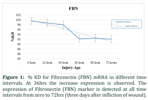
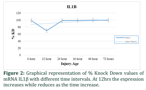
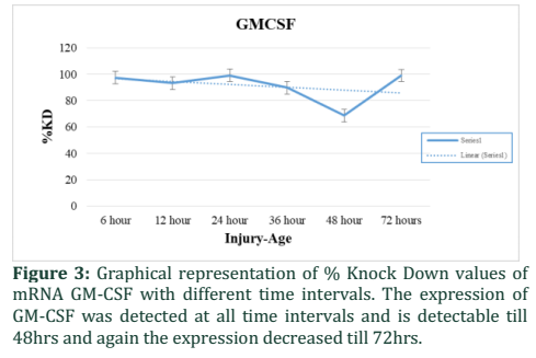
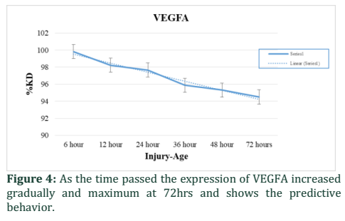
To find out the percent knockdown, the mean and SD of ∆Cq of target mRNAs of each group was calculated. The expression of miRNA-133 was determined with respect to zero-time interval group taken as a control, the expression was found to be varied with time. The molecular markers IL1β, GM-CSF and VEGFA were checked at different time points ranging from 0hrs to 72hrs. Fibronectin (FBN) has increased expression at 24-36hrs (Fig. 1) and contrast behavior at 8hrs interval as compared to the study of [10]. The expression of IL1-β found at 12hrs decreased to a maximum (Fig. 2), which shows that at 12hrs time interval the mRNA of IL1β was expressed maximum and then decreased again at 36 to 72hrs. The expression of GM-CSF was very low at certain time intervals, but at 48hrs interval it was found the expression was increased (Fig. 3). VEGFA showed an increase in its expression at 72hrs interval (Fig. 4).
Figures & Tables
Discussion![]()
This study correlates with the study of Palagummi et al., [1] at 12hrs who studied the expression level of IL1β at different time interval in human dermal lacerated wounds. This study shows contrasting behavior with [3] who studied that the expression of GM-CSF increased at 17hrs, but in this case it was detected at all the points but increased at 48hrs. The possible reason for this contradiction might be the selection of tissue and the technique used. Another possible reason might be the environment of the country. This study showed the contrast behavior in the study of Palagummi et al., [1] at 72hrs interval who showed that the expression of VEGFA is maximum at 5 days interval. But in this study, it is maximum at 3rd day after wound infliction. The possible reason for this contrasting behavior would be that this study limited to 72hrs.
Authors' Contribution
Nusrat Saba: Designed the study, carried out the genomic work. Saadia Noreen: Performed data analysis. Mubarak Ali Anjum: Participated in sample collection. Saqib Ali: Participated in sample collection. Muhammad Jawad: Participated in sample collection. Allah Rakha: Principle Investigator of the research.
The authors declare that there is no conflict of interest regarding the publication of this paper.
References![]()
- Palagummi S, Harbison S, Elliot D, Fleming R. A multiplex analysis of RNA expression during injury healing in human dermal injuries for injury-age estimation. Forensic Science International: Genetics Supplement Series, (2013); 4(1): e17-e18.
- Bauer M. RNA in forensic science. Forensic Science International: Genetics, (2007); 1(1): 69-74.
- Takamiya M, Biwasaka H, Saigusa K, Nakayashiki N, Aoki Y. Wound age estimation by simultaneous detection of 9 cytokines in human dermal wounds with a multiplex bead-based immunoassay: an estimative method using outsourced examinations. Legal Medicine, (2009); 11(4): 186-190.
- Takamiya M, Fujita S, Saigusa K, Aoki Y. Simultaneous detections of 27 cytokines during cerebral wound healing by multiplexed bead-based immunoassay for wound age estimation. Journal of neurotrauma, (2007); 24(12): 1833-1844.
- Saukko P, Knight B Knight's forensic pathology fourth edition. 2015; CRC press.
- Oehmichen M. Vitality and time course of wounds. Forensic science international, (2004); 144(2-3): 221-231.
- Clark RA. Fibronectin (FBN) matrix deposition and Fibronectin (FBN) receptor expression in healing and normal skin. Journal of Investigative Dermatology, (1990); 94(6): s128-s134.
- Grinnell F, Billingham RE, Burgess L. Distribution of Fibronectin (FBN) during wound healing in vivo. Journal of Investigative Dermatology, (1981); 76(3): 181-189.
- Galiano RD, Tepper OM, Pelo CR, Bhatt KA, Callaghan M, et al. Topical vascular endothelial growth factor accelerates diabetic wound healing through increased angiogenesis and by mobilizing and recruiting bone marrow-derived cells. The American journal of pathology, (2004); 164(6): 1935-1947.
- Takamiya M, Kumagai R, Nakayashiki N, Aoki Y. A study on mRNA expressions of Fibronectin (FBN) in dermal and cerebral wound healing for wound age estimation. Legal Medicine, (2006); 8(4): 214-219.
- Shiomi A, Usui T. Pivotal roles of GM-CSF in autoimmunity and inflammation. Mediators of inflammation, (2015); 2015.
- Untergasser A, Cutcutache I, Koressaar T, Ye J, Faircloth BC, et al. Primer3—new capabilities and interfaces. Nucleic acids research, (2012); 40(15): e115-e115.
- Koressaar T, Remm M. Enhancements and modifications of primer design program Primer3. Bioinformatics, (2007); 23(10): 1289-1291.
![]()
This work is licensed under a Creative Commons Attribution-Non Commercial 4.0 International License. To read the copy of this license please visit: https://creativecommons.org/licenses/by-nc/4.0

