

Full Length Research Article
Probiotic potential of encapsulated Lactobacillus species in yogurt formation indigenously isolated from dairy source
Atia Iqbal*, Saba Irshad, Saira Saeed, Aatikah Tareen
Adv. life sci., vol. 8, no. 3, pp. 267-274, July 2021
*– Corresponding Author: Atia Iqbal (Email: atia.iqbal@wum.edu.pk)
Authors' Affiliations
Abstract![]()
Introduction
Methods
Results
Discussion
References
Abstract
Background: Exploration of beneficial bacteria as probiotics inputs are drawing interest in dairy industry but their long-term survival and viability is an important consideration.
Methods: The current work focused on the exploration of probiotic potential of indigenously isolated Lactobacillus strains from dairy products and their encapsulation and utilization in yogurt formation.
Results: These Lactobacillus strains were identified as Lactobacillus fermentum MGA23-1 and Lactobacillus fermentum LMEM19 and found resistant to inhibitory substances like phenol (0.2%), bile salts (0.3%), pancreatin (0.5%) and pepsin (0.3%). The highest antibacterial activity was observed by Lactobacillus fermentum MGA23-1 against Pseudomonas aeruginosa (13mm). Encapsulation experiment showed that the number of bacterial CFU/g increased significantly (p < 0.05) in beads during storage up to 7 days. Chemical characterization of microcapsules was assessed using FTIR and showed characteristics wavelength major at 1541 – 1716 cm-1 and 3336 cm-1. Yogurt was prepared using a single probiotic strain, in a consortium of Lactobacillus fermentum MGA23-1 and Lactobacillus fermentum LMEM19 and in the form of beads. Best results were observed in the case of microencapsulation.
Conclusion: It was concluded that both strains had the potential to be used as a probiotic in the dairy industry.
Keywords: Probiotic; Tolerance to inhibitory substances; Antibacterial activity; Adhesion ability; Safety; Microencapsulation; FTIR
Introduction![]()
Probiotics are defined as nutritious live bacteria that when taken in adequate amounts provide benefit to the health of the host organism. The word probiotic was derived from a Greek word in which pro means promoting and biotic means life [1]. The term probiotic was also defined by World Health Organization as live microorganisms which, when taken in appropriate quantity, provide a health benefit to the host [2].
Probiotic bacteria commonly belong to Lactobacillus and Bifidobacterium species [3]. All of the Lactobacilli spp. are from phylum Firmicutes belongs to class Bacilli and order Lactobacillales. Such strains undergo the process of glycolysis, produce lactic acid, and are known as homo-fermenters [4]. Probiotics are microbes, which are usually present in human digestive tract and are quite valuable to the human health. Lactobacillus is a group of probiotics that have several therapeutic activities [5]. Several research studies have been conducted on gut microbiome and immunity in ancient times, which continuously leads to the increase in usage of probiotics as a functional food [6]. They are usually present with a symbiotic association through different parts of the body and provide a protective role [7]. The basic purpose of using probiotics is the improvement of health by modulating the intestinal microbial balance [8]. They are the group of microbes that are found usually in the human gut and enter into the body through ingestion of different fermented foods like yogurt, various types of cheese and, other fermented cured products [9].
Most of the used probiotic cultures are considered safe and denoted as “generally recognized as safe,” according to the European Food Safety Authority (EFSA). However, there is a lack of apprehension in the use of probiotics through a potential mechanism of action [9].
The effective probiotic should be alive at the time of administration, must be genetically identified and the source of probiotics should be clearly mentioned as they are from an animal source or human source [10]. It should be non-pathogenic, non-toxic, should resist the gastric condition, and have the ability to form strong biofilm in intestinal tissues, should control antimicrobial activity against potentially pathogenic bacteria and have the ability to balance the immune system [11].
Some of the lactobacilli found in foods and supplements are Lactobacillus acidophilus, L. acidophilus DDS-1, Lactobacillus bulgaricus, Lactobacillus rhamnosus GG, Lactobacillus plantarium, Lactobacillus reuteri, Lactobacillus salivarius, Lactobacillus casei, Lactobacillus johnsonii and Lactobacillus gasseri [12].
Recently, Lactobacillus bacteria have been introduced into a universal dimension of commercial products [13]. For the survival of such bacteria in high acidic conditions, a new method has been introduced now a day, called microencapsulation. The encapsulation method involves the entrapment of an active material into other substance wall material that can generate different-sized particles. This method has vast application throughout the food sectors [14]. Different biological materials that are acid liable can be balanced in gastrointestinal fluids by using coating material to pass through the acidic condition of the stomach. Therefore, encapsulated bacteria can survive acidic and alkali conditions during the intestinal transition [14].
Probiotics are live microbes that are intended to have health benefits, but such bacteria may lose their probiotic potential during processing. Therefore, having an ideal probiotic strain with all the required properties is very difficult to obtain. That is why this study was designed to isolate Lactobacillus spp. from different dairy products, to evaluate their survival in low acidic conditions and bile salt presence followed by microencapsulation of selected strains and assessment of their potential survival in probiotic yogurt.
In the future, this type of research will help us to identify minor metabolite proteins and several other compounds that are produced from microorganisms, that are responsible for a specific type of response in the host. Such useful microorganisms improve health and give us knowledge about microbial enzymatic pathways and other useful molecules that are necessary for health promotion [15].
Methods![]()
Isolation and preliminary identification
A total of thirty different dairy samples were used in this study including cow milk, goat milk, buffalo milk and fresh yogurt sample which were collected from different areas of Southern Punjab. All samples were collected in sterilized and tightly packed tubes and kept in storage before further processing. For culturing, serial dilution were prepared in phosphate buffer saline by adding 1g of sample in first tube labeled as 100. From the first tube 1ml was taken and added into the second tube, in this way dilutions were prepared up to 10-5. Later 100ul of the sample was taken from 100 ,10-3 and 10-5 dilution and spread over media. For isolation, all samples were cultured anaerobically on MRS agar (pH 6.4) [16] and incubated at 37°C for 48 hours. Single strains were picked from culture plates and purified on MRS agar using the streaking method. All strains were subcultured three times prior to experimental analysis. Total twelve strains were isolated initially and identified by gram staining, and spore staining. Biochemical tests were performed according to Bergey’s Manual of Determinative Bacteriology. For long-term storage, stock cultures were maintained at -80°C in MRS broth containing 50% glycerol.
Molecular identification
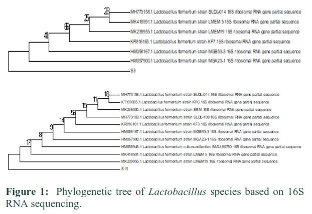
After identification by biochemical testing, only two of the selected strains were identified through 16S rRNA gene analysis commercially (Macrogen, Korea). DNA Dragon software was used to assemble sequences, which were then blast matched in NCBI nucleotide blast and the phylogenetic tree was constructed using software MEGA 7 with neighbor-joining method keeping bootstrap value 1000.
Screening of Lactobacilli for potential probiotic characters
Effect of different temperatures and high salt concentration
The ability of strains to survive at different temperatures and high salt concentrations was evaluated. The different temperature ranges and salt concentrations were used to optimize the maximal growth of Lactobacillus strains. For this, 100ul of 24 h old bacterial culture was inoculated in MRS medium (6.4) and incubated at 19°C, 28°C, 37°C and 50°C for 48h. After incubation, pattern of the growth was determined by taking OD at 600nm in a UV visible spectrophotometer. Similarly, three different NaCl (4%, 8% and 12%) concentrations were used and2 the test strain was being inoculated and incubated at 37°C for 48h anaerobically. Growth pattern was determined by taking OD at 600nm.
Acid and bile salt tolerance
24h old bacterial culture was inoculated into MRS broth adjusted to different pH 3, 5, 7 and 9 and three different (0.05%, 0.1% and 0.3%) concentrations of bile salt (sodium deoxycholate) and incubated at 37°C for 48h anaerobically. MRS broth without bile salts and pH (6.4) served as control. The growth pattern was determined by taking OD at 600nm in a UV visible spectrophotometer. The different pH and bile salt concentrations were used to optimize the maximal growth of Lactobacillus strains to check their viability.
Effect of pancreatin and phenol
MRS broth was prepared with 0.5% pancreatin then inoculated and incubated for 48h at 37°C under anaerobic conditions. The growth rate of bacteria was observed by estimating O.D values at wavelength of 560nm. On the other hand, three different concentration of phenol (0.1%, 0.2% and 0.4%) were used. MRS broth without phenol was used as control. After inoculation, tubes were incubated at 37°C for 24-48 h anaerobically. Optical density (OD) was recorded at 560 nm after incubation and compared with the control.
Exposure to gastric stimulant
This test involves providing the bacteria artificial acidic environment along with the pepsin enzyme. For this purpose, MRS media was prepared to have 0.3% pepsin and 0.5% NaCl. The pH was adjusted to 2 using 0.1M HCl and bacterial culture was inoculated and incubated under anaerobic condition for 24-48 hours at 37⁰C. The growth rate was observed by measuring optical density at 560nm
Auto-aggregation and Co-aggregation property
The 24h old bacterial culture was centrifuged for 5 minutes at 6000 rpm. Pellet was taken in Eppendorf, washed two times with PBS solution and OD was measured at the wavelength of 540nm. The suspension was vortexed for 10 seconds and kept for 5h at room temperature. Then the absorbance was measured at 540nm by collecting the upper suspension. Auto-aggregation was calculated by using the formula
Enzyme Assays
The enzyme assay was performed to observe the ability of selected strains to hydrolyze the enzyme in media. To detect amylase, protease and lipase activity the culture was inoculated into specific media and the zone of clearance was measured. Amylase activity was determined by modified MRS agar supplemented with 0.2% starch. After incubation, iodine solution was added and left for 5-10 minutes and the zone of clearance was observed. For protease activity, Milk Agar supplemented with 1% skim milk was used. Test strains were inoculated and incubated for 48h at 37°C and the zone of clearance was observed. For lipase activity, Tributyrin Agar was used. Test strains were inoculated and incubated at 37⁰C for two days. Then, zone of clearance was observed [17].
Detection of virulence factor
For thermonuclease activity, Brain Heart Infusion broth was used. After inoculation, tubes were incubated for 2h at 50°C. After 2h, pink colored zone were formed around the edges of tubes and was considered as a positive result [18]. For hemolytic activity, Blood Agar was used. Hemolysis was depicted as either α or β or γ after 24h of incubation. Nutrient media supplemented with gelatin was used to determine gelatinase activity. [19]
Antibacterial resistance
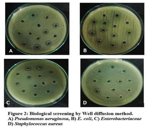
Lactobacilli cultures were analyzed for antibacterial resistance by agar well diffusion method. Pathogenic bacteria used as indicators including Staphylococcus aureus, E.coli, Enterobacteriaceae spp., Pseudomonas aeruginosa, Salmonella typhimurium [19].
Biogenic amine production test
The production of biogenic amines (tyramine and histamine) was assessed using a decarboxylase medium. MRS broth containing 1% amino acid (thiamine, tyrosine) was prepared; test strain was being inoculated and incubated at 37°C for 14h. Culture from this media was then used to inoculate into decarboxylation liquid medium and incubated for 4 h at 37°C and color change of the medium was noticed, while plain MRS broth was used as control [20].
Microencapsulation of Lactobacilli strain
Formation of Beads and Microbiological analysis of Microencapsulated bacteria
Microencapsulation of Lactobacillus strain was done by extrusion techniques [21]. The microencapsulated bacteria were solubilized as described by [21] with some modification. Bacteria that were encapsulated were released by taking 1g of beads and suspended in 9ml of PBS buffer (pH 7.5). The pH of this solution was adjusted to acidic (4.5) and kept for 2h at room temperature. The sample was being diluted to appropriate concentrations and was spread on agar surface. The plates were incubated at 37°C under anaerobic conditions for 24 h and the growth was measured as cfu/ml and was compared with free culture in MRS broth [21-23].
FTIR of beads
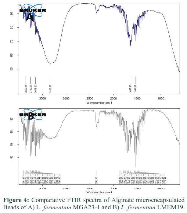
The chemical characteristics of the microcapsules were assessed using an infrared spectrometer (FTIR) between 4000 and 600 cm−1, using 64 scans at a resolution of 4 cm−1. The FTIR spectrum was catalogued separately for free bacterial culture, blank beads and beads along with bacteria [24].
Probiotic Yogurt Preparation
Commercialized, standardized and pasteurized cow milk was heated for 10 min at 90°C. Then, it was cooled at 45°C and separated into six equal portions or sets (100ml each). First set (C: control) was provided with the commercially available yogurt culture, the second and third set was inoculated with free bacterial culture, the fourth and fifth set was inoculated with encapsulated bacteria and the last set was inoculated with a consortium of two strains with equal concentration. All experimental sets were incubated for approximately 24h until a pH of 4.6-4.8 was attained and later placed in cold storage (4°C) where they were stored up to 4 weeks. Each week, samples were collected for interpretation of different parameters. Fermentation time (TpH 4.5) was observed, this is the time in hours necessary to reach pH 4.5. All experiments were done in triplicate and this experiment was repeated three times as well. Following parameters were observed during storage:
Measurement of pH: The pH of the preparations at 0 h and after fermentation was measured using a digital microprocessor pH meter. The pH meter was standardized using reference pH 4.0 and 7.0 buffer solutions.
Survival test of probiotic bacteria: For the survival test, the prepared probiotic yogurt was stored at 4°C for 28 days weekly, the stored product was diluted serially 10-1 to 10-7 in sterile PBS and plated out in an MRS agar plate. Viable numbers were evaluated after 1, 7, 14 and 21 days of storage.
Total solid content: The total solids of samples were determined by drying samples at 110°C overnight to constant weight. First initial weight of sample was taken and then it was placed in a hot dry oven. After drying, samples were cooled at room temperature and the final weight of sample was measured. The final weight of sample after drying was considered as total solid contents [25]. The total solid content was calculated by using the following formula:
Total solid content = 1- dry weight / wet weight × 100
Syneresis: To determine Syneresis, 3 tubes with 2ml of yogurt were weighed and placed in a centrifuge. Tubes were centrifuged at 2000 rpm for 5 min and the separated supernatant was then weighed. Syneresis was calculated using the following equation:
Syneresis % = Ws / Wy × 100
Where Ws is the supernatant weight after centrifugation and Wy is the weight of the yogurt in the tube [26].
Statistical analysis
Data were analyzed using MS office excel package. Results are expressed as mean ± SD and were analyzed with SPSS software by Duncan’s test. Statistical significance was considered at p < 0.05.
Results![]()
The bacteria were isolated from dairy products to evaluate their probiotic properties. The selected strains were identified as Lactobacillus fermentum MGA23-1, Lactobacillus fermentum LMEM19 after the biochemical and molecular characterization. Similitudes were shown by constructing the phylogenetic tree. (Fig 1)
Screening of Lactobacilli for potential probiotic characters
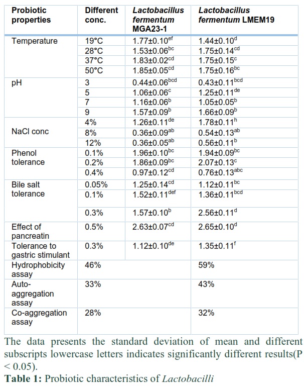
Both Lactobacillus fermentum MGA23-1 and Lactobacillus fermentum LMEM19 demonstrated significant growth (p < 0.05) at all the temperatures. Temperature 28°C and 37°C were considered as the ideal temperatures for both of the strains in light of the fact that the growth rate of bacteria was higher. (Table 1). The selected strains had the ability to survive under low pH. Practicality of Lactobacillus strains would be influenced within the sight of bile salts. So the capacity of strain to endure various groupings of bile salts was assessed. Both Lactobacillus fermentum MGA23-1 and Lactobacillus fermentum LMEM19 endure presence of bile salts and develop best at 0.3% fixation. Resilience of Lactobacillus strains in presence of pancreatin, a compound that is delivered in small digestive organs was assessed. Impact of pancreatin was fundamentally expanding (p < 0.05) by contrasting it and control. Both of the strains fill in presence of 0.5% pancreatin. 0.5% pancreatin is available in digestive organs and a decent probiotic bacterium must be ready to endure this fixation also. 100% endurance was seen, and the optimum growth was shown by all the strains. Resilience of Lactobacillus strains in presence of pepsin compound was assessed and seen that the two strains indicated critical development (p < 0.05) in presence of catalyst. To examine the impact of fomentation on bacterial development, impact of tumult was noticed, and development bend was drawn. Hydrophobicity examine was performed to check the following capacity of Lactobacilli spp. Lactobacillus fermentum MGA23-1 displayed 46% hydrophobicity and Lactobacillus fermentum LMEM19 showed 59% hydrophobicity.
Biological screening
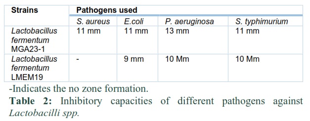
Most elevated zone of hindrance was shown in the presence of Lactobacillus fermentum MGA23-1 against Pseudomonas aeruginosa. While, Lactobacillus fermentum LMEM19 showed the maximum inhibitory zone against Salmonella typhimurium. (Fig.2)
Microencapsulation
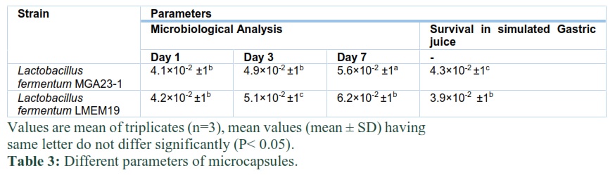
The mean breadth of globules was 2 mm. It was seen that beads had smooth edges. No morphological changes were observed during storage of beads. (Fig.3) Microorganisms stayed alive in dabs during the analysis (7 days) at temperatures of 4°C. Globules of L. fermentum LMEM19 had the most noteworthy measure of CFU/g 4.2 × 10⁻² after 24h of hatching. At day 7, CFU/g were expanded to 5.1×10⁻² to 6.2×10⁻². Also, if there should arise an occurrence of L. fermentum MGA23-1, CFU/g was expanded essentially (P < 0.05) from 4.9×10⁻² to 5.6×10⁻² at day 7. The quantity of bacterial CFU/g were not altogether not quite the same as the number toward the start. However, after day 3 and day 7 was higher (p < 0.05) than toward the start in this way demonstrating great endurance of microorganisms.
Survival of beads under stimulated gastric juice
The endurance pace of microencapsulated probiotic culture under reproduced small digestive tract juice was researched under in vitro conditions. By utilizing microencapsulating procedure societies had the option to endure. This is on the grounds that when microbes are typified they are being shielded by the center material from acidic conditions just as in presence of bile salts. Microencapsulation in alginate arrangements spoke to better well-being against gastric juice and was better ready to ensure cells. Number of microbes in dots increments fundamentally (p < 0.05) in simulated small intestine juice. Most noteworthy CFU/g of 4.3×10⁻² was seen in the event of L. fermentum MGA23-1 and 3.9×10⁻² CFU/g was seen by L. fermentum LMEM19 (Table 3).
FTIR of encapsulated bacteria
Lactobacillus strains were epitomized utilizing sodium alginate as a center material to evaluate the steadiness of probiotic culture in yogurt. FTIR range was confirmed particularly for nothing bacterial culture, clear globules (alginate) and beads alongside microorganisms. It was seen that peak intensity expanded if there should arise an occurrence of embodiment. Sodium alginate introduced a band situated at 2363 cm-1 that ascribes to the Si-H (Silane) practical group. Band at 3336 cm-1 is compared to the class Amide widening of N-H. Pinnacle recognized at 1473 cm-1 highlights the Methylene group. 1716 cm-1 force of pinnacle increments and relates the even spread of COO-. At frequency 1456 and 1473 cm-1 peaks noticed and compared to the deviated extending of the C=C and C-H group. Peaks observed at 1792 and 1844 cm-1 are ascribed to formed corrosive halide and fragrant compound. In L. fermentum LMEM19, the solid absorbance design was seen at 1541, 1557, 1635, 1698 and 1716 cm-1 showing the presence of N-H, N-H, C=C, C=N and C=O bonds at these positions separately. These bonds meant the presence of Amide, Alkene, imine/oxime and carboxylic corrosive. In L. fermentum MGA23-1, the solid absorbance design was seen at 3336 cm-1 demonstrating the presence of N-H bond which signified the presence of Amide group (Fig 4).
Production of novel probiotic yogurt
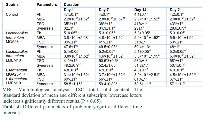
Yogurt was prepared utilizing Lactobacillus fermentum MGA23-1 and Lactobacillus fermentum LMEM19 in free structure just as in mix. pH of yogurt was seen at various time spans (following seven days during 21 days of storage). pH estimations of yogurt diminished during stockpiling because of post-fermentation. Huge contrasts (p < 0.05) in the pH of yogurts during storage was distinguished. Yogurt that included single strain introduced the most elevated acidity esteems. Decrease in pH can be because of the leftover action of microorganisms. Information demonstrated that the yogurt arranged from the blends of Lactobacillus fermentum MGA23-1 and Lactobacillus fermentum LMEM19 yielded a result of good quality and with expanding pH (4.8-4.9) which implies less acidity. A decrease in pH was seen by totally set of arranged yogurt and all the while increment in acidity. During stockpiling, the suitability of cells increments fundamentally. In yogurt with free bacterial culture number of suitable cells was 4.7×10⁻² cfu/ml during storage. Yogurt prepared utilizing probiotic culture in consortium of two strains held probiotic estimations of 4.5×10⁻² cfu/ml.
Yogurt was prepared by the utilization of probiotic culture have number of suitable cells of 5.5×10⁻². Alginate microencapsulated probiotic culture were steadier in the yogurts when contrasted with free probiotic culture. Yogurt samples were analyzed for all out strong substance and its rate changes between 35-73 %. If there should arise an occurrence of microencapsulated probiotic culture complete solid count range went from 41%-51%. If there should be an occurrence of control yogurt all out strong substance increment altogether from 35-43% (Table 4). Total solid content of encapsulated culture was altogether less in contrast with single and blend strains. Assessment of Syneresis have prime significance mostly during storage. Contrasts were determined in yogurt that was made with three distinctive type of societies. The huge decline (p > 0.05) in the syneresis of the probiotic yogurts was seen during capacity. Control yogurts introduced less syneresis than probiotic yogurts. Probiotic yogurts with low estimations of syneresis is might be because of the non-appearance of extra solids.
Figures & Tables

Discussion![]()
A good probiotic must have the capacity to increase the number of beneficial microorganisms in the gastrointestinal tract and at the same time; to decrease the impact of pathogenic microorganisms present without causing alterations that will trigger pathology Current study used the dairy samples for the isolation of lactobacilli with probiotic potential. The present results are in line with the previous studies conducted by [28; 29]. They used the dairy samples to isolate the beneficial bacteria with probiotic potential. The growth of lactobacilli was checked on different temperatures, results indicated that at 37°C bacterial growth was higher than the other temperatures. The study conducted by [18] likewise consider and indicated that Lactobacilli strain endure best at 37°C.
In order to function efficiently lower sections of the intestinal tract, orally delivered bacteria have to survive through the gastrointestinal tract to their site of function. The first major barrier to overcome during this passage is survival in the acidic environment. Lactobacillus strains were grown at different pH levels to see whether they could tolerate low or neutral pH. The selected strains had the ability to survive under low pH, as the strains demonstrated best development at pH 5. The findings of [29 30] presented a similar report and comparable outcomes were obtained like this investigation.
Pancreatic enzymes are secreted into the small intestine through the pancreatic duct and they are involved in digestion of proteins, carbohydrates and fats in foods. Ability to tolerate the presence of pancreatic enzymes is another criterion for selection of probiotic bacteria. 0.5% pancreatin is present in intestines, good probiotic bacteria must able to tolerate this concentration, 100% survival and maximum growth was observed by all the strains. The study of [30] also demonstrated that bacteria can tolerate 0.5% pancreatin but with increasing concentration, growth was decreased. Resilience of Lactobacillus strains in presence of pancreatin, a compound which is delivered in small digestive organs was assessed. Both of the strains survived in presence of 0.5% pancreatin. 0.5% pancreatin is available in digestive organs and gastrointestinal tract. However, with increasing concentration of pancreatin growth was decreased. The [30] reported the similar outcomes, that microscopic organisms can endure 0.5% pancreatin.
In selecting promising probiotic strains, hemolytic activity is considered a safety property. When grown in 5 percent sheep blood agar, none of the strains tested exhibited or -hemolysis behaviors. None of our strains have demonstrated α-hemolysis and β-hemolysis, indicating that they may be used as a probiotic strain. [31]additionally did likewise read for harmfulness factor identification and negative outcome acquired like in our examination. γ hemolysis was seen by all strains. The investigations of [30] additionally studied that non-appearance of hemolysis is viewed as protected and gamma hemolysis was noticed. Biogenic amines creating capacity of all strains was likewise recognized. None of strains indicated positive outcome for this test. The findings were consistent with the [31] likewise performed amine creation test and results were positive when contrasted with our outcomes however low creation was noticed.
One of the beneficial properties of probiotic strains in preventing gastrointestinal infection is their ability to inhibit pathogenic bacterial development in the intestinal tract. The antibacterial activity of selected strains were checked against different human pathogens. The inhibitory effect of lactobacilli was may be due to the production of organic acids. Karami et al., 2017 likewise noticed the inhibitory impact of lactobacilli against S. aureus and P. aeruginosa. In our examination, most noteworthy zone saw by S. aureus was 11mm by Lactobacillus fermentum MGA23-1 (Table 2). However, according to the investigations of [32] most noteworthy zone noticed was 10mm. Essentially, against P. aeruginosa was 13mm though in [32] investigation a zone of 10mm was noticed. The FTIR spectra of microcapsules with and without probiotics were observed in spectral region of 1000 to 3500 cm-1. The findings of the study were in line with [33]. After examining all potential test for an effective probiotic, probiotic yogurt was prepared. Yogurt was stored for a period of 21 days and different parameters were observed. Physical properties of yogurt was then checked. The previously conducted study [34] reported the probiotic yogurt and the outcomes are in line with the present study.
In this investigation, Lactobacillus strains were separated which demonstrated great probiotic attributes: they had the option to endure low pH, had the capacity to survive at viable temperatures of the gastrointestinal tract, and ready to endure bile salts presence. Lactobacillus strains were more stable in alginate beads when contrasted with free culture and microbial count expanded in beads. Besides, the strains were affirmed protected according to the harmfulness factors. Isolated Lactobacillus strains may potentially be used as probiotic culture in this way. The findings suggest that these four strains have probiotic potential; however, further research is needed to determine their true potential, which includes antibiotic resistance, co-aggregation capability with pathogenic bacteria, simulated gastrointestinal juice tolerance, and phenol tolerance.
Author Contributions
Author: one
Conceived idea and design the study, collected the data from experimental methodology and wrote the paper.
Author: two
Design the study; provide analysis tools and other facilities.
Contributed the data.
Author: three & four
Analysis of the study, Proof reading of the final draft
Contributed the data.
All authors read and approved the final version of the manuscript and declared that they have no conflict of interest.
Acknowledgement
The Authors would like to thanks, Department of Microbiology and Molecular Genetics, The Women University Multan, and Higher Education Commission Pakistan for providing support and all the facilities.
References![]()
- de Simone C. The unregulated probiotic market. Clinical Gastroenterology and Hepatology, (2018); 17(5): 809-817.
- Hojsak I. Probiotics in children: what is the evidence?. Pediatric gastroenterology, hepatology& nutrition, (2017); 20(3):139-46.
- Abrahamsson T. Using probiotics to prevent necrotisingenterocolitis. ActaPaediatrica, (2017); 106(11): 1718-1719.
- Özogul F, Hamed I. The importance of lactic acid bacteria for the prevention of bacterial growth and their biogenic amines formation: A review. Critical reviews in food science and nutrition, (2018); 58(10): 1660-1670.
- Al-Dhabi NA, ValanArasu M, Vijayaraghavan P, Esmail GA, Duraipandiyan V, Kim YO, Kim H, Kim HJ. Probiotic and Antioxidant Potential of Lactobacillus reuteriLR12 and Lactobacillus lactisLL10 Isolated from Pineapple Puree and Quality Analysis of Pineapple-Flavored Goat Milk Yoghurt during Storage. Microorganisms, (2020);8(10):1461.
- Ashaolu TJ. Immune boosting functional foods and their mechanisms: A critical evaluation of probiotics and prebiotics. Biomedicine & Pharmacotherapy,(2020);130:110625.
- Reid G. Probiotics: definition, scope and mechanisms of action. Best practice & research Clinical gastroenterology, (2016); 30(1): 17-25.
- Nagpal R, Kumar A, Kumar M, Behare PV, Jain S, Yadav H. Probiotics, their health benefits and applications for developing healthier foods: a review. FEMS microbiology letters, (2012); 334(1): 1-5.
- Plaza-Diaz J, Ruiz-Ojeda FJ, Gil-Campos M, Gil A. Mechanisms of action of probiotics. Advances in nutrition, (2019); 10(1): S49–S66.
- Reid G, Gadir AA, Dhir R. Probiotics: Reiterating what they are and what they are not. Frontiers in Microbiology, (2019); 10(424): 1-6.
- Shewale RN, Sawale PD, Khedkar CD, Singh A. Selection criteria for probiotics: A review. International Journal of Probiotics & Prebiotics, (2019); 9(1/2): 17-22.
- Arshad F, Mehmood R, Hussain S, Khan MA, Khan MS. Lactobacilli as probiotics and their isolation from different sources. British Journal of Research,(2018);5(3):43.
- Song CE, Kuppusamy P, Jeong YI, Shim HH, Lee KD. Microencapsulation of endophytic LAB (KCC-41) and its probiotic and fermentative potential for cabbage kimchi. International Microbiology, (2019); 22(1): 121-30.
- Kavitake D, Kandasamy S, Devi PB, Shetty PH. Recent developments on encapsulation of lactic acid bacteria as potential starter culture in fermented foods–A review. Food Bioscience, (2018); 1(21): 34-44.
- Marco ML. Defining how microorganisms benefit human health. Microbial Biotechnology, (2021);14(1): 35-40.
- De Man, J. C., Rogosa, D., & Sharpe, M. E. (1960). A medium for the cultivation of lactobacilli. Journal of applied Bacteriology, 23(1), 130-135.
- Leighton, H., Gopalakrishnan, S., Zhang, J. A., Rogers, R. F., Zhang, Z., & Tallapragada, V. (2018). Azimuthal distribution of deep convection, environmental factors, and tropical cyclone rapid intensification: A perspective from HWRF ensemble forecasts of Hurricane Edouard (2014). Journal of the Atmospheric Sciences, 75(1): 275-295.
- Moreno I, Marasca E. T. G, de Sá, P. B. Z. R, de Souza Moitinho J, Marquezini M. G, Alves M. R. C, Bromberg R. Evaluation of probiotic potential of bacteriocinogenic lactic acid bacteria strains isolated from meat products. Probiotics and antimicrobial proteins, (2018);10(4): 762-774.
- Cappuccino G, Sherman N. Microbiology: a laboratory manual, 2005; Benjamin/Cummings Publishing Co., Inc., Menlo Park, California
- Feng JR, Wang F, Qiu X, McFarland LV, Chen PF, Zhou R, Liu J, Zhao Q, Li J. Efficacy and safety of probiotic-supplemented triple therapy for eradication of Helicobacter pylori in children: a systematic review and network meta-analysis. European Journal of Clinical Pharmacology, (2017); 73(10):1199-208.
- Musikasang H, Tani A, H-kittikun A, Maneerat S. Probiotic potential of lactic acid bacteria isolated from chicken gastrointestinal digestive tract. World Journal of Microbiology and Biotechnology, (2009); 25(8): 1337-1345.
- CoghettoC, Brinques G, Siqueira N, Pletsch J, Soares R, Ayub M. Electrospraying microencapsulation of Lactobacillus plantarum enhances cell viability under refrigeration storage and simulated gastric and intestinal fluids. Journal Of Functional Foods, (2016); 24: 316-326.
- Zanjani MA, Tarzi BG, Sharifan A, Mohammadi N. Microencapsulation of probiotics by calcium alginate-gelatinized starch with chitosan coating and evaluation of survival in simulated human gastro-intestinal condition. Iranian journal of pharmaceutical research: IJPR, (2014); 13(3): 843.
- Kumar A, Kumar D. Development of antioxidant rich fruit supplemented probiotic yogurts using free and microencapsulated Lactobacillus rhamnosus culture. Journal of food science and technology, (2016); 53(1): 667-75.
- Bosnea L, Kopsahelis N, Kokkali V, Terpou A, Kanellaki M. Production of a novel probiotic yogurt by incorporation of L.casei enriched fresh apple pieces, dried raisins and wheat grains. Food and Bioproducts Processing, (2017); 102: 62-71.
- Mani-López E, Palou E, López-Malo A. Probiotic viability and storage stability of yogurts and fermented milks prepared with several mixtures of lactic acid bacteria. Journal of Dairy Science, (2014); 97(5): 2578-2590.
- Amer E, Mahrous H, Shekib LA. Isolation and identification of Probiotic Lactic Acid Bacteria from dairy products. Biotechnology Research, (2017); 3(3): 65-70.
- Rubbani, Umaima, and Atia Iqbal. "Evaluation of isolated Lactobacillus strains as Probiotics in yogurt preparation." Advancements in Life Sciences 7.2 (2020): 79-85..
- Lee J, Yun HS, Cho KW, Oh S, Kim SH, Chun T, Kim B, Whang KY. Evaluation of probiotic characteristics of newly isolated Lactobacillus spp. immune modulation and longevity. International journal of food microbiology, (2011); 148(2): 80-86.
- Padmavathi, Tallapragada, et al. "Screening of potential probiotic lactic acid bacteria and production of amylase and its partial purification." Journal of Genetic Engineering and Biotechnology 16.2 (2018): 357-362.
- Oh, Nam Su, et al. "Probiotic and anti-inflammatory potential of Lactobacillus rhamnosus 4B15 and Lactobacillus gasseri 4M13 isolated from infant feces." PloS one 13.2 (2018): e0192021.
- Karami S, Roayaei M, Hamzavi H, Bahmani M, Hassanzad-Azar H, Leila M, Rafieian-Kopaei M. Isolation and identification of probiotic Lactobacillus from local dairy and evaluating their antagonistic effect on pathogens. International journal of pharmaceutical investigation, (2017); 7(3): 1-5.
- Rather, Sajad A., et al. "Effect of double alginate microencapsulation on in vitro digestibility and thermal tolerance of Lactobacillus plantarum NCDC201 and L. casei NCDC297." LWT-Food Science and Technology 83 (2017): 50-58.
- Delavenne E, Ismail R, Pawtowski A, Mounier J, Barbier G, Le Blay G. Assessment of lactobacilli strains as yogurt bioprotective cultures. Food Control, (2013); 30(1):206-13.
This work is licensed under a Creative Commons Attribution-Non Commercial 4.0 International License. To read the copy of this license please visit: https://creativecommons.org/licenses/by-nc/4.0