

Full Length Research Article
Harnessing Natural Compounds for Effective Inhibition of HK2 and GLS1 in Rheumatoid Arthritis: A Structure-Based Virtual Screening Approach
Yousef M. Alshehre
Adv. life sci., vol. 11, no. 1, pp. 188-193, February 2024
*– Corresponding Author: Yousef M. Alshehre (yalshehre@ut.edu.sa)
Authors' Affiliations
[Date Received: 08/10/2023; Date Revised: 04/11/2023; Date Published: 25/02/2024]
Editorial Note on Version of Record
28 May 2025: This article has been corrected. See https://doi.org/10.62940/als.v13i0.4285 for more information.
Abstract![]()
Introduction
Methods
Results
Discussion
References
Abstract
Background: Rheumatoid arthritis (RA) is a persistent, inflammatory autoimmune disease that mostly affects joints. Advanced RA may cause irreversible bone damage, limiting joint mobility. RA patients are at higher risk of secondary complications like myocardial infarctions, strokes, pulmonary dysfunction, neurological abnormalities, and depression. Physical therapy is a common arthritis treatment that can reduce symptoms when combined with medications. In RA, metabolic reprogramming is frequently characterized by increased glycolysis and glutaminolysis, which coincide with increased expression of key enzymes, namely hexokinase 2 (HK2) and glutaminase 1 (GLS1).
Methods: Here in this study, 2706 phytochemicals from the Biopurify Phytochemicals subsets of the ZINC database were computationally screened against the active sites of HK2 and GLS1 proteins using Autodock vina. The docking poses and interaction analysis were examined using the Discovery Studio visualizer.
Results: The compounds ZINC8234294, ZINC49888756, ZINC95627883, ZINC4096846, ZINC4098366 and ZINC49823084 interacted strongly with the majority of the active site residues of both HK2 and GLS1 proteins. The interactions of these compounds with HK2 and GLS1 were found to be facilitated by various aspects of the interactions, including the Ligand SASA, the total number of favorable and unfavorable interactions, the charge, and the number of hydrophobic and hydrogen bonds formed during the interaction.
Conclusion: Targeting HK2 and GLS1, these compounds could serve as potential drug candidates for metabolic reprogramming in RA. Given that the current study is based on in silico analysis, experimental validation is necessary.
Keywords: Rheumatoid arthritis; Autoimmune disease; Phytochemicals; HK2; GLS1
Introduction![]()
Rheumatoid arthritis (RA) is a persistent autoimmune disease that causes joint stiffness, tenderness, swelling, eventually leading to bone erosion, joint deformities, and physical impairments [1-4]. The global prevalence of RA is around 1%, with a predilection for middle-aged women [5]. Physical therapy is a common arthritis intervention that, when combined with other modalities such as pharmaceuticals, can significantly improve arthritic symptoms. Nonsteroidal anti-inflammatory drugs, corticosteroids, disease-modifying antirheumatic drugs, and glucocorticoids are the mainstays of RA treatment. Nonetheless, these pharmaceuticals are primarily capable of slowing the progression of RA and may also increase susceptibility to infections. Prolonged use of these pharmaceuticals can lead to drug dependency as well as a slew of side effects such as gastrointestinal irritation, dermatological rashes, hepatic toxicity, and alopecia [6,7].
Metabolic reprogramming (MR) is a complicated biological response that is intricately controlled by increased enzyme expression. It is the basic mechanism that drives both physiological and pathological changes in the RA synovium. RA synovial fibroblasts (RASFs) in particular play a critical role in orchestrating the degenerative processes affecting bone and cartilage in RA. The transformation of RASFs from dormant to metabolically aggressive fibroblasts like synoviocytes (FLS) needs a significant rise in biosynthetic demands. This metabolic shift coincides with changes in cellular metabolism and the bioenergetic network, allowing for rapid cell proliferation and fueling enhanced migratory and invasive capacities, as well as increased pro-inflammatory mediator synthesis within the RA joint [8,9]. Proliferating cells within this context undergo a transition from reliance on oxidative phosphorylation to a preference for aerobic glycolysis and glutaminolysis. This metabolic adaptation increases the breakdown of glucose and glutamine as energy sources dramatically [10,11]. The increased glycolysis levels provide the cells with the means to meet their increased energy requirements. Further, glutaminolysis offers the crucial biosynthetic building blocks required to sustain the increased metabolic demands. These metabolic enhancements are accompanied by elevated expressions of key enzymes, namely hexokinase 2 (HK2) and glutaminase 1 (GLS1) in the RA. HK2 is expressed at a high, yet modulable, level in adipose, skeletal, and cardiac tissues. In the K/BxN arthritis model, suppression of HK2 improved inflammatory responses and reduced the extent of cartilage deterioration [12]. The goal of this study was to find new natural compound inhibitors of HK2 and GLS1 using in silico tools, with the goal of addressing the MR underlying RA.
Methods![]()
Protein preparation
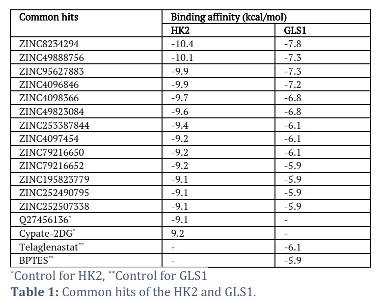
The 3-dimensional (3D) structures of HK2 (PDB ID: 5HEX) and GLS1 (PDB ID: 3VP1) were obtained from the Protein Databank [13]. To ensure the integrity and unobstructed nature of the protein structures used in molecular docking simulations, the HK2 and GLS1 protein structures underwent preprocessing using Discovery Studio (DS) 2021 software. This preprocessing involved the elimination of native ligands (Q27456136 for HK2 and BPTES for GLS1) and water molecules from the structures. The monomeric proteins were subjected to minimization and subsequently converted into the. pdbqt format in order to facilitate subsequent docking analysis.
Natural compound library retrieval and preparation
The Biopurify Phytochemicals subsets were obtained in the .sdf file format from the ZINC database. In the DS 2021 software, the ‘Prepare Ligands module’ was used to prepare a dataset of 2706 phytochemicals for subsequent docking analysis. This preparation involved performing energy minimization and applying a force field to ensure the suitability of the ligands for the docking analysis.
Molecular docking-based virtual screening
Virtual screening (VS) contributes to the expansion of compound databases by identifying active compounds and eliminating inactive ones prior to experimental validation. This method uses computational methods to analyze large datasets of three-dimensional molecule structures [14]. Autodock vina was used to screen the compound library against the enzyme active sites of HK2 and GLS1. DS 2021 accomplished further analyses of compound interactions and visual inspections, focusing on complexes with lower binding energy values to determine the most thermodynamically stable complexes.
Ligand interaction analysis
Following the docking-based VS, the resulting docked complex was subjected to additional analysis to examine the interaction between the ligand and the target proteins (HK2 and GLS1) using the ‘analyze ligand poses’ tool in DS software. The ligands’ favorable, unfavorable, total charge, the total number of hydrophobic, and hydrogen bond interactions with the active site residues of the target proteins were computed.
Results![]()
The main objective of this study was to specifically focus on two key enzymes, HK2 and GLS1, which are associated with RA. The 3D structure of both enzymes was available in the PDB database in complex with certain inhibitors, which helped to find the specific binding sites and screening of the potential ligands. In the search of potential natural inhibitors targeting both the targeted enzymes, ‘Biopurify Phytochemicals’ were utilized for the VS process.
During the screening procedure, the XYZ coordinates of the HK2 (5HEX) were established as 85.678242, 15.727273, and -102.007333, correspondingly. In addition, Q27456136 (co-crystal ligand) and a recognized inhibitor, Cypate-2DG, were employed as positive controls. Likewise, the XYZ coordinates of the GLS1 (3VP1) were determined to be 2.347000, -9.268778, and -0.893333. The co-crystal ligand BPTES and the GLS1 inhibitor Telaglenastat were used as positive controls.
During the VS process, it was observed that 34 phytochemicals exhibited higher binding energy compared to the control in the HK2 case, whereas 72 phytochemicals were identified in the GLS1 case. In the context of visual inspection and interaction analysis, it was observed that 13 compounds exhibited the best effectiveness and prevalence in targeting both HK2 and GLS1 (Table 1).
In this study, six of the 13 common ligands were described in detail with their binding interactions with the receptor molecules (Table 2). The receptor-ligand interactions of the six determined compounds with HK2 and GLS1 were found to facilitate various aspects of the interactions, such as Ligand SASA, total favorable and unfavorable interactions, charge, as well as the total number of hydrophobic and hydrogen bonds formed during the interaction.
Interaction with HK2
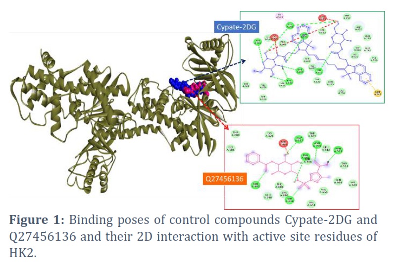
The common ligands that were found to have better efficacy in terms of binding energy were examined using 2D and 3D interaction analysis, as well as an examination of the ligand poses during receptor-ligand interactions. Figure 1 depicts the binding poses of the control compounds for HK2 enzyme, namely Cypate-2DG and Q27456136, which show that some of the residues such as ASN706, SER614, ASN683, ASP657, PRO513, PHE604, PHE602, THR680, ASN656, and others are the main binding residues in the active site of HK2.
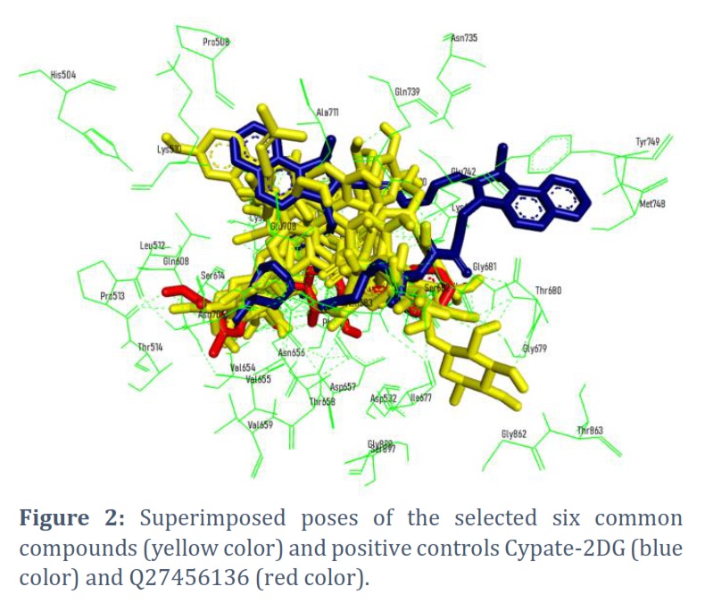
The selected six ligands, namely, ZINC8234294, ZINC49888756, ZINC95627883, ZINC4096846, ZINC4098366, and ZINC49823084 and their superimposed interaction with active site residues and the positive controls Cypate-2DG and Q27456136 were displayed in figure 2.
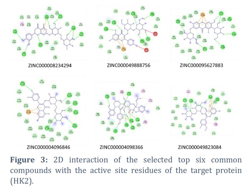
Figure 2 illustrates that all selected compounds and positive controls are located within the identical binding pocket of the HK2 enzyme. Further, each of the selected common compounds and its 2D interaction with the active site residues of the HK2 is displayed in figure 3. It is glaringly obvious that the majority of active site residues interact with the six compounds via hydrogen bonds, hydrophobic interactions, van der Waals interactions, and other types of interactions.
Interaction with GLS1
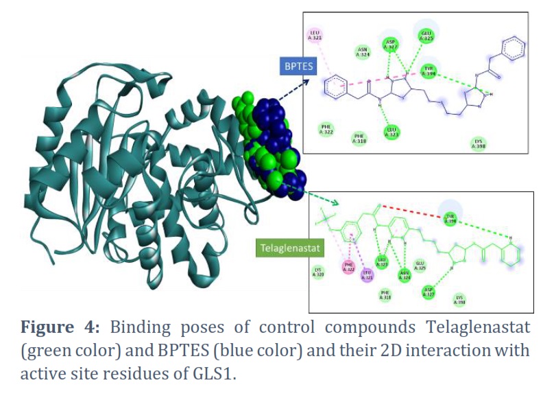
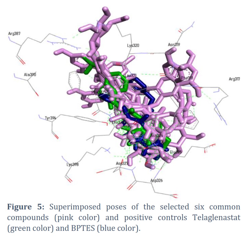
Using 2D and 3D interaction analysis, as well as an examination of the ligand poses during receptor-ligand interactions, six common ligands with greater binding energy efficiency with both receptors (HK2 and GLS1) were investigated. Figure 4 illustrates the binding conformations of the control compounds, Telaglenastat and BPTES, with the GLS1 enzyme. The binding poses reveal that specific residues, such as LEU323, ASP327, GLU325, TYR94, PHE322, LEU321, and ASN324, among others, serve as the primary binding residues within the active site of GLS1.
All six of the specified compounds—ZINC000008234294, ZINC000049888756, ZINC000095627883, ZINC000004096846, ZINC000004098366, and ZINC000049823084 as well as their superimposed interactions with GLS1’s active site residues and the positive controls Telaglenastat (green color) and BPTES were shown in Figure 5.
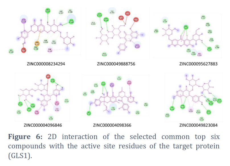
Similar to the case of HK2, Figure 6 depicts the 2D interaction between the active site residues of GLS1 and the selected common compounds. The six compounds (ZINC000008234294, ZINC000049888756, ZINC000095627883, ZINC000004096846, ZINC000004098366, and ZINC000049823084) establish interactions with most active site residues through various mechanisms, including hydrogen bonding, hydrophobic interactions, van der Waals interactions, and other means.
Figures & Tables

RA is an inflammatory disease that causes symmetrical joint inflammation, discomfort, and substantial stiffness at rest [3]. Currently, the major goal of RA therapy is to reduce inflammatory reactions and relieve pain [15]. A synergistic approach combining physical therapy and pharmaceutical therapies has shown significant success in alleviating arthritic symptoms. While NSAIDs, corticosteroids, and disease-modifying antirheumatic drugs are the cornerstones of RA treatment, assistive equipment such as braces and splints are also used to improve overall body function [16]. In extreme situations, surgical procedures aimed at the affected joints can effectively slow disease development. Nonetheless, continuous use of the aforementioned pharmacological agents has the potential for side effects such as gastrointestinal bleeding, dyspepsia, and other NSAID-related adverse reactions [17]. Therefore, finding anti-RA treatments from natural sources becomes critical in order to avoid the adverse effects associated with present medications. With this purpose this study screened the natural compounds against the HK2 and GLS1 enzyme to address the MR underlying RA.
Thirteen compounds were identified as having the highest efficacy and prevalence in simultaneously targeting both HK2 and GLS1. Six compounds from the common pool (ZINC000008234294, ZINC000049888756, ZINC000095627883, ZINC000004096846, ZINC000004098366, and ZINC000049823084) were examined in detail to determine their binding interactions with the receptor molecules (HK2 and GLS1). These compounds interact with both HK2 and GLS1 proteins, primarily at active site residues, using a variety of mechanisms including hydrogen bonding, hydrophobic interactions, van der Waals interactions, and other molecular affinities.
Natural products and their derivatives have played an important role in the discovery of therapeutic agents throughout history [18]. These natural compounds are distinguished by their inherent ability to interact with biologically significant protein targets. Their remarkable structural diversity and biological activities continue to inspire the development of both small-molecule [19] and macrocyclic drugs [20]. Natural products have had a significant impact on the pharmaceutical landscape, serving as a primary source of novel therapeutics in the drug development pipeline since the mid-1970s. Specifically, from the 1980s to the 2010s, approximately two-thirds of pharmaceutical drugs were derived from unmodified natural products (5%), derivatives of natural products (28%), or compounds containing pharmacophores derived from natural products (35%) [21].
RA is a chronic, inflammatory autoimmune disease that primarily affects joints. In RA, MR increases glycolysis and glutaminolysis, as well as HK2 and GLS1 expression. This study tested phytochemicals from the ZINC database against the HK2 and GLS1 proteins. The compounds ZINC000008234294, ZINC000049888756, ZINC000095627883, ZINC000004096846, ZINC000004098366, and ZINC000049823084 interacted strongly with the majority of the active site residues of the HK2 and GLS1 proteins. By targeting the HK2 and GLS1, these compounds could be potential drug candidates for MR in RA.
Acknowledgement
The author wishes to thank the Physiotherapy Clinic at the General Administration of Medical Services at the University of Tabuk for their support.
Conflict of Interest
The authors declare that there is no conflict of interest.
YMA: Conceptualization, Data collection/curation, reviewing, editing and writing the manuscript.
![]() References
References
- Bullock J, Rizvi SAA, Saleh AM, Ahmed SS, Do DP, et al. Rheumatoid Arthritis: A Brief Overview of the Treatment. Medical Principles and Practice, (2018); 27(6): 501-507.
- Li TP, Zhang AH, Miao JH, Sun H, Yan GL, et al. Applications and potential mechanisms of herbal medicines for rheumatoid arthritis treatment: a systematic review. RSC Advances, (2019); 9(45): 26381-26392.
- Angelotti F, Parma A, Cafaro G, Capecchi R, Alunno A, Puxeddu I. One year in review 2017: pathogenesis of rheumatoid arthritis. Clinical and Experimental Rheumatology, (2017); 35(3): 368-378.
- Gibofsky A. Overview of epidemiology, pathophysiology, and diagnosis of rheumatoid arthritis. American Journal of Managed Care, (2012); 18(13 Suppl): S295-302.
- Nemtsova MV, Zaletaev DV, Bure IV, Mikhaylenko DS, Kuznetsova EB, et al. Epigenetic Changes in the Pathogenesis of Rheumatoid Arthritis. Frontiers in Genetics, (2019); 10570.
- Kahlenberg JM, Fox DA. Advances in the medical treatment of rheumatoid arthritis. Hand Clinics, (2011); 27(1): 11-20.
- Conigliaro P, Triggianese P, De Martino E, Fonti GL, Chimenti MS, et al. Challenges in the treatment of Rheumatoid Arthritis. Autoimmun Reviews, (2019); 18(7): 706-713.
- Sanchez-Lopez E, Cheng A, Guma M. Can Metabolic Pathways Be Therapeutic Targets in Rheumatoid Arthritis? Journal of Clinical Medicine, (2019); 8(5).
- Biniecka M, Canavan M, McGarry T, Gao W, McCormick J, et al. Dysregulated bioenergetics: a key regulator of joint inflammation. Annals of the Rheumatic Diseases, (2016); 75(12): 2192-2200.
- Araujo L, Khim P, Mkhikian H, Mortales CL, Demetriou M. Glycolysis and glutaminolysis cooperatively control T cell function by limiting metabolite supply to N-glycosylation. Elife, (2017); 6.
- Garcia-Carbonell R, Divakaruni AS, Lodi A, Vicente-Suarez I, Saha A, et al. Critical Role of Glucose Metabolism in Rheumatoid Arthritis Fibroblast-like Synoviocytes. Arthritis & Rheumatology, (2016); 68(7): 1614-1626.
- Bustamante MF, Oliveira PG, Garcia-Carbonell R, Croft AP, Smith JM, et al. Hexokinase 2 as a novel selective metabolic target for rheumatoid arthritis. Annals of the Rheumatic Diseases, (2018); 77(11): 1636-1643.
- Berman HM, Westbrook J, Feng Z, Gilliland G, Bhat TN, et al. The Protein Data Bank. Nucleic Acids Research, (2000); 28(1): 235-242.
- Maia EHB, Assis LC, de Oliveira TA, da Silva AM, Taranto AG. Structure-Based Virtual Screening: From Classical to Artificial Intelligence. Frontiers in Chemistry, (2020); 8343.
- Sun H, Zhang AH, Song Q, Fang H, Liu XY, et al. Functional metabolomics discover pentose and glucuronate interconversion pathways as promising targets for Yang Huang syndrome treatment with Yinchenhao Tang. RSC Advances, (2018); 8(64): 36831-36839.
- Allen A, Carville S, McKenna F, Guideline Development G. Diagnosis and management of rheumatoid arthritis in adults: summary of updated NICE guidance. BMJ, (2018); 362k3015.
- Mota LM, Cruz BA, Brenol CV, Pereira IA, Rezende-Fronza LS, et al. Guidelines for the drug treatment of rheumatoid arthritis. Revista Brasileira de Reumatologia, (2013); 53(2): 158-183.
- Calixto JB. The role of natural products in modern drug discovery. Annals of the Brazilian Academy of Sciences, (2019); 91 Suppl 3e20190105.
- Rodrigues T, Reker D, Schneider P, Schneider G. Counting on natural products for drug design. Nature Chemistry, (2016); 8(6): 531-541.
- Driggers EM, Hale SP, Lee J, Terrett NK. The exploration of macrocycles for drug discovery–an underexploited structural class. Nature Reviews Drug Discovery, (2008); 7(7): 608-624.
- Newman DJ, Cragg GM. Natural Products as Sources of New Drugs over the Nearly Four Decades from 01/1981 to 09/2019. Journal of Natural Products, (2020); 83(3): 770-803.
This work is licensed under a Creative Commons Attribution-Non Commercial 4.0 International License. To read the copy of this license please visit: https://creativecommons.org/licenses/by-nc/4.0