

Full Length Research Article
Sphingomyelin, Plasminogen, and Docosahexaenoic in Sera of Autism Spectrum Disorder Children
Ali Fadheel Hamoud1, Narjis Hadi Al-Saadi2*
Adv. life sci., vol. 11, no. 1, pp. 226-232, February 2024
*– Corresponding Author: Narjis Hadi Al-Saadi (narjis.h@uokerbala.edu.iq)
Authors' Affiliations
2. Department of Chemistry, College of Science, Karbala University, Karbala – Iraq
[Date Received: 03/08/2023; Date Revised: 09/02/2024; Date Published: 25/02/2024]
Abstract![]()
Introduction
Methods
Results
Discussion
References
Abstract
Background: Autism spectrum disorder (ASD), more commonly referred to as autism, is a neurodevelopmental disorder that is pervasive, highly heritable, and extremely variable. It is characterized by underlying cognitive features that frequently co-occur with other conditions. Since ASD is a multifactor disease, genetics, and environmental factors can play crucial roles in its progression. However, very few biological parameters can be used as a prediction for ASD which can help in diagnosis and starting the treatment early. Given the rapidly increasing prevalence of ASD, there is an urgent need to identify related diagnostic biomarkers. This study aims to investigate the association between some blood parameters that can be used to predict ASD and classify the severity, which were the main aims of the current inquiry.
Method: A case-control study was conducted on children with ASD, 37 Kids with ASD participated in the current investigation and 46 kids as the control group, their ages were between 3-12 years. Children with ASD were divided into two subgroups depending on the severity of ASD using the Gilliam scale. Competitive and sandwich ELISA were used to measure the biochemical markers of this study.
Result: After blood samples were collected three parameters were measured (sphingomyelin, plasminogen, and docosahexaenoic acid). In medium ASD cases, the results display that there is a significant increase in all parameters (sphingomyelin, plasminogen, and docosahexaenoic acid) respectively [(OR:4.691, CI:1.289~17.068, p=0.014), (OR:7.5, CI:1.844~30.509, p=0.001), (OR:5.156, CI:1.412~18.831, p=0.001)]. On other hand, in under medium cases of ASD, there is a significant decrease in Sphingomyelin levels (OR:0.97, CI:0.356~0.836, p=0.001), plasminogen (OR: 0.5, CI: 0.169~0.560, p=0.05), and docosahexaenoic (OR: 0.22, CI: 0.63~1.771, p=0.003) when compared with the control group.
Conclusion: In sum, our results showed that these noninvasive parameters can be used as biomarkers for ASD diagnosis and disease propagation. More research needs to be done to cover other pathophysiology parameters with genetics analysis for ASD that can be used as prediction biomarkers.
Keywords: Autism spectrum disorder; Sphingomyelin; Plasminogen; Docosahexaenoic acid
Introduction![]()
referred to as autism, is a neurodevelopmental spectrum disorder (NDSD) characterized by a wide range of symptoms and underlying cognitive features that frequently co-occur with other diseases[1]. The strengths and weaknesses of autistic persons have captivated academics and doctors for at least 500 years. Since its inclusion as a subtype within the general diagnostic category of ‘pervasive developmental disorders (PDDs),’ the term autism has been used in a variety of ways to represent both a broader presentation and a specific diagnosis. In 1980, the DSM-III expanded its list of mental disorders to include pervasive developmental disorders (PDDs), which encompass a wider spectrum of issues with social communication[2]. Recent diagnostic systems like ICD-11 and DSM-5 use the umbrella term ‘ASD’ and differentiate individuals using additional clinical specifiers and modifiers because there are no clear borders between the PDDs and it is difficult to reliably distinguish them [3].
Autistic individuals may have varying degrees of intellectual disability, problems with social communication and interaction, sensory abnormalities, repetitive habits, and more [4]. It is not uncommon for people with autism to also suffer from a mental or neurological problem, such as epilepsy, anxiety, depression, or a hyperactivity/attention disorder like attention-deficit/hyperactivity disorder (ADHD)[5]. Diagnosis of autism requires gathering information about a person’s development, usually from the parents, and seeing how they interact with others, both in their immediate environment and in social situations1,2. Early intervention is essential for children with autism due to the prevalence of common communication difficulties [6]. Interventions are used at all stages of life, from parent-mediated and/or therapist-delivered interventions in childhood to strategies and procedures learned in school that foster independence in adulthood. Medications help alleviate some of autism’s secondary symptoms, like irritability and comorbidities like anxiety [7].
Human brain development is said to be at its peak between the ages of 0 and 2. However, not all areas of the brain are fully developed by age two and continue to grow and change throughout childhood and adolescence.[8]. Docosahexaenoic acid (DHA, 22:6n-3) is a long-chain omega-3 polyunsaturated fatty acid (n-3 LC-PUFA) that is essential for normal brain development in humans. The frontal lobes, where DHA is highly concentrated, are responsible for executive function and higher-order cognitive activities including planning, problem solving, and paying attention. [9]. High-level cognitive development in children is closely correlated with their emotional, social, and behavioral maturation, a phenomenon that has been the subject of numerous studies [10, 11]. Because of its unusual structure and numerous double bonds, DHA plays a crucial function in signaling in nerve cells [12]. DHA also features six double bonds among its 22 carbon atoms. Instead of having double bonds and methylene units (-CH-) dispersed randomly along the carbon chain, more than 95% of PUFA in mammalian tissue is homoallylic [13].
The most abundant eukaryotic sphingolipid and cell membrane component is sphingomyelin (SM) [14]. It is prevalent in the central nervous system, and the myelin sheath that surrounds neuronal axons has a unique structure [15]. The SM has been reported to play an important role in cell processes [16] and signal transduction [17]. Finally, it has a critical effect on brain development in human infants [18]. Sphingolipid metabolism is assumed to be a crucial pathway in neurodegeneration and neuroinflammation due to the roles of Sphingolipids in many vital biological processes and their large abundance in the central nervous system as major components of oligodendrocytes and myelin sheaths [19].
Plasminogen (PLG) is another component that can influence the course of ASD. Several enzymes first synthesize PLG in the liver, and then they activate it. Interestingly, PLG has been linked to fibrinolysis, wound healing, cell signaling, and inflammatory regulation [20].
Methods![]()
Study Design
A case-control study was carried out on the ASD cohort. After excluding other mental disorders (Down syndrome, obsessive-compulsive disorder, mental disorders, or any other psychiatric or neurological diseases), thirty-seven children with autism, aged 3-12 years (mean ± SD = 6.35±2.37 years) who trained at Hamaem Al-Salam Center in Iraq/Najaf, and 46 healthy children as the control group involved in current study. The ages were matched in the two groups (p =0.929). The Gilliam scale was used to determine the severity of ASD.
Ethics
After the parents signed the consent form, all samples were collected from the cohorts. Furthermore, the Karbala University Ethics Committee process was followed in current project under the form number 6633257/KUEC.
Blood specimen collection
Under aseptic technique, 5 mL of venous blood samples were collected from children with ASD and healthy children, then placed in gel-tubes at room temperature to clot before being centrifuged at 3000 g for 10 minutes. The serum was extracted and stored in an Eppendorf tube at – 60°C until use.
Evaluation of biochemical markers
The concentration of serum sphingomyelin, plasminogen, and DHA were measured by using a competitive and sandwich ELISA kit (BT LB Company, China).
Statistical analysis
The Statistical Package of Social Sciences (SPSS) version 26 (IBM) was used to examine all of the data. The Mann-Whitney U test was employed for abnormal distribution and an independent t-test was utilized for normal distribution of variables based on normality distribution. The Spearman's rank correlation coefficient (r) was utilized to determine the relationship between the factors under consideration. For all statistical tests employed in this investigation, a p-value of 0.05 or less was considered statistically significant.
Results![]()
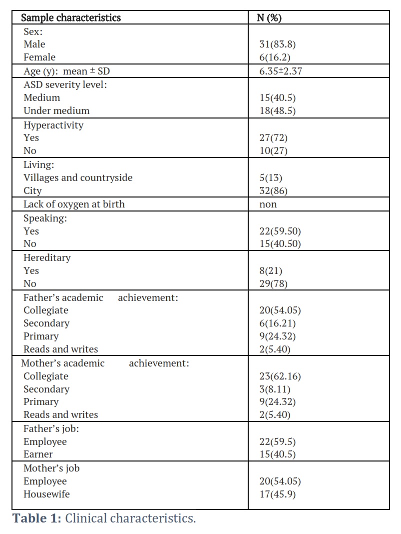
Characteristic features:
First, the clinical characteristics included sex, age, ASD level, hyperactivity, living, speaking, and family history of ASD summarized in (Table 1).
Blood parameters under study
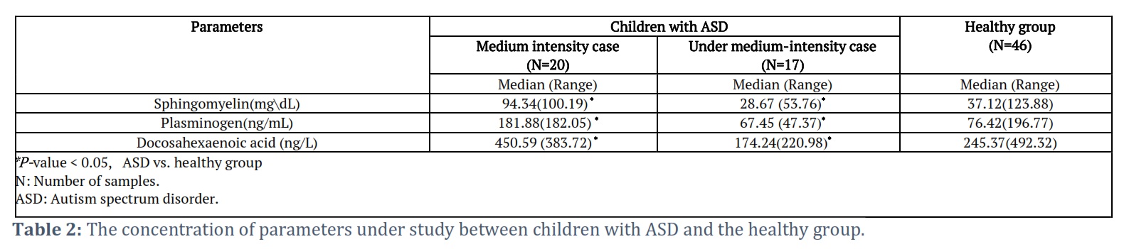
Second, the three biomarkers measured in the serum that been collected from ASD and control groups. In details, medium-intensity cases of children with ASD classification, the results that presented in Table 2 show a significant increase in levels of sphingomyelin (OR:4.691, CI:1.289~17.068, p=0.014), plasminogen (OR:7.5, CI:1.844~30.509, p=0.001), and docosahexaenoic acid (OR:5.156, CI:1.412~18.831, p=0.001) when compared them with control group. In contrast, the level of these biomarkers in under medium-intensity group, showed a significant decrease in levels of sphingomyelin (OR:0.97, CI:0.356~0.836, p=0.001), plasminogen (OR: 0.5, CI: 0.169~0.560, p=0.05), and docosahexaenoic acid (OR: 0.22, CI: 0.63~1.771, p=0.003).
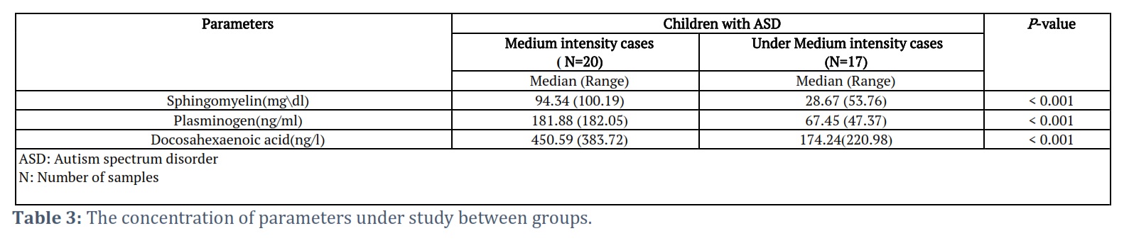
Biomarker variations across ASD subtypes have been evaluated by comparing two groups (Medium intensity and under medium-intensity group). According to the findings, all parameters in the medium intensity group are significantly higher than those in the low intensity group (p<0.001) (Table 3).
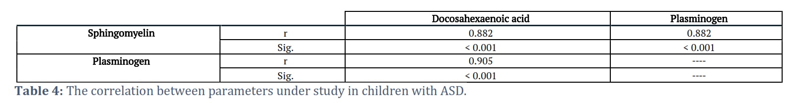
To examine if there is a link between the blood biomarkers. Table (4) shows the Spearman's correlation coefficient (r) used to determine the associations between the variables (sphingomyelin, plasminogen, and docosahexaenoic acid). Surprisingly, the results revealed a significant strong association between these variables (p< 0.001).
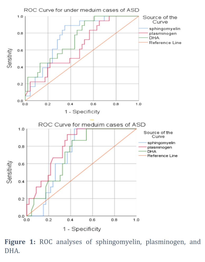
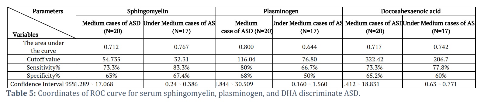
The optimum cutoff points for serum Sphingomyelin, Plasminogen, and Docosahexaenoic acid levels were presented in Table (5) along with the area under the ROC curve (AUC) and sensitivity, and specificity shown in Figure (1). The value of the area under the curve for most parameters under study ranged between (0.7 to 0.8), also the percentage of sensitivity and specificity for most parameters ranged between 70% to 80% may be used this parameter as a biomarker to diagnose ASD in children after further study.
Figures & Tables
Autism spectrum disorder (ASD) has been one of the most puzzling disorders of the previous decade. Sphingomyelin, plasminogen, and docosahexaenoic acid (DHA) are three characteristics that have received little attention while being highlighted as biomarkers for ASD.
The primary goal of this study is to establish a connection between these variables and two forms of autism spectrum disorder. Initiate with When comparing the ASD medium group with the control group, the results show that the ASD medium group has a higher concentration of Sphingomyelin, which may lead to hyperactivity in children. These findings are supported by a very interesting Vivo experiment in which a mouse model was used to evaluate the effect of different doses of propionic acid (PPA). After receiving the medication, these mice became extremely active [21]. Alterations in brain lipid profiles are thought to be a risk factor for autism spectrum disorder. Consequently, alterations in brain lipids have been linked to ASD. The brain’s lipid profile can be altered by two different routes: an increase in dietary and enteric short-chain fatty acids. Therefore, using a rodent model, we may link alterations or increases in brain sphingomyelin to the development of ASD-like behaviors. Additionally, ASD may be influenced by a change in the way the brain processes fatty acids [22]. The particular sphingomyelin found in brain lipids is crucial to the growth and maturation of the brain and the mind. Thus, any reduction may have an effect on mental growth. Sphingomyelin concentration was low in the ASD medium group when compared to the control group. Because sphingomyelin supports brain myelination, a process strongly related with cognitive maturation and growth in the brain [23], decreased sphingomyelin concentration has been shown to play a role in ASD development [24].
Plasminogen, via increasing the activity of tissue plasminogen activators, is a second important factor in the development of ASD. The results demon start that plasminogen levels are higher in children with ASD than in control group. Furthermore, Hasan Bozkurt's 2021 found that any increase in blood Plasminogen in male ASD children will co-incidence with increase in tissue Plasminogen activator (tPA) [25]. In other words, any elevation of blood Plasminogen led to increase is tPA.
Inside the blood vessel, the main function of tPA is a thrombolytic enzyme, and its main star get is plasminogen. However, By contrast, in the brain parenchyma, tPA has been associated with multiple physiological and pathological events including synaptic plasticity and cell death[26]. Also, Tsirka et al. reported that t-PA-deficient mice are resistant to neural degeneration [27]. In a recent study, t-PA is involved in social behavior[28]. The results obtained support Bauman & Kemper, 1994 hypothesis about the overgrowth in limbic structures, the overgrowth, and decreased growth of the cerebellum in male ASD patients [29].
N-3 fatty acids have various advantages, but they can also have negative side effects, such as enhanced lipid peroxidation [30]. Docosahexaenoic acid (DHA) is highly oxidized when it is found in high concentration, and the presence of its six double bonds in the 22-carbon fatty acyl chain increases its susceptibility to oxidation [31].
The quantity of pentadiene moieties contained in polyunsaturated fatty acids (PUFAs), on the other hand, is widely known to be strongly connected to the rate of oxidation. As a result, membranes enriched in DHA are more vulnerable to oxidative damage, and membrane oxidative changes have numerous potential repercussions in human pathophysiology. DHA levels were significantly higher in the ASD medium group than in the control group. DHA may fulfill a vital role in antioxidant pathways under certain conditions. Because DHA exhibits double bonds, three elements may mitigate oxidative stress by binding with it. Phosphatidylethanolamine plasmalogens (PE plasmalogens), diacyl-glycerophosphoethanolamine (diacyl-GPE), and phosphatidylcholine (PC) are the chemical structures. remarkably, any rise in DHA blood levels means an increase in the affinity of binding between those elements, and hence a reduction in DHA's oxidation. DHA has already been shown to accumulate in overabundance and it has been proposed that DHA when bound to PC, is related to diminished cell growth and enhanced cytotoxicity [32].
Any fluctuations in plasma N-3 fatty acid levels, particularly DHA, have catastrophic consequences for the human brain. Furthermore, epidemiological data suggest that populations with lower levels of n-3 fatty acids, notably DHA, in plasma and brain tissue have greater prevalence of psychiatric disorders, such as depression and bipolar disorder [33, 34]. The plasma level of DHA was low in ASD under medium group compared with control group. In addition, children with hyperactivity disorder and adults with schizophrenia have been shown to have lower plasma phospholipid and DHA contents than the control[35].
In summary, the current study found that all the parameters have a good sensitivity and specificity for ASD diagnosis when compared to the control group. In addition, these biomarkers consider risk factors for children with ASD. To unravel all the mysteries of ASD, Genetic approaches and other biochemical tests need to be developed.
Author Contributions
Study design and conception, Proofreading, and Statistical analysis by Narjis Hadi Al-Saadi
Material preparation, Data collection and analysis, and a first draft was written by: Ali Fadheel Hamoud
Interpretation of the results by Narjis Hadi Al-Saadi & Ali Fadheel Hamoud
The authors declare that there is no conflict of interest.
![]() References
References
- Gontard A Von, Hussong J, Yang SS, Chase J, Franco I, Wright A. Aims Mathematics, (2021);41(1):1–9.
- Rosen NE, Lord C, Volkmar FR. The Diagnosis of Autism: From Kanner to DSM-III to DSM-5 and Beyond. Journal of autism and developmental disorders,(2021);51(12):4253–70.
- Lord C, Brugha TS, Charman T, Cusack J, Dumas G, Jones EJH, et al. Autism spectrum disorder Access options Access Nature and 54 other Nature Portfolio journals, (2020);6(1):1–46.
- Zhu J, Guo M, Yang T, Lai X, Tang T, Chen J, et al. Nutritional Status and Symptoms in Preschool Children With Autism Spectrum Disorder: A Two-Center Comparative Study in Chongqing and Hainan Province, China.Frontiers in Pediatrics, (2020);8(469):1–28.
- Patel, Goyena R. Prevalence of Depression in Children with Dyslexia, Attention Deficit Hyperactivity Disorder, and High Functioning Autism Spectrum Disorder. Journal of Chemical Information and Modeling, (2019);15(2):9–25.
- Okoye C, Obialo-Ibeawuchi CM, Obajeun OA, Sarwar S, Tawfik C, Waleed MS, et al. Early Diagnosis of Autism Spectrum Disorder: A Review and Analysis of the Risks and Benefits. Cureus, (2023);15(8):1–14.
- Bradshaw J, Steiner AM, Gengoux G, Koegel LK. Feasibility and Effectiveness of Very Early Intervention for Infants At-Risk for Autism Spectrum Disorder: A Systematic Review. Journal of autism and developmental disorders, (2015);45(3):778–94.
- Emekli-Alturfan E, Alturfan AA. The emerging relationship between vitamin K and neurodegenerative diseases: a review of current evidence. Molecular Biology Reports, (2023);50(1):815–28.
- Brown TT, Jernigan TL. Brain development during the preschool years. Neuropsychology Review, (2012);22(4): 313–333.
- Anderson V, Fenwick T, Manly T, Robertson I. Attentional skills following traumatic brain injury in childhood: A componential analysis, Brain Injury, (1998);12(11):937–49.
- Barkley RA. The Executive Functions and Self-Regulation: An Evolutionary Neuropsychological Perspective. Neuropsychol Review, (2001);11(1):1–29.
- Khalid W, Gill P, Arshad MS, Ali A, Ranjha MMAN, Mukhtar S, et al. Functional behavior of DHA and EPA in the formation of babies brain at different stages of age, and protect from different brain-related diseases. International Journal of Food Properties, (2022);25(1):1021–44.
- Sun GY, Simonyi A, Fritsche KL, Chuang DY, Hannink M, Gu Z, et al. Docosahexaenoic acid (DHA): An essential nutrient and a nutraceutical for brain health and diseases. Prostaglandins, Leukotrienes and Essential Fatty Acids, (2018);136(September):3–13.
- Brenna JT, Carlson SE. Docosahexaenoic acid and human brain development: Evidence that adietary supply is needed for optimal development. Journal of Human Evolution, (2014);77(December):99–106.
- Gault CR, Obeid LM, Hannun YA. An overview of sphingolipid metabolism: From synthesis to breakdown. Advances in Experimental Medicine and Biology , (2010);688(April):1–23.
- Babin F, Sarda P, Limasset B, Descomps B, Rieu D, Mendy F, et al. Nervonic acid in red blood cell sphingomyelin in premature infants: An index of myelin maturation? National Center for Biotechnology Information, (1993);28(7):627–30.
- Smith SM, Nichols TE. Threshold-free cluster enhancement: Addressing problems of smoothing, threshold dependence and localisation in cluster inference. Neuroimage, (2009);44(1):83–98.
- Merrill AH, Sandhoff K. Chapter 14 Sphingolipids: metabolism and cell signaling. New Comprehensive Biochemistry, (2002);36(02):373–407.
- Emekli-Alturfan E, Alturfan AA. The emerging relationship between vitamin K and neurodegenerative diseases: a review of current evidence, Molecular Biology Reports, (2023);50(1):815–28.
- Lingwood D, Simons K. Lipid rafts as a membrane-organizing principle. Science (80- ), (2010);327(5961):46–50.
- Schneider N, Hauser J, Oliveira M, Cazaubon E, Mottaz SC, O’Neill B V., et al. Sphingomyelin in brain and cognitive development: Preliminary data. eNeuro, (2019);6(4):1–27.
- Baker SK, Chen ZL, Norris EH, Revenko AS, MacLeod AR, Strickland S. Blood-derived plasminogen drives brain inflammation and plaque deposition in a mouse model of Alzheimer’s disease. Proceedings of the National Academy of Sciences,(2018);115(41):E9687–96.
- Schneider N, Hauser J, Oliveira M, Cazaubon E, Mottaz SC, O’Neill B V., et al. Sphingomyelin in brain and cognitive development: Preliminary data. eNeuro, (2019);6(4):1–27.
- Ju J, Yang X, Jiang J, Wang D, Zhang Y, Zhao X, et al. Structural and Lipidomic Alterations of Striatal Myelin in 16p11.2 Deletion Mouse Model of Autism Spectrum Disorder. Frontiers in Cellular Neuroscience, (2021);15(August):1–25.
- Dong MX, Li CM, Shen P, Hu QC, Wei YD, Ren YF, et al. Recombinant tissue plasminogen activator induces long-term anxiety-like behaviors via the ERK1/2-GAD1-GABA cascade in the hippocampus of a rat model. Neuropharmacology, (2018);128(5):119–31.
- Bozkurt H, Şimşek Ş, Şahin S. Elevated levels of cortisol, brain-derived neurotropic factor and tissue plasminogen activator in male children with autism spectrum disorder. Autism Research, (2021);14(10):2078–84.
- Yepes M, Roussel BD, Ali C, Vivien D. Tissue-type plasminogen activator in the ischemic brain: more than a thrombolytic.Trends in Neurosciences, (2009) ;32 (1) : 48–55.
- Tsirka SE, Gualandris A, Amaral DG, Strickland S. Excitotoxin-induced neuronal degeneration and seizure are mediated by tissue plasminogen activator. Nature, (1995);377(6547):340–4.
- Şimşek Ş, Çetin İ, Çim A, Kaya S. Elevated levels of tissue plasminogen activator and E-selectin in male children with autism spectrum disorder. Autism Research, (2016);9(12):1241–7.
- Wagner BA, Burns CP, Buettner GR. Free Radical-Mediated Lipid Peroxidation in Cells: Oxidizability Is a Function of Cell Lipid bis-Allylic Hydrogen Content. Biochemistry, (1994);33(15):4449–53.
- Field CJ, Johnson IR, Schley PD. Nutrients and their role in host resistance to infection. Journal of Leukocyte Biology, (2002);71(1):16–32.
- Véricel E, Polette A, Bacot S, Calzada C, Lagarde M. Pro- and antioxidant activities of docosahexaenoic acid on human blood platelets. Thrombosis and Haemostasis, (2003);1(3):566–72.
- Cai J, Leung PS. Unlocking the potential of aquatic foods in global food security and nutrition: A missing piece under the lens of seafood liking index. Global Food Security, (2022);33(14):1–67.
- Bragg MG, Prado EL, Stewart CP. Choline and docosahexaenoic acid during the first 1000 days and children’s health and development in low- and middle-income countries. Nutrition Reviews, (2022);80(4):656–76.
- Song W, Zhang K, Xue T, Han J, Peng F, Ding C, et al. Cognitive improvement effect of nervonic acid and essential fatty acids on rats ingesting Acer truncatum Bunge seed oil revealed by lipidomics approach. Food & Function, (2022);13(5):2475–90.
This work is licensed under a Creative Commons Attribution-Non Commercial 4.0 International License. To read the copy of this license please visit: https://creativecommons.org/licenses/by-nc/4.0